
¿Cuál es su diagnóstico?
What should the diagnosis be?
Paciente mujer de 52 años, sin antecedentes de interés, que acude a nuestra consulta, remitida por su odontólogo del Centro de Salud, por presentar una tumoración a nivel mandibular derecho, que no produce dolor ni alteraciones de la sensibilidad. En la
radiografía panorámica se observa una radiolucidez a nivel mandibular, que ocupa desde pieza 4,1 hasta la 4,5. A la exploración física se observa el abombamiento a nivel mandibular, sin movilidad dentaria, ni dolor a la percusión dental, ni aparente alteración de la mucosa oral.
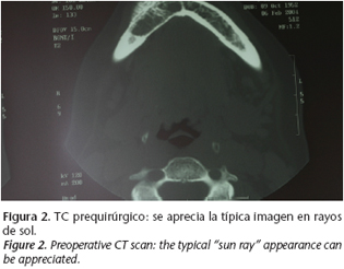
En la TC se aprecia una lesión en la parte anterior derecha mandibular, en la región basal, de 3 x 1,7 centímetros. Los márgenes internos con aparente patrón geográfico de bordes mal definidos que disminuyen el córtex adyacente. En el interior hay imágenes de trabeculación. Reacción perióstica en el borde anterior de la lesión mandibular, similar a rayos de sol. No se observan masa en las partes blandas ni alteraciones en las raíces dentales. El diagnóstico diferencial no incluye las lesiones óseas quísticas, debido a la reacción perióstica. No impresiona de malignidad por la nula afectación de las partes blandas.
Intraosseous hemangioma of the mandible. An intraoral approach
R. Luaces Rey1, , A. García-Rozado González2, J.L. López-Cedrún Cembranos3, J. Ferreras Granado2, , E Charro Huerga1
1 Médico Residente
2 Médico Adjunto
3 Jefe de Servicio ]]>
Dirección para correspondencia
El diagnóstico de sospecha radiológico es el de hemangioma intramandibular, por lo anteriormente descrito, y por presentar el canal vascular en la proximidad y aumentado de tamaño, en comparación con el contralateral.
Se realiza un estudio preoperatorio, que no contraindica la intervención quirúrgica. Bajo anestesia general, mediante un abordaje intraoral, con incisión en fondo de vestíbulo inferior derecho, identificando y preservando el nervio mentoniano derecho, se realiza exéresis de la tumoración, ligando con seda la arteria nutricia, y aplicando en el lecho quirúrgico Tissucol® y Surgicel®. La paciente presentó un postoperatorio sin complicaciones. El informe anatomopatológico de la pieza fue de hemangioma cavernoso de mandíbula y lleva un seguimiento en consultas externas de 13 meses, con buena evolución. A los 9 meses tras la intervención se le ha realizado una TC de control, en la que se aprecia el defecto quirúrgico en proceso de regeneración, sin signos de recidiva local.
Discusión
Los hemangiomas intraóseos son lesiones benignas, debidos a la proliferación de vasos sanguíneos, muy poco frecuentes. Suponen un 0,2% de neoplasias óseas, siendo las vértebras y el cráneo sus principales lugares de asiento.1 La mandíbula y el maxilar son los siguientes lugares de aparición. Más de la mitad de los hemangiomas centrales de los maxilares ocurren en la mandíbula, siendo la región posterior su asiento más frecuente.2,3
Su etiología es desconocida, pero se sospecha que hay algunas que son neoplasias reales, y otras que tienen un origen traumático. 2 La distribución por sexos presenta el doble de frecuencia en mujeres que en hombres, siendo el pico de incidencia la 2ª década de la vida.2, 4
]]> La mayoría son hallazgos casuales radiográficos,5 sin dar clínica alguna,2-4 si bien es cierto que en otras ocasiones produce erosión y reabsorción de los dientes,5 (produciendo movilidad de los dientes afectados), desfiguración facial por la expansión asimétrica de lento crecimiento del maxilar o de la mandíbula, dolor o parestesias. Está descrito que pueda detectarse la pulsación del hemangioma a la auscultación o a la palpación de las tablas óseas adelgazadas, sangrado gingival espontáneo alrededor de los dientes de la zona o bien incluso puede debutar como un sangrado catastrófico tras una extracción de un diente,6 asociado a un hemangioma o por la realización de una biopsia.7 Para evitar estos últimos accidentes es muy importante tener un diagnóstico de sospecha, apoyado en las pruebas de imagen.24Teniendo en cuenta que las biopsias están contraindicadas debido al alto riesgo de sangrado, su diagnóstico de sospecha se realiza mediante las pruebas radiológicas y la clínica, si bien es muy importante tener en cuenta que su presentación radiográfica es muy variable,2-4 (hay descritas hasta doce apariencias distintas)8 e inespecífica. El ensanchamiento del canal del nervio dentario inferior sugiere un origen vascular de la lesión8. Hay autores que sostienen que en la mitad de los casos se observa un margen esclerótico. 9 Pueden aparecer como radiolucencia multiloculada, con apariencia similar a pompas de jabón. También como una lesión radiolúcida redondeada en la que se observa el hueso trabecular en el centro. Es menos común su aparición como radiolucidez similar a un quiste.10
El diagnostico diferencial debe de hacerse con otras entidades con apariencia radiológica compatible, como el quiste dental residual, queratoquiste odontogénico, quiste óseo aneurismático, ameloblastoma, mixoma odontogénico, granuloma de células gigantes, y otras patologías similares.10
Previamente al tratamiento, es muy importante el evaluar tres factores: el tamaño, la localización, y el aporte vascular. A partir de ahí comienza el conflicto, puesto que están descritos distintos procedimientos terapéuticos: la cirugía, la ligadura de la arteria carótida externa, la radioterapia,11 la inoculación de agentes esclerosantes, 12 la crioterapia y la embolización.13 En ocasiones resulta muy útil la realización de una angiografía antes de decidirnos por alguno de ellos.
Aunque está descrita con éxito la utilización de la embolización de forma aislada13 (que en ocasiones es necesario repetir para una completa devascularización), tal vez el tratamiento óptimo consista en la realización de una angiografía superselectiva con embolización, y si es necesario seguido de una escisión quirúrgica, si bien no debe de trascurrir más de dos semanas entre ambas para evitar la apertura de colaterales.14 La embolización no es un proceso inocuo, estando descritos entre sus complicaciones la suelta de ateromas, la rotura de catéteres, la rotura de vasos,15 hemiplejía, ceguera, parálisis facial,16 reacciones alérgicas Está documentado el riesgo de necrosis avascular de la mandíbula tras la opción terapéutica de embolización seguido de cirugía.17 Sería muy interesante durante la cirugía se pudiese conseguir una hipotensión controlada mantenida, por parte del anestesista, y el mantenimiento de hipotermia regulada, con el fin de reducir las perdidas sanguíneas.18
Por lo general, las arterias de las que depende el hemangioma proceden principalmente, aunque no de forma exclusiva, de la arteria carótida externa. A pesar de eso, hay que tener muy en cuenta que al ligar la carótida externa, esto provoca la apertura de colaterales provenientes de la arteria carótida externa contralateral,5,16,19 (que pueden resultar inaccesibles a la embolización con catéteres), o en la arteria carótida interna ipsilateral,5 (lo que podría producir un aumento de la lesión, debido al aumento de presión). Es decir que de la ligadura de la A.C.E. no puede esperarse que controle un sangrado agudo o que produzca una adecuada devascularización de una lesión vascular.20-22
Aunque está descrita la utilización de agentes esclerosantes inyectados, pueden resultar ciertamente muy peligrosos, debido a la proximidad del sistema vascular intracraneal.12 La radioterapia puede resultar fallida debido al nivel de maduración de las células de la malformación, y al riesgo de degenerar en sarcoma, producir osteoradionecrosis, retraso en el desarrollo de huesos y dientes y al complicar una posible posterior intervención quirúrgica.23
El diagnóstico definitivo nos lo da el análisis anatomopatológico de la tumoración. La mayoría son de tipo cavernoso, en los que los espacios vasculares dilatados, de paredes finas, son rodeados de células endoteliales benignas. También es posible que sean canales vasculares numerosos, y pequeños, del tamaño de capilar. Si bien, puede ser una mezcla de ambos subtipos.2-4
A pesar de que se trata de una entidad con características de benignidad, está aconsejado su tratamiento precoz para evitar posibles complicaciones derivadas de su crecimiento local, que podrían incluso ensombrecer su pronóstico por un sangrado catastrófico. Con un correcto tratamiento, el pronóstico de estas lesiones es bueno, con regeneración ósea de la zona afectada en los siguientes meses. Las recidivas son muy raras.
]]> Conclusiones
Los hemangiomas intramandibulares son una patología poco habitual, frecuentemente diagnosticados como hallazgos casuales tras pruebas radiográficas por otro motivo. No suelen dar síntomas, a menos que se dejen evolucionar, y su pronóstico es bueno.
En cuanto al tratamiento tenemos varias posibilidades, si bien la opción del tratamiento quirúrgico con embolización previa se presenta como muy válida. Puede resultar útil la realización prequirúrgica de una angiografía superselectiva.
Bibliografía
1. Faerber T, Hiatt WR. Haemagioma of the frontal bone. J Oral Maxillofac Surg 1991;49:1018. [ Links ]
2. Yih WI, Ma GS, Merril RG, y cols. Central Hemangioma of the jaws. J Oral Maxillofac Surg 1989;47:1154. [ Links ]
3. Princ, Gasnier F, Naman N, y cols. Angiomes de Maxillaires. Rev Stomatol Chir Maxillofac 1985;86:295. [ Links ]
4. Loche MJ, Cock H, Friedlander A. Central arteriovenous malformation on the maxillofacial skeleton. J Oral Maxillofac Surg 1991;49:759. [ Links ]
5. Guibert-Trainer F, Piton J, Riche MC, Merland JJ, Caille JM. Vascular malformation of the mandibuke (intraosseus haemengioma). The importance of preoperative embolization . A study of nine cases. Eur J Radiol 1982;2:257-72. [ Links ]
6. Taylor BG, Etheridge SN. Hemangiomas of the mandibule and the maxilla presenting as surgical emergencies. Am J Surg 1964;108:574. [ Links ]
7. Lamberg MA, Tasanen A, Jaaskelainen J. Fatality fron central hemangioma of the mandibule. J Oral Surg 1979;37:578. [ Links ]
8. Worth HM, Stoneman DW. Radiology of vascular abnormalities in and about the jaws. Dent Radiography Photography 1979;52:1-23. [ Links ]
9. Langlais RP, Langland OE, Nortje CJ. Multilocular radiolucencies. Diagnostic Imaging of the Jaws. Malern: Williams and Wilkins 1995;327- 384. [ Links ]
10. Beziat JL, Marcelino JP, Bascoulergue Y, Vitrey D. Central vascular malformation of the mandibule: a case report. J Oral Maxillofac Surg 1997;55:415-9. [ Links ]
11. Laws IM. Pulsating Hemangima of the jaws. Br J Oral Surg 1968;5:223. [ Links ]
12. Clin DC,. Treatment of maxillary hemangioma with a sclerosing agent. Oral Surg Oral Med Oral Pathol 1983;55:247. [ Links ]
13. Bien S, Garcia-Cervignon E, Laurent A , y cols. Guerison par traitment endovasculaire transcutane dùn angiome de la mandibule et des parties molles adjacentes. Rev Stomatol Chir Maxillofac 1988;89:148. [ Links ]
14. Hupp JR. Superselective angiography with distal subtraction and embolization of a maxillary haemangioma in a patient? [ Links ]
15. McKenna SJ, Roddy SC. Delayed Management of a Mandibular Vascular Malformation. J Oral Maxillofacial Surg 1989;47:517-22. 16. Braun IF, Levy S. The use of transarterial microembolization in the management of hemangiomas of the perioral region. J Oral Maxillofac Surg 1985;43:239. [ Links ] [ Links ]
17. Gallagher DM, Hilley D, Epker BN. Surgical treatment of an arteriovenous malformation of the mandibule in a child. A case report. Maxillofac Surg 1983;11:279. [ Links ]
18. Mulliken JB, Murray JE, Castaneda AR, et al. Management of vascular malformations of the face using total circulatory arrest. Surg Gynecol Obstet 1978;146:168. [ Links ]
19. Yeoman CM. Management of hemangioma involving facial, mandibular and pharyngeal structures. Br J Oral Maxillofac Surg 1987;825:195. [ Links ]
20. Schindel J, Matz S, Edlan A, y cols. Central cavernous hemangioma of the jaws. J Oral Surg 1978;36:803. [ Links ]
21. Spatz S, Kaltman S, Fraber S. Vascular malformation: Report of a case with eight year follow-up. J Oral Surg 1985;43:381. [ Links ]
22. Selfe RW, Sherman M, Miller TF. Arteriovenous malformations of the mandibule. Otolaryngology 1978;86:659. [ Links ]
23. Perriman A, Uthman A, Kuzair KY. Central hemangioma of the jaws. Oral Surg 1974;37:502. [ Links ]
24. Drage N, Whaites E, Hussain K. Haemangioma of the body of the mandibule : a case report. Br J Oral Maxillofac Surg 2003;41:112-4. [ Links ]
]]>
![]() Dirección para correspondencia:
Dirección para correspondencia:
Ramon Luaces Rey
Centro Hospitalario Universitario Juan Canalejo
Xubias de Arriba 84, 15006 A Coruña, España.
Email. rluarey@canalejo.org