<?xml version="1.0" encoding="ISO-8859-1"?><article xmlns:mml="http://www.w3.org/1998/Math/MathML" xmlns:xlink="http://www.w3.org/1999/xlink" xmlns:xsi="http://www.w3.org/2001/XMLSchema-instance">
<front>
<journal-meta>
<journal-id>1130-0558</journal-id>
<journal-title><![CDATA[Revista Española de Cirugía Oral y Maxilofacial]]></journal-title>
<abbrev-journal-title><![CDATA[Rev Esp Cirug Oral y Maxilofac]]></abbrev-journal-title>
<issn>1130-0558</issn>
<publisher>
<publisher-name><![CDATA[ Sociedad Española de Cirugía Oral y Maxilofacial y de Cabeza y Cuello]]></publisher-name>
</publisher>
</journal-meta>
<article-meta>
<article-id>S1130-05582015000200005</article-id>
<article-id pub-id-type="doi">10.1016/j.maxilo.2014.07.010</article-id>
<title-group>
<article-title xml:lang="en"><![CDATA[Maxillary sinus augmentation with bovine hydroxyapatite alone: a safe technique with predictable outcomes in patients with severe maxillary atrophy]]></article-title>
<article-title xml:lang="es"><![CDATA[Elevación del seno maxilar con hidroxiapatita bovina sola: una técnica segura con resultados predecibles en pacientes con atrofia maxilar grave]]></article-title>
</title-group>
<contrib-group>
<contrib contrib-type="author">
<name>
<surname><![CDATA[López López]]></surname>
<given-names><![CDATA[Ana María]]></given-names>
</name>
<xref ref-type="aff" rid="A01"/>
</contrib>
<contrib contrib-type="author">
<name>
<surname><![CDATA[Planells del Pozo]]></surname>
<given-names><![CDATA[Paloma]]></given-names>
</name>
<xref ref-type="aff" rid="A02"/>
</contrib>
<contrib contrib-type="author">
<name>
<surname><![CDATA[Maza Muela]]></surname>
<given-names><![CDATA[Cristina]]></given-names>
</name>
<xref ref-type="aff" rid="A01"/>
</contrib>
<contrib contrib-type="author">
<name>
<surname><![CDATA[Ochandiano Caicoya]]></surname>
<given-names><![CDATA[Santiago]]></given-names>
</name>
<xref ref-type="aff" rid="A01"/>
</contrib>
<contrib contrib-type="author">
<name>
<surname><![CDATA[Navarro Cuéllar]]></surname>
<given-names><![CDATA[Carlos]]></given-names>
</name>
<xref ref-type="aff" rid="A01"/>
</contrib>
<contrib contrib-type="author">
<name>
<surname><![CDATA[Salmerón Escobar]]></surname>
<given-names><![CDATA[José Ignacio]]></given-names>
</name>
<xref ref-type="aff" rid="A01"/>
</contrib>
</contrib-group>
<aff id="A01">
<institution><![CDATA[,Gregorio Marañón University Hospital Oral and Maxillofacial Surgery ]]></institution>
<addr-line><![CDATA[ ]]></addr-line>
<country>Spain</country>
</aff>
<aff id="A02">
<institution><![CDATA[,Complutense University Madrid  ]]></institution>
<addr-line><![CDATA[Madrid ]]></addr-line>
<country>Spain</country>
</aff>
<pub-date pub-type="pub">
<day>00</day>
<month>06</month>
<year>2015</year>
</pub-date>
<pub-date pub-type="epub">
<day>00</day>
<month>06</month>
<year>2015</year>
</pub-date>
<volume>37</volume>
<numero>2</numero>
<fpage>87</fpage>
<lpage>92</lpage>
<copyright-statement/>
<copyright-year/>
<self-uri xlink:href="http://scielo.isciii.es/scielo.php?script=sci_arttext&amp;pid=S1130-05582015000200005&amp;lng=en&amp;nrm=iso"></self-uri><self-uri xlink:href="http://scielo.isciii.es/scielo.php?script=sci_abstract&amp;pid=S1130-05582015000200005&amp;lng=en&amp;nrm=iso"></self-uri><self-uri xlink:href="http://scielo.isciii.es/scielo.php?script=sci_pdf&amp;pid=S1130-05582015000200005&amp;lng=en&amp;nrm=iso"></self-uri><abstract abstract-type="short" xml:lang="en"><p><![CDATA[Sinus augmentation is a preprosthetic technique for rehabilitating posterior sector of the atrophied maxilla with implant-supported prosthesis. We retrospectively analyzed 34 consecutive sinus augmentations performed using only bovine hydroxyapatite. The presurgical height in 92 % of the cases was 4 mm or less. The success rate of the maxillary sinus augmentation was 100 % for this technique. 13.4 % of the implants were placed immediately with a success rate of implants placement of 93.9 %. The non-osseointegrated implants were all successfully replaced. Follow-up period was 1268 days. The success rate obtained using bovine hydroxyapatite alone is similar to that using other types of materials, while avoids morbidity of the autologous bone donor area.]]></p></abstract>
<abstract abstract-type="short" xml:lang="es"><p><![CDATA[La elevación del seno es una técnica preprotésica para la rehabilitación del sector posterior atrofiado del maxilar con una prótesis de implantación. Analizamos retrospectivamente 34 casos consecutivos de elevación del seno maxilar con el empleo de tan solo hidroxiapatita bovina sola. La altura prequirúrgica era de 4 mm o menos en el 92 % de los casos. El porcentaje de éxitos de la elevación del seno maxilar con esta técnica fue del 100 %. Un 13,4 % de los implantes se colocaron inmediatamente, con un porcentaje de éxitos de la implantación del 93,9 %. Los implantes no osteointegrados se sustituyeron todos con éxito. El periodo de seguimiento fue de 1268 días. El porcentaje de éxitos obtenido con el empleo de hidroxiapatita bovina sola es similar al de otros tipos de materiales, al tiempo que se evita la morbilidad en el área donante de hueso autólogo.]]></p></abstract>
<kwd-group>
<kwd lng="en"><![CDATA[Maxillary sinus augmentation]]></kwd>
<kwd lng="en"><![CDATA[Severe atrophy]]></kwd>
<kwd lng="en"><![CDATA[Dental implants]]></kwd>
<kwd lng="en"><![CDATA[Bovine hydroxyapatite]]></kwd>
<kwd lng="es"><![CDATA[Elevación del seno maxilar]]></kwd>
<kwd lng="es"><![CDATA[Atrofia grave]]></kwd>
<kwd lng="es"><![CDATA[Implantes dentales]]></kwd>
<kwd lng="es"><![CDATA[Hidroxiapatita bovina]]></kwd>
</kwd-group>
</article-meta>
</front><body><![CDATA[ <p><a name="top"></a></p>     <p><font face="Verdana" size="2"><b>ORIGINAL</b></font></p>     <p>&nbsp;</p>     <p><font face="Verdana" size="4"><b>Maxillary sinus augmentation with bovine hydroxyapatite alone:  a safe technique with predictable outcomes in patients with severe maxillary atrophy</b></font></p>     <p><font face="Verdana" size="4"><b>Elevaci&oacute;n del seno maxilar con hidroxiapatita bovina sola:  una t&eacute;cnica segura con resultados predecibles en pacientes con atrofia maxilar grave</b></font></p>     <p>&nbsp;</p>     <p>&nbsp;</p>     <p><font face="Verdana" size="2"><b>Ana Mar&iacute;a L&oacute;pez L&oacute;pez<sup>a</sup>, Paloma Planells del Pozo<sup>b</sup>, Cristina Maza Muela<sup>a</sup>, Santiago Ochandiano Caicoya<sup>a</sup>, Carlos Navarro Cu&eacute;llar<sup>a</sup> and Jos&eacute; Ignacio Salmer&oacute;n Escobar<sup>a</sup></b></font></p>     <p><font face="Verdana" size="2"><sup>a</sup>Oral and Maxillofacial Surgery, Gregorio Mara&ntilde;&oacute;n University Hospital, Spain    <br><sup>b</sup>Complutense University Madrid, Spain</font></p>     ]]></body>
<body><![CDATA[<p><font face="Verdana" size="2"><a href="#bajo">Correspondence</a></font></p>     <p>&nbsp;</p>     <p>&nbsp;</p> <hr size="1">    <p><font face="Verdana" size="2"><b>ABSTRACT</b></font></p>     <p><font face="Verdana" size="2">Sinus augmentation is a preprosthetic technique for rehabilitating posterior sector of the atrophied maxilla with implant-supported prosthesis. We retrospectively analyzed 34 consecutive sinus augmentations performed using only bovine hydroxyapatite.    <br>The presurgical height in 92% of the cases was 4 mm or less.    <br>The success rate of the maxillary sinus augmentation was 100% for this technique. 13.4% of the implants were placed immediately with a success rate of implants placement of 93.9%. The non-osseointegrated implants were all successfully replaced. Follow-up period was 1268 days.    <br>The success rate obtained using bovine hydroxyapatite alone is similar to that using other types of materials, while avoids morbidity of the autologous bone donor area.</font></p>     <p><font face="Verdana" size="2"><b>Key words:</b> Maxillary sinus augmentation. Severe atrophy. Dental implants. Bovine hydroxyapatite.</font></p> <hr size="1">    <p><font face="Verdana" size="2"><b>RESUMEN</b></font></p>     ]]></body>
<body><![CDATA[<p><font face="Verdana" size="2">La elevaci&oacute;n del seno es una t&eacute;cnica preprot&eacute;sica para la rehabilitaci&oacute;n del sector posterior atrofiado del maxilar con una pr&oacute;tesis de implantaci&oacute;n. Analizamos retrospectivamente 34 casos consecutivos de elevaci&oacute;n del seno maxilar con el empleo de tan solo hidroxiapatita bovina sola.    <br>La altura prequir&uacute;rgica era de 4 mm o menos en el 92% de los casos.    <br>El porcentaje de &eacute;xitos de la elevaci&oacute;n del seno maxilar con esta t&eacute;cnica fue del 100%. Un 13,4% de los implantes se colocaron inmediatamente, con un porcentaje de &eacute;xitos de la implantaci&oacute;n del 93,9%. Los implantes no osteointegrados se sustituyeron todos con &eacute;xito. El periodo de seguimiento fue de 1268 d&iacute;as.    <br>El porcentaje de &eacute;xitos obtenido con el empleo de hidroxiapatita bovina sola es similar al de otros tipos de materiales, al tiempo que se evita la morbilidad en el &aacute;rea donante de hueso aut&oacute;logo.</font></p>     <p><font face="Verdana" size="2"><b>Palabras clave:</b> Elevaci&oacute;n del seno maxilar. Atrofia grave. Implantes dentales. Hidroxiapatita bovina.</font></p> <hr size="1">     <p>&nbsp;</p>     <p><font face="Verdana" size="2"><b>Introduction</b></font></p>     <p><font face="Verdana" size="2">Sinus augmentation was first reported by Tatum in 1976 and subsequently described by Boyne and James in 1980.<sup>1,2</sup> It is a preprosthetic technique to increase the height of the lateral and posterior sectors of atrophied maxillas for their rehabilitation with implant-supported prostheses. It is a predictable technique to restore sufficient bone volume for implant placement.<sup>3</sup></font></p>     <p><font face="Verdana" size="2">The etiology of maxillary bone resorption is multifactorial and influenced by age, bone diseases, and tooth extractions. Thus, bone resorption continues after tooth extraction, producing a decrease in height and width. With the decrease in bone height, the remnant bone narrows and becomes closer to the nasal cavity, maxillary sinuses, and the group of nerves of the incisive canal.<sup>4</sup> Patients with severe maxillary atrophy are subject to changes in masticatory, swallowing and speech function, and these changes can often result in psychological problems.</font></p>     <p><font face="Verdana" size="2">During the maxillary sinus floor elevation procedure, the space created between the residual maxillary ridge and the elevated Schneiderian membrane is usually filled with grafting material. In this way, a bone fraction is created that may allow for reliable implant placement, either simultaneously with the elevation procedure when the residual ridge allows for primary implant stability or as a second stage after healing of the grafted site.<sup>5</sup></font></p>     ]]></body>
<body><![CDATA[<p><font face="Verdana" size="2">The utilization of different filling materials has been reported after augmentation. The autologous graft is the gold standard, given its osteogenic, osteoinductive, and osteoconductive properties. It can be obtained from different areas, including retromolar trigone, chin, or iliac crest. Other options, such as allografts, xenografts, and alloplastic materials, have also been widely reported in the literature.</font></p>     <p><font face="Verdana" size="2">Recent studies analyzed maxillary sinus filling with periosteal cell grafts from the lateral jaw.<sup>6</sup> Some authors have also supported the non-utilization of cavity filling.<sup>7</sup></font></p>     <p><font face="Verdana" size="2">Other available techniques to increase the maxillary height include bone splitting, bone sandwich graft after LeFort-type osteotomy, and onlay grafts.</font></p>     <p><font face="Verdana" size="2">The objective of this study was to retrospectively analyze sinus augmentations performed with biomaterial alone as well as the implants success rate. We also describe different complications that were observed during the procedure.</font></p>     <p>&nbsp;</p>     <p><font face="Verdana" size="2"><b>Materials and methods</b></font></p>     <p><font face="Verdana" size="2">This retrospective study included 34 consecutive sinus augmentations performed with biomaterial alone from 2008 through 2011.</font></p>     <p><font face="Verdana" size="2">All surgeries were performed by the same surgeon. Outcomes were evaluated by a maxillofacial surgeon not connected with the clinic, based on radiological images and clinical records. In all patients, the bone quality of the posterior maxilla was type 4 according to Misch's classification.<sup>8</sup></font></p>     <p><font face="Verdana" size="2">Out of 95 sinus augmentations performed with autologous bone filling, a mixture of bone with hydroxyapatite, or bovine hydroxyapatite alone, we selected 34 augmentations that used bovine hydroxyapatite alone. 61 sinus augmentation were rejected due to the use of a mixture of materials. In these patients, we inserted 84 titanium implants with RBM-treated surface, external hexagon and standard platform.</font></p>     <p><font face="Verdana" size="2">Data were gathered on the sex, age, concomitant diseases, pre- and post-augmentation height, time interval between sinus augmentation and implant placement, timing of prosthetic loading, complications during surgery, and implant losses.</font></p>     ]]></body>
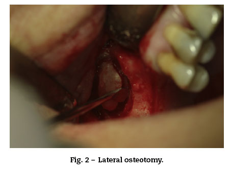
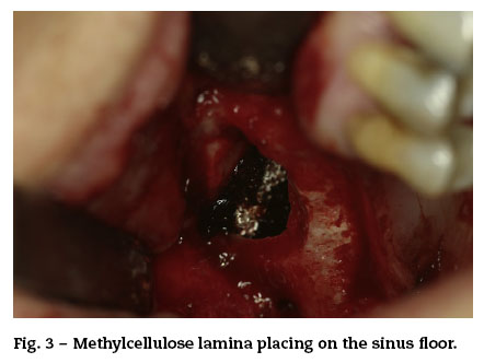
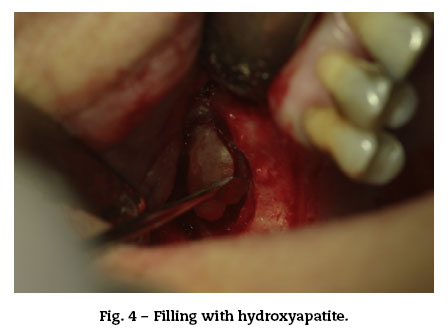
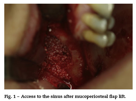
<body><![CDATA[<p><font face="Verdana" size="2">In all patients, an initial panoramic X-ray and CT scan were performed to yield the maximum possible information on the sinus anatomy and to rule out associated disease. All patients subsequently underwent another panoramic X-ray and/or CT scan.</font></p>     <p><font face="Verdana" size="2">Bone gain was measured manually in the panoramic X-ray at 180 days (the magnification was taken into account).</font></p>     <p><i><font face="Verdana" size="2"><b>Surgical protocol</b></font></i></p>     <p><font face="Verdana" size="2">All procedures were conducted under local anesthesia (articaine with adrenalin).</font></p>     <p><font face="Verdana" size="2">The surgery consisted of a crestal incision with anterior and posterior release incisions. The mucoperiosteal flap was lifted and the Caldwell-Luc procedure was performed for lateral access (<a href="#f1">Fig. 1</a>), preparing an oval osteotomy (<a href="#f2">Fig. 2</a>) with a tungsten bur and irrigating with saline solution; perforation of Schneider's membrane was avoided by carefully detaching it from the sinus walls with membrane elevators.</font></p>     <p>&nbsp;</p>     <p align="center"><font face="Verdana" size="2"><a name="f1"><img src="/img/revistas/maxi/v37n2/original4_f1.jpg"></a></font></p>     <p align="center"><font face="Verdana" size="2"><a name="f2"><img src="/img/revistas/maxi/v37n2/original4_f2.jpg"></a></font></p>     <p>&nbsp;</p>     <p><font face="Verdana" size="2">A oxidized cellulose polymer layer was placed on the augmented sinus floor in order to isolate possible undetected microperforations (<a href="#f3">Fig. 3</a>), and the cavity was then filled with two bovine hydroxyapatite vials and compacted (<a href="#f4">Fig. 4</a>). A resorbable collagen membrane of equine origin was placed for the correct isolation of the area. No periodontal dressing material was used.</font></p>     ]]></body>
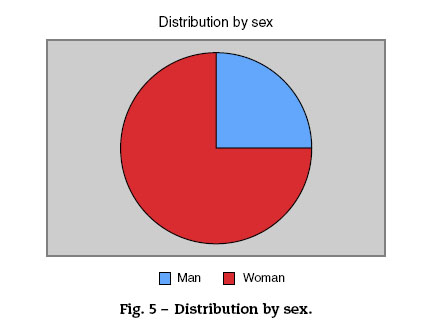
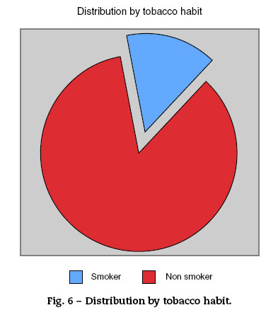
<body><![CDATA[<p>&nbsp;</p>     <p align="center"><font face="Verdana" size="2"><a name="f3"><img src="/img/revistas/maxi/v37n2/original4_f3.jpg"></a></font></p>     <p align="center"><font face="Verdana" size="2"><a name="f4"><img src="/img/revistas/maxi/v37n2/original4_f4.jpg"></a></font></p>     <p>&nbsp;</p>     <p><font face="Verdana" size="2">None of the patients had sinus pathology.</font></p>     <p><font face="Verdana" size="2">All patients were treated with amoxicillin-clavulanic acid (clindamycin for allergic patients) before and after the procedure.<sup>9</sup> They started the treatment an hour before the surgery with amoxicillin 875 mg-clavulanic acid 125 every 8 h and they completed 7 days of treatment. 0.12% chlorhexidine as an oral rinse for ten days was used as well. Sutures were removed at one week post-surgery.</font></p>     <p>&nbsp;</p>     <p><font face="Verdana" size="2"><b>Results</b></font></p>     <p><font face="Verdana" size="2">The study included 34 patients, in whom 37 sinus augmentations were conducted; 75% were female (<a href="#f5">Fig. 5</a>), the mean age was 54 years, and 14.7% of patients were smokers (<a href="#f6">Fig. 6</a>). One patient was diabetic and one had a history of bisphosphonate treatment that had been suspended more than six months before the surgery.</font></p>     <p>&nbsp;</p>     ]]></body>
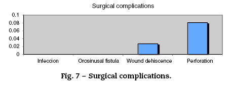
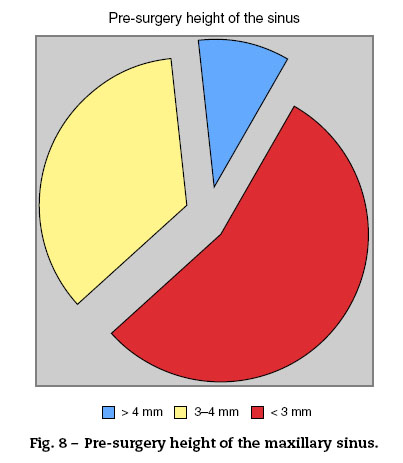
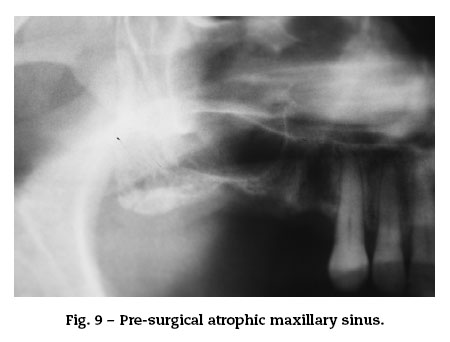
<body><![CDATA[<p align="center"><font face="Verdana" size="2"><a name="f5"><img src="/img/revistas/maxi/v37n2/original4_f5.jpg"></a></font></p>     <p align="center"><font face="Verdana" size="2"><a name="f6"><img src="/img/revistas/maxi/v37n2/original4_f6.jpg"></a></font></p>     <p>&nbsp;</p>     <p><font face="Verdana" size="2">Three Schneider membranes were perforated (8.1%) during the sinus augmentation procedure, and one implant was lost during the osseointegration period. There was no relation between the perforation cases and the implant lost.</font></p>     <p><font face="Verdana" size="2">There were no cases of postsurgical infection and no oral-sinus communication (<a href="#f7">Fig. 7</a>).</font></p>     <p>&nbsp;</p>     <p align="center"><font face="Verdana" size="2"><a name="f7"><img src="/img/revistas/maxi/v37n2/original4_f7.jpg"></a></font></p>     <p>&nbsp;</p>     <p><font face="Verdana" size="2">The mean sinus bone height was 3.1 mm at baseline (i.e., most sinuses were severely resorbed) (Figs. <a href="#f8">8</a> and <a href="#f9">9</a>) and 11.2 mm post-treatment. A success rate of 100% was obtained for this technique.</font></p>     <p>&nbsp;</p>     ]]></body>
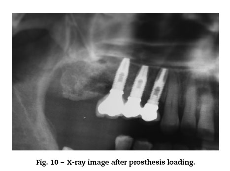
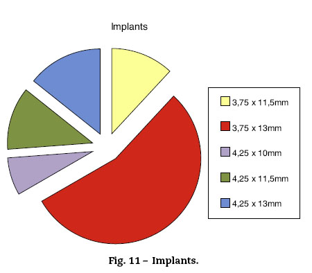
<body><![CDATA[<p align="center"><font face="Verdana" size="2"><a name="f8"><img src="/img/revistas/maxi/v37n2/original4_f8.jpg"></a></font></p>     <p align="center"><font face="Verdana" size="2"><a name="f9"><img src="/img/revistas/maxi/v37n2/original4_f9.jpg"></a></font></p>     <p>&nbsp;</p>     <p><font face="Verdana" size="2">Eighty-two implants were placed (all of them were from the same trademark); only 13.4% were placed immediately (height of &gt;4 mm), given the severe maxillary atrophy (92%) in the majority of the patients.</font></p>     <p><font face="Verdana" size="2">Five implants were lost during the osseointegration period, giving a success rate of 93.9%; these non-osseointegrated implants were all successfully replaced. The implants were loaded at a mean of 143 days after their placement (<a href="#f10">Fig. 10</a>), and the mean follow-up period was 1268 days (range, 396-2778 days). The different sizes places are seen in the chart (<a href="#f11">Fig. 11</a>).</font></p>     <p>&nbsp;</p>     <p align="center"><font face="Verdana" size="2"><a name="f10"><img src="/img/revistas/maxi/v37n2/original4_f10.jpg"></a></font></p>     <p align="center"><font face="Verdana" size="2"><a name="f11"><img src="/img/revistas/maxi/v37n2/original4_f11.jpg"></a></font></p>     <p>&nbsp;</p>     <p><font face="Verdana" size="2">No implant failure rate was observed in patients with comorbidity or tobacco habit.</font></p>     ]]></body>
<body><![CDATA[<p>&nbsp;</p>     <p><font face="Verdana" size="2"><b>Discussion</b></font></p>     <p><font face="Verdana" size="2">The aim of sinus augmentation is to increase the height of the posterior and lateral sectors of the maxilla to permit implant placement for the rehabilitation of patients with atrophic maxilla. Given the concomitant morbidity and rapid resorption of autogenous bone, usually endochondral bone, various bone substitutes have been studied,<sup>10</sup> including a mixture of autologous bone with hydroxyapatite, hydroxyapatite alone, ceramic materials, and methylcellulose, among others.</font></p>     <p><font face="Verdana" size="2">Some authors found that the use of bovine hydroxyapatite avoids donor area morbidity while offering a similar implant success rate to that obtained with other materials; these studies, published in 2005, found no significant differences between sinus augmentation with tricalcium phosphate or autologous bone grafts.<sup>10,11</sup> Bovine hydroxyapatite, which has osteoconductive properties, is obtained from bovine bone in which the organic component has been completely removed. It is believed that the thermal and chemical treatment used to extract this component produces losses in antigenicity and in the potential inflammatory response.</font></p>     <p><font face="Verdana" size="2">In comparison to autologous bone grafts (e.g., from iliac crest or parietal bone), the use of bovine hydroxyapatite avoids the need for general anesthesia during surgery and precludes donor area morbidity.</font></p>     <p><font face="Verdana" size="2">Thus, there are authors who believe that there is no reason to prefer autogenous bone over bone substitutes.<sup>12</sup></font></p>     <p><font face="Verdana" size="2">It has been studied the maxillary sinus lift with out any graft material, but in critic defects, the Oliveira et al. conclude that the poor bone gain results are mainly associated with insufficient bone quality and the large scope of the defects.<sup>13</sup></font></p>     <p><font face="Verdana" size="2">It is remarkable that in Rickerts' metaanalyses published in 2011 comparing the bone fraction after applying: autogenous bone and bone substitutes (bovine hydroxyapatite, bioactive glass, corticocancellous pig bone) revealed no significant differences in bone formation after five months.<sup>14</sup> Zijderveld et al.<sup>12</sup> concluded that the sinus augmented with autogenous bone, the newly formed bone was significantly higher than in the b-tricalciumphosphate group after the same healing period.</font></p>     <p><font face="Verdana" size="2">Membrane perforation was observed in 8.1% of our cases with size less than 2 mm. This is the most frequent complication,<sup>15</sup> and was recorded in 10.1% of cases by Cho-Lee et al.<sup>15</sup> and in 18.42% of cases by Herrero et al.<sup>16</sup> The placement of an oxidized cellulose polymer layer beneath the lifted membrane can be beneficial for isolating the bovine hydroxyapatite. In addition, oxidized cellulose polymer layer adheres to and seals the sinus membrane, which may explain the absence of infections in the present patients, but further studies are needed.</font></p>     <p><font face="Verdana" size="2">There are no major differences between our implant success rate and those obtained by other authors. Thus, the global success rate reported by Cho-Lee et al. in their retrospective study of different filling materials was 96.91%, in comparison to 93.1% in the present study. However, in contrast to our series, the mean height before treatment was 6.59 mm, enabling immediate implant placement in 64.7% of the patients due to the higher primary stability that could be achieved. In the present study, in which the mean height was much lower (3.1 mm), only 13.4% of implants could be placed in the same surgical act. Sakka et al. obtained a 94.8% success rate in patients undergoing sinus augmentation with parietal bone filling.<sup>17</sup> In 2011, Herrero et al.<sup>16</sup> achieved an implant success rate 96.4% but the mean presurgical height, which was not reported, was likely much greater than in the present study, given that an immediate implant could be placed in 89.53% of their cases.</font></p>     ]]></body>
<body><![CDATA[<p><font face="Verdana" size="2">Chen only places immediate dental implants in a sinus with at least 5 mm.<sup>18</sup></font></p>     <p><font face="Verdana" size="2">Our implant success in severely resorbed sinuses using bovine hydroxyapatite alone is within the range reported by other authors. This procedure does not require general anesthesia and avoids patient morbidity.</font></p>     <p><font face="Verdana" size="2">According to these study findings, sinus augmentation with bovine hydroxyapatite alone is a safe surgical technique that yields a very high implant success rate. This approach is not associated with any donor morbidity and achieves the predictable rehabilitation of patients with absence of posterior sectors in a severely atrophied maxilla. Histological and histomorphometrical analysis would be helpful in order to obtain the bone gain.</font></p>     <p>&nbsp;</p>     <p><font face="Verdana" size="2"><b>Ethical disclosures</b></font></p>     <p><font face="Verdana" size="2"><b>Protection of human and animal subjects.</b> The authors declare that the procedures followed were in accordance with the regulations of the relevant clinical research ethics committee and with those of the Code of Ethics of the World Medical Association (Declaration of Helsinki).</font></p>     <p><font face="Verdana" size="2"><b>Confidentiality of data.</b> The authors declare that they have followed the protocols of their work center on the publication of patient data.</font></p>     <p><font face="Verdana" size="2"><b>Right to privacy and informed consent.</b> The authors have obtained the written informed consent of the patients or subjects mentioned in the article. The corresponding author is in possession of this document.</font></p>     <p>&nbsp;</p>     <p><font face="Verdana" size="2"><b>Funding</b></font></p>     ]]></body>
<body><![CDATA[<p><font face="Verdana" size="2">The authors declare that there is no external funding.</font></p>     <p>&nbsp;</p>     <p><font face="Verdana" size="2"><b>Conflicts of interest</b></font></p>     <p><font face="Verdana" size="2">The authors declare that they have no conflicts of interests.</font></p>     <p>&nbsp;</p>     <p><font face="Verdana" size="2"><b>References</b></font></p>     <!-- ref --><p><font face="Verdana" size="2">1. Tatum Jr. H. Maxillary and sinus implant reconstructions. Dent Clin N Am. 1986;30:207-29.    &nbsp;&nbsp;&nbsp;&nbsp;&nbsp;&nbsp;&nbsp;&nbsp;[&#160;<a href="javascript:void(0);" onclick="javascript: window.open('/scielo.php?script=sci_nlinks&ref=5189389&pid=S1130-0558201500020000500001&lng=','','width=640,height=500,resizable=yes,scrollbars=1,menubar=yes,');">Links</a>&#160;]<!-- end-ref --></font></p>    <!-- ref --><p><font face="Verdana" size="2">2. Boyne P.J., James R.A. Grafting of the maxillary sinus floor with autogenous marrow and bone. J Oral Surg. 1980;38:613-6.    &nbsp;&nbsp;&nbsp;&nbsp;&nbsp;&nbsp;&nbsp;&nbsp;[&#160;<a href="javascript:void(0);" onclick="javascript: window.open('/scielo.php?script=sci_nlinks&ref=5189391&pid=S1130-0558201500020000500002&lng=','','width=640,height=500,resizable=yes,scrollbars=1,menubar=yes,');">Links</a>&#160;]<!-- end-ref --></font></p>    ]]></body>
<body><![CDATA[<!-- ref --><p><font face="Verdana" size="2">3. Acocella A., Bertolai R., Nissan J., Sacco R. Clinical, histological and histomorphometrical study of maxillary sinus augmentation using cortico-cancellous fresh frozen bone chips. J Craniomaxillofac Surg. 2011;39:192-9.    &nbsp;&nbsp;&nbsp;&nbsp;&nbsp;&nbsp;&nbsp;&nbsp;[&#160;<a href="javascript:void(0);" onclick="javascript: window.open('/scielo.php?script=sci_nlinks&ref=5189393&pid=S1130-0558201500020000500003&lng=','','width=640,height=500,resizable=yes,scrollbars=1,menubar=yes,');">Links</a>&#160;]<!-- end-ref --></font></p>    <!-- ref --><p><font face="Verdana" size="2">4. Hobo S., Ichida E., Garc&iacute;a L.T. Planificaci&oacute;n del tratamiento En Osteointegraci&oacute;n y rehabilitaci&oacute;n oclusal.  Madrid: Marban, 1997. pp. 55-86.    &nbsp;&nbsp;&nbsp;&nbsp;&nbsp;&nbsp;&nbsp;&nbsp;[&#160;<a href="javascript:void(0);" onclick="javascript: window.open('/scielo.php?script=sci_nlinks&ref=5189395&pid=S1130-0558201500020000500004&lng=','','width=640,height=500,resizable=yes,scrollbars=1,menubar=yes,');">Links</a>&#160;]<!-- end-ref --></font></p>    <!-- ref --><p><font face="Verdana" size="2">5. Rickert D., Slater J.J., Meijer H.J., Vissink A., Raghoebar G.M. Maxillary sinus lift with solely autogenous bone compared to a combination of autogenous bone and growth factors or (solely) bone substitutes. A systematic review. Int J Oral Maxillofac Surg. 2012;41:160-7.    &nbsp;&nbsp;&nbsp;&nbsp;&nbsp;&nbsp;&nbsp;&nbsp;[&#160;<a href="javascript:void(0);" onclick="javascript: window.open('/scielo.php?script=sci_nlinks&ref=5189397&pid=S1130-0558201500020000500005&lng=','','width=640,height=500,resizable=yes,scrollbars=1,menubar=yes,');">Links</a>&#160;]<!-- end-ref --></font></p>    <!-- ref --><p><font face="Verdana" size="2">6. Schmelzeisen R., Schimming R., Sittinger M. Making bone: implant insertion into tissue-engineered bone for maxillary sinus floor augmentation - a preliminary report. J Craniomaxillofac Surg. 2003;31:34-9.    &nbsp;&nbsp;&nbsp;&nbsp;&nbsp;&nbsp;&nbsp;&nbsp;[&#160;<a href="javascript:void(0);" onclick="javascript: window.open('/scielo.php?script=sci_nlinks&ref=5189399&pid=S1130-0558201500020000500006&lng=','','width=640,height=500,resizable=yes,scrollbars=1,menubar=yes,');">Links</a>&#160;]<!-- end-ref --></font></p>    <!-- ref --><p><font face="Verdana" size="2">7. Cricchio G., Palma V.C., Faria P.E., de J.A., Lundgren S., Sennerby L.,  et al. Histological outcomes on the development of new space-making devices for maxillary sinus floor augmentation. Clin Implant Dent Relat Res. 2011;13:224-30.    &nbsp;&nbsp;&nbsp;&nbsp;&nbsp;&nbsp;&nbsp;&nbsp;[&#160;<a href="javascript:void(0);" onclick="javascript: window.open('/scielo.php?script=sci_nlinks&ref=5189401&pid=S1130-0558201500020000500007&lng=','','width=640,height=500,resizable=yes,scrollbars=1,menubar=yes,');">Links</a>&#160;]<!-- end-ref --></font></p>    ]]></body>
<body><![CDATA[<!-- ref --><p><font face="Verdana" size="2">8. Misch C.E. Bone density: a key determinant for treatment planning.  In: Contemporary implant dentistry. Missouri: Mosby Elsevier; 2008.    &nbsp;&nbsp;&nbsp;&nbsp;&nbsp;&nbsp;&nbsp;&nbsp;[&#160;<a href="javascript:void(0);" onclick="javascript: window.open('/scielo.php?script=sci_nlinks&ref=5189403&pid=S1130-0558201500020000500008&lng=','','width=640,height=500,resizable=yes,scrollbars=1,menubar=yes,');">Links</a>&#160;]<!-- end-ref --></font></p>    <!-- ref --><p><font face="Verdana" size="2">9. Villarreal P.M., Fern&aacute;ndez-Bustillo &Aacute;., Acero J., Arruti J.A., Baladr&oacute;n J., Bilbao A. I Conferencia Nacional de Consenso sobre el Injerto &Oacute;seo del Seno Maxilar. Rev Esp Cir Oral Maxilofac. 2010;32:41-63.    &nbsp;&nbsp;&nbsp;&nbsp;&nbsp;&nbsp;&nbsp;&nbsp;[&#160;<a href="javascript:void(0);" onclick="javascript: window.open('/scielo.php?script=sci_nlinks&ref=5189405&pid=S1130-0558201500020000500009&lng=','','width=640,height=500,resizable=yes,scrollbars=1,menubar=yes,');">Links</a>&#160;]<!-- end-ref --></font></p>    <!-- ref --><p><font face="Verdana" size="2">10. Mu&ntilde;oz M., Trullenque A. Comparaci&oacute;n entre distintos sustitutos &oacute;seos utilizados para procedimientos de elevaci&oacute;n de seno maxilar previo a la colocaci&oacute;n de implantes dentales. Av Periodoncia. 2008;20:155-64.    &nbsp;&nbsp;&nbsp;&nbsp;&nbsp;&nbsp;&nbsp;&nbsp;[&#160;<a href="javascript:void(0);" onclick="javascript: window.open('/scielo.php?script=sci_nlinks&ref=5189407&pid=S1130-0558201500020000500010&lng=','','width=640,height=500,resizable=yes,scrollbars=1,menubar=yes,');">Links</a>&#160;]<!-- end-ref --></font></p>    <!-- ref --><p><font face="Verdana" size="2">11. Szabo G., Huys L., Coulthard P., Maiorana C., Garagiola U., Barabas J.,  et al. A prospective multicenter randomized clinical trial of autogenous bone versus beta-tricalcium phosphate graft alone for bilateral sinus elevation: histologic and histomorphometric evaluation. Int J Oral Maxillofac Implants. 2005;20:371-81.    &nbsp;&nbsp;&nbsp;&nbsp;&nbsp;&nbsp;&nbsp;&nbsp;[&#160;<a href="javascript:void(0);" onclick="javascript: window.open('/scielo.php?script=sci_nlinks&ref=5189409&pid=S1130-0558201500020000500011&lng=','','width=640,height=500,resizable=yes,scrollbars=1,menubar=yes,');">Links</a>&#160;]<!-- end-ref --></font></p>    <!-- ref --><p><font face="Verdana" size="2">12. Zijderveld S.A., Zerbo I.R., van J.P., Schulten E.A., ten C.M. Maxillary sinus floor augmentation using a beta-tricalcium phosphate (Cerasorb) alone compared to autogenous bone grafts. Int J Oral Maxillofac Implants. 2005;20:432-40.    &nbsp;&nbsp;&nbsp;&nbsp;&nbsp;&nbsp;&nbsp;&nbsp;[&#160;<a href="javascript:void(0);" onclick="javascript: window.open('/scielo.php?script=sci_nlinks&ref=5189411&pid=S1130-0558201500020000500012&lng=','','width=640,height=500,resizable=yes,scrollbars=1,menubar=yes,');">Links</a>&#160;]<!-- end-ref --></font></p>    ]]></body>
<body><![CDATA[<!-- ref --><p><font face="Verdana" size="2">13. Nkenke E., Stelzle F. Clinical outcomes of sinus floor augmentation for implant placement using autogenous bone or bone substitutes: a systematic review. Clin Oral Implants Res. 2009;20:124-33.    &nbsp;&nbsp;&nbsp;&nbsp;&nbsp;&nbsp;&nbsp;&nbsp;[&#160;<a href="javascript:void(0);" onclick="javascript: window.open('/scielo.php?script=sci_nlinks&ref=5189413&pid=S1130-0558201500020000500013&lng=','','width=640,height=500,resizable=yes,scrollbars=1,menubar=yes,');">Links</a>&#160;]<!-- end-ref --></font></p>    <!-- ref --><p><font face="Verdana" size="2">14. de  Oliveira G.R., Olate S., Cavalieri-Pereira L., Pozzer L., Asprino L., de Moraes M.,  et al. Maxillary sinus floor augmentation using blood without graft material. Preliminary results in 10 patients. J Oral Maxillofac Surg. 2013;71:1670-5.    &nbsp;&nbsp;&nbsp;&nbsp;&nbsp;&nbsp;&nbsp;&nbsp;[&#160;<a href="javascript:void(0);" onclick="javascript: window.open('/scielo.php?script=sci_nlinks&ref=5189415&pid=S1130-0558201500020000500014&lng=','','width=640,height=500,resizable=yes,scrollbars=1,menubar=yes,');">Links</a>&#160;]<!-- end-ref --></font></p>    <!-- ref --><p><font face="Verdana" size="2">15. Cho-Lee G.-Y., Naval-G&iacute;as L., Mancha M., Sastre-P&eacute;rez J., Capote-Moreno A.L., Mu&ntilde;oz-Guerra M.F.,  et al. Elevaci&oacute;n de seno maxilar: An&aacute;lisis cl&iacute;nico de nuestra experiencia en m&aacute;s de 100 casos. Rev Esp Cir Oral Maxilofac. 2009;31:223-30.    &nbsp;&nbsp;&nbsp;&nbsp;&nbsp;&nbsp;&nbsp;&nbsp;[&#160;<a href="javascript:void(0);" onclick="javascript: window.open('/scielo.php?script=sci_nlinks&ref=5189417&pid=S1130-0558201500020000500015&lng=','','width=640,height=500,resizable=yes,scrollbars=1,menubar=yes,');">Links</a>&#160;]<!-- end-ref --></font></p>    <!-- ref --><p><font face="Verdana" size="2">16. Herrero M., Pic&oacute;n M., Almeida D., Trujillo L., N&uacute;&ntilde;ez J.,  Rieto P.A. 382 elevaciones de seno con t&eacute;cnica de ventana lateral y uso de biomaterial de relleno. Rev Esp Cir Oral Maxilofac. 2011;33:109-14.    &nbsp;&nbsp;&nbsp;&nbsp;&nbsp;&nbsp;&nbsp;&nbsp;[&#160;<a href="javascript:void(0);" onclick="javascript: window.open('/scielo.php?script=sci_nlinks&ref=5189419&pid=S1130-0558201500020000500016&lng=','','width=640,height=500,resizable=yes,scrollbars=1,menubar=yes,');">Links</a>&#160;]<!-- end-ref --></font></p>    <!-- ref --><p><font face="Verdana" size="2">17. Sakka S., Krenkel C. Simultaneous maxillary sinus lifting and implant placement with autogenous parietal bone graft: outcome of 17 cases. J Craniomaxillofac Surg. 2011;39:187-91.    &nbsp;&nbsp;&nbsp;&nbsp;&nbsp;&nbsp;&nbsp;&nbsp;[&#160;<a href="javascript:void(0);" onclick="javascript: window.open('/scielo.php?script=sci_nlinks&ref=5189421&pid=S1130-0558201500020000500017&lng=','','width=640,height=500,resizable=yes,scrollbars=1,menubar=yes,');">Links</a>&#160;]<!-- end-ref --></font></p>    ]]></body>
<body><![CDATA[<!-- ref --><p><font face="Verdana" size="2">18. Chen T.W., Chang H.S., Leung K.W., Lai Y.L., Kao S.Y. Implant placement immediately after the lateral approach of the trap door window procedure to create a maxillary sinus lift without bone grafting: a 2-year retrospective evaluation of 47 implants in 33 patients. J Oral Maxillofac Surg. 2007;65:2324-8.    &nbsp;&nbsp;&nbsp;&nbsp;&nbsp;&nbsp;&nbsp;&nbsp;[&#160;<a href="javascript:void(0);" onclick="javascript: window.open('/scielo.php?script=sci_nlinks&ref=5189423&pid=S1130-0558201500020000500018&lng=','','width=640,height=500,resizable=yes,scrollbars=1,menubar=yes,');">Links</a>&#160;]<!-- end-ref --></font></p>     <p>&nbsp;</p>     <p>&nbsp;</p>     <p><font face="Verdana" size="2"><a href="#top"><img border="0" src="/img/revistas/maxi/v37n2/seta.gif" width="15" height="17"></a><a name="bajo"></a><b>Correspondence:</b>    <br>E-mail addresses: <a href="mailto:dra.amlopezlopez@gmail.com">dra.amlopezlopez@gmail.com</a>,    <br><a href="mailto:anitalopez@gmail.com">anitalopez@gmail.com</a>    <br>(A.M. López López).</font></p>     <p><font face="Verdana" size="2">Received: 5 March 2014     <br>Accepted: 1 July 2014</font></p>     ]]></body>
<body><![CDATA[ ]]></body><back>
<ref-list>
<ref id="B1">
<label>1</label><nlm-citation citation-type="journal">
<person-group person-group-type="author">
<name>
<surname><![CDATA[Tatum]]></surname>
<given-names><![CDATA[Jr. H]]></given-names>
</name>
</person-group>
<article-title xml:lang="en"><![CDATA[Maxillary and sinus implant reconstructions]]></article-title>
<source><![CDATA[Dent Clin N Am.]]></source>
<year>1986</year>
<volume>30</volume>
<page-range>207-29</page-range></nlm-citation>
</ref>
<ref id="B2">
<label>2</label><nlm-citation citation-type="journal">
<person-group person-group-type="author">
<name>
<surname><![CDATA[Boyne]]></surname>
<given-names><![CDATA[P.J.]]></given-names>
</name>
<name>
<surname><![CDATA[James]]></surname>
<given-names><![CDATA[R.A]]></given-names>
</name>
</person-group>
<article-title xml:lang="en"><![CDATA[Grafting of the maxillary sinus floor with autogenous marrow and bone]]></article-title>
<source><![CDATA[J Oral Surg.]]></source>
<year>1980</year>
<volume>38</volume>
<page-range>613-6</page-range></nlm-citation>
</ref>
<ref id="B3">
<label>3</label><nlm-citation citation-type="journal">
<person-group person-group-type="author">
<name>
<surname><![CDATA[Acocella]]></surname>
<given-names><![CDATA[A.]]></given-names>
</name>
<name>
<surname><![CDATA[Bertolai]]></surname>
<given-names><![CDATA[R.]]></given-names>
</name>
<name>
<surname><![CDATA[Nissan]]></surname>
<given-names><![CDATA[J.]]></given-names>
</name>
<name>
<surname><![CDATA[Sacco]]></surname>
<given-names><![CDATA[R]]></given-names>
</name>
</person-group>
<article-title xml:lang="en"><![CDATA[Clinical, histological and histomorphometrical study of maxillary sinus augmentation using cortico-cancellous fresh frozen bone chips]]></article-title>
<source><![CDATA[J Craniomaxillofac Surg.]]></source>
<year>2011</year>
<volume>39</volume>
<page-range>192-9</page-range></nlm-citation>
</ref>
<ref id="B4">
<label>4</label><nlm-citation citation-type="book">
<person-group person-group-type="author">
<name>
<surname><![CDATA[Hobo]]></surname>
<given-names><![CDATA[S.]]></given-names>
</name>
<name>
<surname><![CDATA[Ichida]]></surname>
<given-names><![CDATA[E.]]></given-names>
</name>
<name>
<surname><![CDATA[García]]></surname>
<given-names><![CDATA[L.T]]></given-names>
</name>
</person-group>
<article-title xml:lang="es"><![CDATA[Planificación del tratamiento]]></article-title>
<source><![CDATA[Osteointegración y rehabilitación oclusal]]></source>
<year>1997</year>
<page-range>55-86</page-range><publisher-loc><![CDATA[Madrid ]]></publisher-loc>
<publisher-name><![CDATA[Marban]]></publisher-name>
</nlm-citation>
</ref>
<ref id="B5">
<label>5</label><nlm-citation citation-type="journal">
<person-group person-group-type="author">
<name>
<surname><![CDATA[Rickert]]></surname>
<given-names><![CDATA[D.]]></given-names>
</name>
<name>
<surname><![CDATA[Slater]]></surname>
<given-names><![CDATA[J.J.]]></given-names>
</name>
<name>
<surname><![CDATA[Meijer]]></surname>
<given-names><![CDATA[H.J.]]></given-names>
</name>
<name>
<surname><![CDATA[Vissink]]></surname>
<given-names><![CDATA[A.]]></given-names>
</name>
<name>
<surname><![CDATA[Raghoebar]]></surname>
<given-names><![CDATA[G.M]]></given-names>
</name>
</person-group>
<article-title xml:lang="en"><![CDATA[Maxillary sinus lift with solely autogenous bone compared to a combination of autogenous bone and growth factors or (solely) bone substitutes: A systematic review]]></article-title>
<source><![CDATA[Int J Oral Maxillofac Surg.]]></source>
<year>2012</year>
<volume>41</volume>
<page-range>160-7</page-range></nlm-citation>
</ref>
<ref id="B6">
<label>6</label><nlm-citation citation-type="journal">
<person-group person-group-type="author">
<name>
<surname><![CDATA[Schmelzeisen]]></surname>
<given-names><![CDATA[R.]]></given-names>
</name>
<name>
<surname><![CDATA[Schimming]]></surname>
<given-names><![CDATA[R.]]></given-names>
</name>
<name>
<surname><![CDATA[Sittinger]]></surname>
<given-names><![CDATA[M]]></given-names>
</name>
</person-group>
<article-title xml:lang="en"><![CDATA[Making bone: implant insertion into tissue-engineered bone for maxillary sinus floor augmentation - a preliminary report]]></article-title>
<source><![CDATA[J Craniomaxillofac Surg.]]></source>
<year>2003</year>
<volume>31</volume>
<page-range>34-9</page-range></nlm-citation>
</ref>
<ref id="B7">
<label>7</label><nlm-citation citation-type="journal">
<person-group person-group-type="author">
<name>
<surname><![CDATA[Cricchio]]></surname>
<given-names><![CDATA[G.]]></given-names>
</name>
<name>
<surname><![CDATA[Palma]]></surname>
<given-names><![CDATA[V.C.]]></given-names>
</name>
<name>
<surname><![CDATA[Faria]]></surname>
<given-names><![CDATA[P.E.]]></given-names>
</name>
<name>
<surname><![CDATA[de]]></surname>
<given-names><![CDATA[J.A.]]></given-names>
</name>
<name>
<surname><![CDATA[Lundgren]]></surname>
<given-names><![CDATA[S.]]></given-names>
</name>
<name>
<surname><![CDATA[Sennerby]]></surname>
<given-names><![CDATA[L.]]></given-names>
</name>
</person-group>
<article-title xml:lang="en"><![CDATA[Histological outcomes on the development of new space-making devices for maxillary sinus floor augmentation]]></article-title>
<source><![CDATA[Clin Implant Dent Relat Res.]]></source>
<year>2011</year>
<volume>13</volume>
<page-range>224-30</page-range></nlm-citation>
</ref>
<ref id="B8">
<label>8</label><nlm-citation citation-type="book">
<person-group person-group-type="author">
<name>
<surname><![CDATA[Misch]]></surname>
<given-names><![CDATA[C.E]]></given-names>
</name>
</person-group>
<article-title xml:lang="en"><![CDATA[Bone density: a key determinant for treatment planning]]></article-title>
<source><![CDATA[Contemporary implant dentistry]]></source>
<year>2008</year>
<publisher-loc><![CDATA[^eMissouri Missouri]]></publisher-loc>
<publisher-name><![CDATA[Mosby Elsevier]]></publisher-name>
</nlm-citation>
</ref>
<ref id="B9">
<label>9</label><nlm-citation citation-type="journal">
<person-group person-group-type="author">
<name>
<surname><![CDATA[Villarreal]]></surname>
<given-names><![CDATA[P.M.]]></given-names>
</name>
<name>
<surname><![CDATA[Fernández-Bustillo]]></surname>
<given-names><![CDATA[Á.]]></given-names>
</name>
<name>
<surname><![CDATA[Acero]]></surname>
<given-names><![CDATA[J.]]></given-names>
</name>
<name>
<surname><![CDATA[Arruti]]></surname>
<given-names><![CDATA[J.A.]]></given-names>
</name>
<name>
<surname><![CDATA[Baladrón]]></surname>
<given-names><![CDATA[J.]]></given-names>
</name>
<name>
<surname><![CDATA[Bilbao]]></surname>
<given-names><![CDATA[A]]></given-names>
</name>
</person-group>
<article-title xml:lang="es"><![CDATA[I Conferencia Nacional de Consenso sobre el Injerto Óseo del Seno Maxilar]]></article-title>
<source><![CDATA[Rev Esp Cir Oral Maxilofac.]]></source>
<year>2010</year>
<volume>32</volume>
<page-range>41-63</page-range></nlm-citation>
</ref>
<ref id="B10">
<label>10</label><nlm-citation citation-type="journal">
<person-group person-group-type="author">
<name>
<surname><![CDATA[Muñoz]]></surname>
<given-names><![CDATA[M.]]></given-names>
</name>
<name>
<surname><![CDATA[Trullenque]]></surname>
<given-names><![CDATA[A]]></given-names>
</name>
</person-group>
<article-title xml:lang="es"><![CDATA[Comparación entre distintos sustitutos óseos utilizados para procedimientos de elevación de seno maxilar previo a la colocación de implantes dentales]]></article-title>
<source><![CDATA[Av Periodoncia]]></source>
<year>2008</year>
<volume>20</volume>
<page-range>155-64</page-range></nlm-citation>
</ref>
<ref id="B11">
<label>11</label><nlm-citation citation-type="journal">
<person-group person-group-type="author">
<name>
<surname><![CDATA[Szabo]]></surname>
<given-names><![CDATA[G.]]></given-names>
</name>
<name>
<surname><![CDATA[Huys]]></surname>
<given-names><![CDATA[L.]]></given-names>
</name>
<name>
<surname><![CDATA[Coulthard]]></surname>
<given-names><![CDATA[P.]]></given-names>
</name>
<name>
<surname><![CDATA[Maiorana]]></surname>
<given-names><![CDATA[C.]]></given-names>
</name>
<name>
<surname><![CDATA[Garagiola]]></surname>
<given-names><![CDATA[U.]]></given-names>
</name>
<name>
<surname><![CDATA[Barabas]]></surname>
<given-names><![CDATA[J.]]></given-names>
</name>
</person-group>
<article-title xml:lang="en"><![CDATA[A prospective multicenter randomized clinical trial of autogenous bone versus beta-tricalcium phosphate graft alone for bilateral sinus elevation: histologic and histomorphometric evaluation]]></article-title>
<source><![CDATA[Int J Oral Maxillofac Implants]]></source>
<year>2005</year>
<volume>20</volume>
<page-range>371-81</page-range></nlm-citation>
</ref>
<ref id="B12">
<label>12</label><nlm-citation citation-type="journal">
<person-group person-group-type="author">
<name>
<surname><![CDATA[Zijderveld]]></surname>
<given-names><![CDATA[S.A.]]></given-names>
</name>
<name>
<surname><![CDATA[Zerbo]]></surname>
<given-names><![CDATA[I.R.]]></given-names>
</name>
<name>
<surname><![CDATA[van]]></surname>
<given-names><![CDATA[J.P.]]></given-names>
</name>
<name>
<surname><![CDATA[Schulten]]></surname>
<given-names><![CDATA[E.A.]]></given-names>
</name>
<name>
<surname><![CDATA[ten]]></surname>
<given-names><![CDATA[C.M]]></given-names>
</name>
</person-group>
<article-title xml:lang="en"><![CDATA[Maxillary sinus floor augmentation using a beta-tricalcium phosphate (Cerasorb) alone compared to autogenous bone grafts]]></article-title>
<source><![CDATA[Int J Oral Maxillofac Implants]]></source>
<year>2005</year>
<volume>20</volume>
<page-range>432-40</page-range></nlm-citation>
</ref>
<ref id="B13">
<label>13</label><nlm-citation citation-type="journal">
<person-group person-group-type="author">
<name>
<surname><![CDATA[Nkenke]]></surname>
<given-names><![CDATA[E.]]></given-names>
</name>
<name>
<surname><![CDATA[Stelzle]]></surname>
<given-names><![CDATA[F]]></given-names>
</name>
</person-group>
<article-title xml:lang="en"><![CDATA[Clinical outcomes of sinus floor augmentation for implant placement using autogenous bone or bone substitutes: a systematic review]]></article-title>
<source><![CDATA[Clin Oral Implants Res.]]></source>
<year>2009</year>
<volume>20</volume>
<page-range>124-33</page-range></nlm-citation>
</ref>
<ref id="B14">
<label>14</label><nlm-citation citation-type="journal">
<person-group person-group-type="author">
<name>
<surname><![CDATA[de Oliveira]]></surname>
<given-names><![CDATA[G.R.]]></given-names>
</name>
<name>
<surname><![CDATA[Olate]]></surname>
<given-names><![CDATA[S.]]></given-names>
</name>
<name>
<surname><![CDATA[Cavalieri-Pereira]]></surname>
<given-names><![CDATA[L.]]></given-names>
</name>
<name>
<surname><![CDATA[Pozzer]]></surname>
<given-names><![CDATA[L.]]></given-names>
</name>
<name>
<surname><![CDATA[Asprino]]></surname>
<given-names><![CDATA[L.]]></given-names>
</name>
<name>
<surname><![CDATA[de Moraes]]></surname>
<given-names><![CDATA[M.]]></given-names>
</name>
</person-group>
<article-title xml:lang="en"><![CDATA[Maxillary sinus floor augmentation using blood without graft material: Preliminary results in 10 patients]]></article-title>
<source><![CDATA[J Oral Maxillofac Surg.]]></source>
<year>2013</year>
<volume>71</volume>
<page-range>1670-5</page-range></nlm-citation>
</ref>
<ref id="B15">
<label>15</label><nlm-citation citation-type="journal">
<person-group person-group-type="author">
<name>
<surname><![CDATA[Cho-Lee]]></surname>
<given-names><![CDATA[G.-Y.]]></given-names>
</name>
<name>
<surname><![CDATA[Naval-Gías]]></surname>
<given-names><![CDATA[L.]]></given-names>
</name>
<name>
<surname><![CDATA[Mancha]]></surname>
<given-names><![CDATA[M.]]></given-names>
</name>
<name>
<surname><![CDATA[Sastre-Pérez]]></surname>
<given-names><![CDATA[J.]]></given-names>
</name>
<name>
<surname><![CDATA[Capote-Moreno]]></surname>
<given-names><![CDATA[A.L.]]></given-names>
</name>
<name>
<surname><![CDATA[Muñoz-Guerra]]></surname>
<given-names><![CDATA[M.F.]]></given-names>
</name>
</person-group>
<article-title xml:lang="es"><![CDATA[Elevación de seno maxilar: Análisis clínico de nuestra experiencia en más de 100 casos]]></article-title>
<source><![CDATA[Rev Esp Cir Oral Maxilofac.]]></source>
<year>2009</year>
<volume>31</volume>
<page-range>223-30</page-range></nlm-citation>
</ref>
<ref id="B16">
<label>16</label><nlm-citation citation-type="journal">
<person-group person-group-type="author">
<name>
<surname><![CDATA[Herrero]]></surname>
<given-names><![CDATA[M.]]></given-names>
</name>
<name>
<surname><![CDATA[Picón]]></surname>
<given-names><![CDATA[M.]]></given-names>
</name>
<name>
<surname><![CDATA[Almeida]]></surname>
<given-names><![CDATA[D.]]></given-names>
</name>
<name>
<surname><![CDATA[Trujillo]]></surname>
<given-names><![CDATA[L.]]></given-names>
</name>
<name>
<surname><![CDATA[Núñez]]></surname>
<given-names><![CDATA[J.]]></given-names>
</name>
<name>
<surname><![CDATA[Rieto]]></surname>
<given-names><![CDATA[P.A]]></given-names>
</name>
</person-group>
<article-title xml:lang="es"><![CDATA[382 elevaciones de seno con técnica de ventana lateral y uso de biomaterial de relleno]]></article-title>
<source><![CDATA[Rev Esp Cir Oral Maxilofac.]]></source>
<year>2011</year>
<volume>33</volume>
<page-range>109-14</page-range></nlm-citation>
</ref>
<ref id="B17">
<label>17</label><nlm-citation citation-type="journal">
<person-group person-group-type="author">
<name>
<surname><![CDATA[Sakka]]></surname>
<given-names><![CDATA[S.]]></given-names>
</name>
<name>
<surname><![CDATA[Krenkel]]></surname>
<given-names><![CDATA[C]]></given-names>
</name>
</person-group>
<article-title xml:lang="en"><![CDATA[Simultaneous maxillary sinus lifting and implant placement with autogenous parietal bone graft: outcome of 17 cases]]></article-title>
<source><![CDATA[J Craniomaxillofac Surg.]]></source>
<year>2011</year>
<volume>39</volume>
<page-range>187-91</page-range></nlm-citation>
</ref>
<ref id="B18">
<label>18</label><nlm-citation citation-type="journal">
<person-group person-group-type="author">
<name>
<surname><![CDATA[Chen]]></surname>
<given-names><![CDATA[T.W.]]></given-names>
</name>
<name>
<surname><![CDATA[Chang]]></surname>
<given-names><![CDATA[H.S.]]></given-names>
</name>
<name>
<surname><![CDATA[Leung]]></surname>
<given-names><![CDATA[K.W.]]></given-names>
</name>
<name>
<surname><![CDATA[Lai]]></surname>
<given-names><![CDATA[Y.L.]]></given-names>
</name>
<name>
<surname><![CDATA[Kao]]></surname>
<given-names><![CDATA[S.Y]]></given-names>
</name>
</person-group>
<article-title xml:lang="en"><![CDATA[Implant placement immediately after the lateral approach of the trap door window procedure to create a maxillary sinus lift without bone grafting: a 2-year retrospective evaluation of 47 implants in 33 patients]]></article-title>
<source><![CDATA[J Oral Maxillofac Surg.]]></source>
<year>2007</year>
<volume>65</volume>
<page-range>2324-8</page-range></nlm-citation>
</ref>
</ref-list>
</back>
</article>