<?xml version="1.0" encoding="ISO-8859-1"?><article xmlns:mml="http://www.w3.org/1998/Math/MathML" xmlns:xlink="http://www.w3.org/1999/xlink" xmlns:xsi="http://www.w3.org/2001/XMLSchema-instance">
<front>
<journal-meta>
<journal-id>1137-6627</journal-id>
<journal-title><![CDATA[Anales del Sistema Sanitario de Navarra]]></journal-title>
<abbrev-journal-title><![CDATA[Anales Sis San Navarra]]></abbrev-journal-title>
<issn>1137-6627</issn>
<publisher>
<publisher-name><![CDATA[Gobierno de Navarra. Departamento de Salud]]></publisher-name>
</publisher>
</journal-meta>
<article-meta>
<article-id>S1137-66272013000100020</article-id>
<article-id pub-id-type="doi">10.4321/S1137-66272013000100020</article-id>
<title-group>
<article-title xml:lang="en"><![CDATA[Gastric lipoma and pyloric obstruction in a 51-year-old woman]]></article-title>
<article-title xml:lang="es"><![CDATA[Lipoma gástrico y obstrucción pilórica en una mujer de 51 años]]></article-title>
</title-group>
<contrib-group>
<contrib contrib-type="author">
<name>
<surname><![CDATA[Furtado]]></surname>
<given-names><![CDATA[W.S.]]></given-names>
</name>
<xref ref-type="aff" rid="A01"/>
</contrib>
<contrib contrib-type="author">
<name>
<surname><![CDATA[Mello]]></surname>
<given-names><![CDATA[D.A.C.P.G.]]></given-names>
</name>
<xref ref-type="aff" rid="A01"/>
</contrib>
<contrib contrib-type="author">
<name>
<surname><![CDATA[Santos]]></surname>
<given-names><![CDATA[V.M.]]></given-names>
</name>
<xref ref-type="aff" rid="A02"/>
<xref ref-type="aff" rid="A03"/>
</contrib>
<contrib contrib-type="author">
<name>
<surname><![CDATA[Bringel]]></surname>
<given-names><![CDATA[T.L.]]></given-names>
</name>
<xref ref-type="aff" rid="A01"/>
</contrib>
<contrib contrib-type="author">
<name>
<surname><![CDATA[Oliveira Junior]]></surname>
<given-names><![CDATA[W.P.]]></given-names>
</name>
<xref ref-type="aff" rid="A01"/>
</contrib>
<contrib contrib-type="author">
<name>
<surname><![CDATA[Moura]]></surname>
<given-names><![CDATA[H.J.L.]]></given-names>
</name>
<xref ref-type="aff" rid="A01"/>
</contrib>
</contrib-group>
<aff id="A01">
<institution><![CDATA[,Armed Forces Hospital General Surgery Division ]]></institution>
<addr-line><![CDATA[Brasília DF]]></addr-line>
<country>Brazil</country>
</aff>
<aff id="A02">
<institution><![CDATA[,Catholic University Medical Course ]]></institution>
<addr-line><![CDATA[ ]]></addr-line>
</aff>
<aff id="A03">
<institution><![CDATA[,Armed Forces Hospital Internal Medicina Department ]]></institution>
<addr-line><![CDATA[Brasília DF]]></addr-line>
<country>Brazil</country>
</aff>
<pub-date pub-type="pub">
<day>00</day>
<month>04</month>
<year>2013</year>
</pub-date>
<pub-date pub-type="epub">
<day>00</day>
<month>04</month>
<year>2013</year>
</pub-date>
<volume>36</volume>
<numero>1</numero>
<fpage>145</fpage>
<lpage>148</lpage>
<copyright-statement/>
<copyright-year/>
<self-uri xlink:href="http://scielo.isciii.es/scielo.php?script=sci_arttext&amp;pid=S1137-66272013000100020&amp;lng=en&amp;nrm=iso"></self-uri><self-uri xlink:href="http://scielo.isciii.es/scielo.php?script=sci_abstract&amp;pid=S1137-66272013000100020&amp;lng=en&amp;nrm=iso"></self-uri><self-uri xlink:href="http://scielo.isciii.es/scielo.php?script=sci_pdf&amp;pid=S1137-66272013000100020&amp;lng=en&amp;nrm=iso"></self-uri><abstract abstract-type="short" xml:lang="en"><p><![CDATA[Gastric lipoma is considered a rare condition that may constitute a challenging diagnosis. A 51-year-old woman presented dysphagia and abdominal pain, and an upper digestive endoscopic study disclosed a gastric tumor located in the submucosa of the pyloric antrum. Conclusive diagnosis was established after repeated endoscopic biopsies, and the patient was subjected to an atypical gastrectomy, which evolved into a pyloric stenosis. This complication was appropriately corrected and the patient remains symptomless, under outpatient surveillance.]]></p></abstract>
<abstract abstract-type="short" xml:lang="es"><p><![CDATA[Lipoma gástrico es un hallazgo muy raro que suele constituir un desafío diagnóstico. Una mujer con 51 años presentó disfagia y dolor abdominal; en una endoscopia digestiva superior se encontró un tumor gástrico submucoso localizado en el antro pilórico. Se hicieron biopsias endoscópicas para establecer el diagnóstico. Esta paciente se sometió a una gastrectomía atípica, que ocasionó estenosis del píloro. Esta complicación fue adecuadamente tratada, y la paciente se mantiene sin síntomas con control ambulatorio.]]></p></abstract>
<kwd-group>
<kwd lng="en"><![CDATA[Atypical gastrectomy]]></kwd>
<kwd lng="en"><![CDATA[Benign tumor]]></kwd>
<kwd lng="en"><![CDATA[Gastric lipoma]]></kwd>
<kwd lng="en"><![CDATA[Pyloric obstruction]]></kwd>
<kwd lng="es"><![CDATA[Gastrectomía atípica]]></kwd>
<kwd lng="es"><![CDATA[Lipoma gástrico]]></kwd>
<kwd lng="es"><![CDATA[Obstrucción pilórica]]></kwd>
<kwd lng="es"><![CDATA[Tumor gástrico benigno]]></kwd>
</kwd-group>
</article-meta>
</front><body><![CDATA[ <p><a name="top"></a><font face="Verdana" size="2"><b>NOTAS CLÍNICAS</b></font></p>     <p>&nbsp;</p>     <p><font face="Verdana" size="4"><b>Gastric lipoma and pyloric obstruction in a 51-year-old woman</b></font></p>     <p><font face="Verdana" size="4"><b>Lipoma gástrico y obstrucción pilórica en una mujer de 51 años</b></font></p>     <p>&nbsp;</p>     <p>&nbsp;</p>     <p><font face="Verdana" size="2"><b>W.S. Furtado<sup>1</sup><sub>, </sub>D.A.C.P.G. Mello<sup>1</sup>, V.M. Santos<sup>2</sup>, T.L. Bringel<sup>1</sup>, W.P. Oliveira Junior<sup>1</sup>, H.J.L. Moura<sup>1</sup></b></font></p>     <p><font face="Verdana" size="2">1. General Surgery Division of Armed Forces Hospital, Brasília-DF, Brazil    <br>2. Catholic University Medical Course and Internal Medicina Department of Armed Forces Hospital, Brasília-DF, Brazil</font></p>     <p><font face="Verdana" size="2"><a href="#bajo">Dirección para correspondencia</a></font></p>     ]]></body>
<body><![CDATA[<p>&nbsp;</p>     <p>&nbsp;</p> <hr size="1">     <p><font face="Verdana" size="2"><b>ABSTRACT</b></font></p>     <p><font face="Verdana" size="2">Gastric lipoma is considered a rare condition that may constitute a challenging diagnosis. A 51-year-old woman presented dysphagia and abdominal pain, and an upper digestive endoscopic study disclosed a gastric tumor located in the submucosa of the pyloric antrum. Conclusive diagnosis was established after repeated endoscopic biopsies, and the patient was subjected to an atypical gastrectomy, which evolved into a pyloric stenosis. This complication was appropriately corrected and the patient remains symptomless, under outpatient surveillance.</font></p>     <p><font face="Verdana" size="2"><b>Key words:</b> Atypical gastrectomy. Benign tumor. Gastric lipoma. Pyloric obstruction.</font></p> <hr size="1">     <p><font face="Verdana" size="2"><b>RESUMEN</b></font></p>     <p><font face="Verdana" size="2">Lipoma gástrico es un hallazgo muy raro que suele constituir un desafío diagnóstico. Una mujer con 51 años presentó disfagia y dolor abdominal; en una endoscopia digestiva superior se encontró un tumor gástrico submucoso localizado en el antro pilórico. Se hicieron biopsias <i>endoscópicas</i> para establecer el diagnóstico. Esta paciente se sometió a una gastrectomía atípica, que ocasionó estenosis del píloro. Esta complicación fue adecuadamente tratada, y la paciente se mantiene sin síntomas con control ambulatorio.</font></p>     <p><font face="Verdana" size="2"><b>Palabras clave:</b> Gastrectomía atípica. Lipoma gástrico. Obstrucción pilórica. Tumor gástrico benigno.</font></p> <hr size="1">     <p>&nbsp;</p>     <p><font face="Verdana" size="2"><b>Introduction</b></font></p>     ]]></body>
<body><![CDATA[<p><font face="Verdana" size="2">Gastrointestinal lipomas more frequently occur in the colon (60-75%) and the small intestine (up to 31,2%)<sup>1-7</sup>. Only 5% of them are found in the stomach, which accounts for less than 1% of the gastric tumors, and 2% to 3% of benign tumors of this organ<sup>2,4-10</sup>. They can be under recognized or misdiagnosed by more common masses<sup>5</sup> like gastrointestinal stromal tumor, leiomyoma, fibroma, neurilemmoma, adenomyoma, Brunner's gland adenoma, and heterotopic pancreas<sup>2</sup>. The first description of gastric lipoma is attributed to Cruveilhier (1842)<sup>8</sup>. There are only about 220 cases of gastric lipomas described in the literature<sup>2,5,8</sup>, around 10% are intramural or subserosal, while the vast majority have origin in the submucosa<sup>5,7-10</sup>. The etiology of lipoma remains unknown<sup>2,6,8</sup>, and it may constitute an acquired condition or an embryological misplacement<sup>6</sup>. The tumor is constituted by well differentiated adipocytes with a fibrous capsule, and if sectioned it grossly appears as an yellowish tissue<sup>2,3,5,7,10</sup>. Although more commonly located in the gastric antrum (75%)<sup>2</sup>, these tumors may be found at any site of the stomach<sup>4-10</sup>. Gastric lipoma is a rare benign condition, which may mimic malignancy of the stomach.</font></p>     <p>&nbsp;</p>     <p><font face="Verdana" size="2"><b>Case report</b></font></p>     <p><font face="Verdana" size="2">
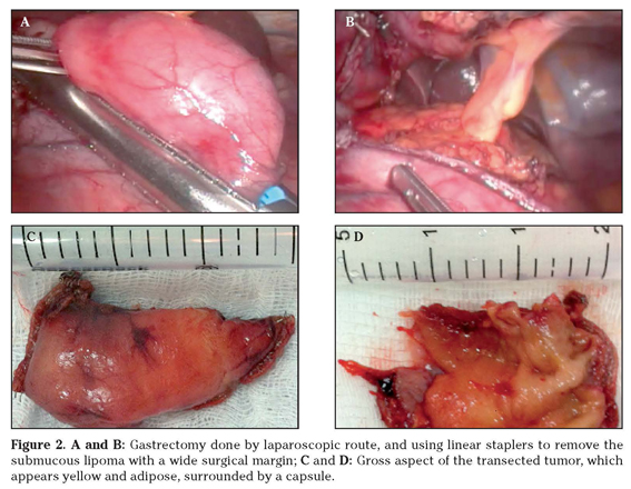
A 51-year-old Brazilian woman was admitted because of a severe epigastric pain associated with episodes of vomiting, dyspepsia, dysphagia and weight loss of 5 kg during 8 months. Her body mass index was 19.81Kg/m<sup>2</sup>, and the routine laboratory determinations were unremarkable. She underwent three endoscopy studies that disclosed an antral mass with around 3 cm (<a href="#f1">Fig. 1A-B</a>). PET/CT images revealed a spherical hypoattenuating mass (31 mm x 20 mm) with fat density, without enhanced metabolism, at the antrum; and the ecoendoscopy study found a submucosal homogeneous hyperecogenic mass (27 mm x 20 mm) on the same site. The specimens obtained from two routine endoscopic biopsies revealed normal mucosa. Another biopsy was performed - guided by endoscopic ultrasound, and revealed mature fat tissue at the submucosa, and diagnosis of submucosal lipoma was characterized (<a href="#f1">Fig. 1C-D</a>). She underwent an atypical gastrectomy by laparoscopic route, and employing linear staplers (<a href="#f2">Fig. 2A-B</a>) to remove the gastric mass along with a wide free surgical margin. Grossly, the aspect of transected tumor was yellow and adipose (<a href="#f2">Fig. 2C-D</a>), with a capsule. On the fifth postoperative day, the patient presented with vomiting and dysphagia, and the endoscopic evaluation revealed a pyloric substenosis, corrected by dilation. After an uneventful evolution, she was discharged to home; nevertheless, five days later, she claimed of anorexia and recurrent vomiting, and another endoscopy detected pyloric obstruction. Therefore, the patient was submitted to a pylorotomy, and was discharged to outpatient surveillance on the third postoperative day, asymptomatic and accepting solid foods.</font></p>     <p>&nbsp;</p>     <p align="center"><font face="Verdana" size="2"><a name="f1"><img src="/img/revistas/asisna/v36n1/nota2_f1.jpg"></a></font></p>     <p>&nbsp;</p>     <p align="center"><font face="Verdana" size="2"><a name="f2"><img src="/img/revistas/asisna/v36n1/nota2_f2.jpg"></a></font></p>     <p>&nbsp;</p>     <p>&nbsp;</p>     ]]></body>
<body><![CDATA[<p><font face="Verdana" size="2"><b>Discussion</b></font></p>     <p><font face="Verdana" size="2">This 51-year-old female presented with clinical features typical of gastrointestinal obstruction and her age was in accordance with the mean age of patients at diagnosis of gastric lipomas<sup>5,8</sup>. However, no specific symptoms could hardly contribute to raise suspicion about gastric lipoma, and the final diagnosis was based on imaging investigation and further histopathology studies. The upper digestive endoscopy showed a submucosal mass measuring about 3 cm in diameter. The first two biopsy procedures did not furnished enough tissue to histopathology analysis, phenomenon that is frequently reported in literature<sup>3-5,8,9</sup>. The tissue samples from the third biopsy, which was guided by endoscopic ultrasound, revealed the origin of the tumor - a classical lipoma. Surgical approach was done, as most of the authors recommend for symptomatic patients<sup>3-5,7-9,11</sup>. The occurrence of pyloric stenosis was an expected complication of the atypical gastrectomy, because the tumor resection was performed at the antrum, and with a large free surgical margin. Gastric lipomas predominate in people over than 50 years of age<sup>2,5,8,11</sup>, and appear as solitary and asymptomatic masses<sup>5-10</sup>. However, patients with gastric lipomatosis have been reported as an exceeding rare condition<sup>12</sup>. The symptoms depend on localization and size of the tumor and of associated entities<sup>2,7-10</sup>. Those lipomas developed at the antrum and the body of the stomach may origin intussusceptions into the pylorus causing obstruction and pain; while voluminous tumors may be associated with venous stasis, resulting in ulceration and hemorrhage<sup>6-10</sup>. Lipomas larger than 2 cm usually cause symptoms (diarrhea, obstruction, intussusception, hemorrhage)<sup>2,4,5,7,10</sup>. The most common symptoms are dyspepsia, epigastric pain, upper gastrointestinal bleeding (usually by ulceration and necrosis), obstruction and intussusception<sup>3-11</sup>. Because of the absence of symptoms, in the vast majority of cases gastric lipoma constitutes an endoscopic finding<sup>3,5,6,8,10</sup>, and the tumor often appears as a smooth, yellowish submucosal mass with or without ulceration<sup>3-5,10</sup>. Classical imaging aspects like «tenting», «cushion sign» and «naked fat sign» are useful to diagnose submucosal lipomas.<sup>2-7,11</sup> To perform a successful endoscopic biopsy of submucosal lipomas can be a challenging task<sup>2,4,5,9</sup>. In general, these biopsies only reveal a normal gastric mucosa<sup>2,3,6,8,9</sup>. The use of electrocautery to elicit a local mucosal ulceration, before the biopsy procedure, might be of some utility<sup>5</sup>. Computerized tomography (CT) is a highly specific tool that can contribute to diagnosis<sup>2,3-9,10</sup>. In CT images, the lipoma typically appears as a mass that is isodense (-70H to -120H) with fat<sup>3,4,6,7,9,10</sup>. Endoscopic ultrasound is useful to diagnosis of gastric lipoma, showing the hyperecoic density of the tumor in the submucosa<sup>2,3-10</sup>. Gastric lipomas do not have malignant potential<sup>2,4-6,9</sup>, however, synchronous gastric carcinomas have been very rarely described<sup>6,9</sup>. Treatment of gastric lipoma, is controversial<sup>4,9</sup>, but resection is the best choice for symptomatic tumors<sup>2-5,8,10</sup>. Laparoscopic route is used for tumors up to 6 cm in diameter<sup>2,7</sup>. Pedunculated lipomas smaller than 3 cm may be excised by upper digestive endoscopy route<sup>4,7,8,11</sup>, but mere observation is an alternative<sup>2,4,7,10,11</sup>. Hemorrhages can be treated by endoscopic clipping<sup>5</sup>.</font></p>     <p>&nbsp;</p>     <p><font face="Verdana" size="2"><b>References</b></font></p>     <!-- ref --><p><font face="Verdana" size="2">1. Calvo AM, Montón S, Rubio T, Repiso M, Sarasibar H. Invaginación intestinal en el adulto secundaria a lipoma yeyunal. An Sist Sanit Navar 2005; 28: 115-118.    &nbsp;&nbsp;&nbsp;&nbsp;&nbsp;&nbsp;&nbsp;&nbsp;[&#160;<a href="javascript:void(0);" onclick="javascript: window.open('/scielo.php?script=sci_nlinks&ref=401073&pid=S1137-6627201300010002000001&lng=','','width=640,height=500,resizable=yes,scrollbars=1,menubar=yes,');">Links</a>&#160;]<!-- end-ref --></font></p>    <!-- ref --><p><font face="Verdana" size="2">2. Neto FA, Ferreira MC, Bertoncello LC, Neto AA, De Aveiro WC, Bento CA, et al. Gastric lipoma presenting as a giant bulging mass in an oligosymptomatic patient: a case report. J Med Case Rep 2012;6:317.    &nbsp;&nbsp;&nbsp;&nbsp;&nbsp;&nbsp;&nbsp;&nbsp;[&#160;<a href="javascript:void(0);" onclick="javascript: window.open('/scielo.php?script=sci_nlinks&ref=401075&pid=S1137-6627201300010002000002&lng=','','width=640,height=500,resizable=yes,scrollbars=1,menubar=yes,');">Links</a>&#160;]<!-- end-ref --></font></p>    <!-- ref --><p><font face="Verdana" size="2">3. Penston J, Penston V. Gastric lipoma: a rare cause of iron-deficiency anaemia. BMJ Case Rep 2009; 2009. pii: bcr09.2008.0954. Epub 2009 Mar 24.    &nbsp;&nbsp;&nbsp;&nbsp;&nbsp;&nbsp;&nbsp;&nbsp;[&#160;<a href="javascript:void(0);" onclick="javascript: window.open('/scielo.php?script=sci_nlinks&ref=401077&pid=S1137-6627201300010002000003&lng=','','width=640,height=500,resizable=yes,scrollbars=1,menubar=yes,');">Links</a>&#160;]<!-- end-ref --></font></p>    ]]></body>
<body><![CDATA[<!-- ref --><p><font face="Verdana" size="2">4. Sadio A, Peixoto P, Castanheira A, Cancela E, Ministro P, Casimiro C, et al. Gastric lipoma - an unusual cause of upper gastrointestinal bleeding. Rev Esp Enferm Dig 2010: 102: 398-400.    &nbsp;&nbsp;&nbsp;&nbsp;&nbsp;&nbsp;&nbsp;&nbsp;[&#160;<a href="javascript:void(0);" onclick="javascript: window.open('/scielo.php?script=sci_nlinks&ref=401079&pid=S1137-6627201300010002000004&lng=','','width=640,height=500,resizable=yes,scrollbars=1,menubar=yes,');">Links</a>&#160;]<!-- end-ref --></font></p>    <!-- ref --><p><font face="Verdana" size="2">5. Saltzman JR, Carr-Locke DL, Fink SA. Lipoma case report. Med Gen Med 2005; 7: 16.    &nbsp;&nbsp;&nbsp;&nbsp;&nbsp;&nbsp;&nbsp;&nbsp;[&#160;<a href="javascript:void(0);" onclick="javascript: window.open('/scielo.php?script=sci_nlinks&ref=401081&pid=S1137-6627201300010002000005&lng=','','width=640,height=500,resizable=yes,scrollbars=1,menubar=yes,');">Links</a>&#160;]<!-- end-ref --></font></p>    <!-- ref --><p><font face="Verdana" size="2">6. Yamamoto T, Imakiire K, Hashiguchi S, Matsumoto J, Kadono J, Hamada N, et al. A rare case of gastric lipoma with early gastric cancer. Intern Med 2004;43: 1039-1041.    &nbsp;&nbsp;&nbsp;&nbsp;&nbsp;&nbsp;&nbsp;&nbsp;[&#160;<a href="javascript:void(0);" onclick="javascript: window.open('/scielo.php?script=sci_nlinks&ref=401083&pid=S1137-6627201300010002000006&lng=','','width=640,height=500,resizable=yes,scrollbars=1,menubar=yes,');">Links</a>&#160;]<!-- end-ref --></font></p>    <!-- ref --><p><font face="Verdana" size="2">7. Zameer M, Kanojia RP, Rao KL, Menon P, Samujh R, Thapa BR. Gastric lipoma. J Indian Assoc Pediatr Surg 2010; 15: 64-66.    &nbsp;&nbsp;&nbsp;&nbsp;&nbsp;&nbsp;&nbsp;&nbsp;[&#160;<a href="javascript:void(0);" onclick="javascript: window.open('/scielo.php?script=sci_nlinks&ref=401085&pid=S1137-6627201300010002000007&lng=','','width=640,height=500,resizable=yes,scrollbars=1,menubar=yes,');">Links</a>&#160;]<!-- end-ref --></font></p>    <!-- ref --><p><font face="Verdana" size="2">8. Kim DD, Tsai AI, Otani AR, Puglia CR, Malheiros CA. Lipoma gástrico: case report. Rev Col Bras Cir 2011; 38: 205-206.    &nbsp;&nbsp;&nbsp;&nbsp;&nbsp;&nbsp;&nbsp;&nbsp;[&#160;<a href="javascript:void(0);" onclick="javascript: window.open('/scielo.php?script=sci_nlinks&ref=401087&pid=S1137-6627201300010002000008&lng=','','width=640,height=500,resizable=yes,scrollbars=1,menubar=yes,');">Links</a>&#160;]<!-- end-ref --></font></p>    ]]></body>
<body><![CDATA[<!-- ref --><p><font face="Verdana" size="2">9. Krasniqi AS, Hoxha FT, Bicaj BX, Hashani SI, Hasimja SM, Kelmendi SM, et al. Symptomatic subserosal gastric lipoma successfully treated with enucleation. World J Gastroenterol 2008; 14: 5930-5932.    &nbsp;&nbsp;&nbsp;&nbsp;&nbsp;&nbsp;&nbsp;&nbsp;[&#160;<a href="javascript:void(0);" onclick="javascript: window.open('/scielo.php?script=sci_nlinks&ref=401089&pid=S1137-6627201300010002000009&lng=','','width=640,height=500,resizable=yes,scrollbars=1,menubar=yes,');">Links</a>&#160;]<!-- end-ref --></font></p>    <!-- ref --><p><font face="Verdana" size="2">10. Thompson WM, Kende AI, Levy AD. Imaging characteristics of g 10. DONE HJ. Lipoma of the stomach. Br Med J 1961; 1: 183.    &nbsp;&nbsp;&nbsp;&nbsp;&nbsp;&nbsp;&nbsp;&nbsp;[&#160;<a href="javascript:void(0);" onclick="javascript: window.open('/scielo.php?script=sci_nlinks&ref=401091&pid=S1137-6627201300010002000010&lng=','','width=640,height=500,resizable=yes,scrollbars=1,menubar=yes,');">Links</a>&#160;]<!-- end-ref --></font></p>    <!-- ref --><p><font face="Verdana" size="2">11. Bijlani RS, Kulkarni VM, Shahani RB, Shah HK, Dalvi A, Samsi AB. Gastric lipoma presenting as obstruction and hematemesis. J Postgrad Med 1993; 39: 42-43.    &nbsp;&nbsp;&nbsp;&nbsp;&nbsp;&nbsp;&nbsp;&nbsp;[&#160;<a href="javascript:void(0);" onclick="javascript: window.open('/scielo.php?script=sci_nlinks&ref=401093&pid=S1137-6627201300010002000011&lng=','','width=640,height=500,resizable=yes,scrollbars=1,menubar=yes,');">Links</a>&#160;]<!-- end-ref --></font></p>     <p>&nbsp;</p>     <p>&nbsp;</p>     <p><font face="Verdana" size="2"><a href="#top"><img border="0" src="/img/revistas/asisna/v36n1/seta.gif" width="15" height="17"></a><a name="bajo"></a><b>Dirección para correspondencia:</b>    <br>Vitorino Modesto dos Santos    ]]></body>
<body><![CDATA[<br>Hospital das Forças Armadas    <br>Estrada do contorno do bosque, s/n    <br>Cruzeiro Novo, 70658-900    <br>Brasilia-DF    <br>E-mail:  <a href="mailto:vitorinomodestos@gmail.com">vitorinomodestos@gmail.com</a></font></p>     <p><font face="Verdana" size="2">Recepción: 30 de octubre de 2012    <br>Aceptación definitiva: 21 de noviembre de 2012</font></p>      ]]></body><back>
<ref-list>
<ref id="B1">
<label>1</label><nlm-citation citation-type="journal">
<person-group person-group-type="author">
<name>
<surname><![CDATA[Calvo]]></surname>
<given-names><![CDATA[AM]]></given-names>
</name>
<name>
<surname><![CDATA[Montón]]></surname>
<given-names><![CDATA[S]]></given-names>
</name>
<name>
<surname><![CDATA[Rubio]]></surname>
<given-names><![CDATA[T]]></given-names>
</name>
<name>
<surname><![CDATA[Repiso]]></surname>
<given-names><![CDATA[M]]></given-names>
</name>
<name>
<surname><![CDATA[Sarasibar]]></surname>
<given-names><![CDATA[H]]></given-names>
</name>
</person-group>
<article-title xml:lang="es"><![CDATA[Invaginación intestinal en el adulto secundaria a lipoma yeyunal]]></article-title>
<source><![CDATA[An Sist Sanit Navar]]></source>
<year>2005</year>
<volume>28</volume>
<page-range>115-118</page-range></nlm-citation>
</ref>
<ref id="B2">
<label>2</label><nlm-citation citation-type="journal">
<person-group person-group-type="author">
<name>
<surname><![CDATA[Neto]]></surname>
<given-names><![CDATA[FA]]></given-names>
</name>
<name>
<surname><![CDATA[Ferreira]]></surname>
<given-names><![CDATA[MC]]></given-names>
</name>
<name>
<surname><![CDATA[Bertoncello]]></surname>
<given-names><![CDATA[LC]]></given-names>
</name>
<name>
<surname><![CDATA[Neto]]></surname>
<given-names><![CDATA[AA]]></given-names>
</name>
<name>
<surname><![CDATA[De Aveiro]]></surname>
<given-names><![CDATA[WC]]></given-names>
</name>
<name>
<surname><![CDATA[Bento]]></surname>
<given-names><![CDATA[CA]]></given-names>
</name>
</person-group>
<article-title xml:lang="en"><![CDATA[Gastric lipoma presenting as a giant bulging mass in an oligosymptomatic patient: a case report]]></article-title>
<source><![CDATA[J Med Case Rep]]></source>
<year>2012</year>
<volume>6</volume>
<page-range>317</page-range></nlm-citation>
</ref>
<ref id="B3">
<label>3</label><nlm-citation citation-type="journal">
<person-group person-group-type="author">
<name>
<surname><![CDATA[Penston]]></surname>
<given-names><![CDATA[J]]></given-names>
</name>
<name>
<surname><![CDATA[Penston]]></surname>
<given-names><![CDATA[V]]></given-names>
</name>
</person-group>
<article-title xml:lang="en"><![CDATA[Gastric lipoma: a rare cause of iron-deficiency anaemia]]></article-title>
<source><![CDATA[BMJ Case Rep]]></source>
<year>2009</year>
</nlm-citation>
</ref>
<ref id="B4">
<label>4</label><nlm-citation citation-type="journal">
<person-group person-group-type="author">
<name>
<surname><![CDATA[Sadio]]></surname>
<given-names><![CDATA[A]]></given-names>
</name>
<name>
<surname><![CDATA[Peixoto]]></surname>
<given-names><![CDATA[P]]></given-names>
</name>
<name>
<surname><![CDATA[Castanheira]]></surname>
<given-names><![CDATA[A]]></given-names>
</name>
<name>
<surname><![CDATA[Cancela]]></surname>
<given-names><![CDATA[E]]></given-names>
</name>
<name>
<surname><![CDATA[Ministro]]></surname>
<given-names><![CDATA[P]]></given-names>
</name>
<name>
<surname><![CDATA[Casimiro]]></surname>
<given-names><![CDATA[C]]></given-names>
</name>
</person-group>
<article-title xml:lang="en"><![CDATA[Gastric lipoma: an unusual cause of upper gastrointestinal bleeding]]></article-title>
<source><![CDATA[Rev Esp Enferm Dig]]></source>
<year>2010</year>
<volume>102</volume>
<page-range>398-400</page-range></nlm-citation>
</ref>
<ref id="B5">
<label>5</label><nlm-citation citation-type="journal">
<person-group person-group-type="author">
<name>
<surname><![CDATA[Saltzman]]></surname>
<given-names><![CDATA[JR]]></given-names>
</name>
<name>
<surname><![CDATA[Carr-Locke]]></surname>
<given-names><![CDATA[DL]]></given-names>
</name>
<name>
<surname><![CDATA[Fink]]></surname>
<given-names><![CDATA[SA]]></given-names>
</name>
</person-group>
<article-title xml:lang="en"><![CDATA[Lipoma case report]]></article-title>
<source><![CDATA[Med Gen Med]]></source>
<year>2005</year>
<volume>7</volume>
<page-range>16</page-range></nlm-citation>
</ref>
<ref id="B6">
<label>6</label><nlm-citation citation-type="journal">
<person-group person-group-type="author">
<name>
<surname><![CDATA[Yamamoto]]></surname>
<given-names><![CDATA[T]]></given-names>
</name>
<name>
<surname><![CDATA[Imakiire]]></surname>
<given-names><![CDATA[K]]></given-names>
</name>
<name>
<surname><![CDATA[Hashiguchi]]></surname>
<given-names><![CDATA[S]]></given-names>
</name>
<name>
<surname><![CDATA[Matsumoto]]></surname>
<given-names><![CDATA[J]]></given-names>
</name>
<name>
<surname><![CDATA[Kadono]]></surname>
<given-names><![CDATA[J]]></given-names>
</name>
<name>
<surname><![CDATA[Hamada]]></surname>
<given-names><![CDATA[N]]></given-names>
</name>
</person-group>
<article-title xml:lang="en"><![CDATA[A rare case of gastric lipoma with early gastric cancer]]></article-title>
<source><![CDATA[Intern Med]]></source>
<year>2004</year>
<volume>43</volume>
<page-range>1039-1041</page-range></nlm-citation>
</ref>
<ref id="B7">
<label>7</label><nlm-citation citation-type="journal">
<person-group person-group-type="author">
<name>
<surname><![CDATA[Zameer]]></surname>
<given-names><![CDATA[M]]></given-names>
</name>
<name>
<surname><![CDATA[Kanojia]]></surname>
<given-names><![CDATA[RP]]></given-names>
</name>
<name>
<surname><![CDATA[Rao]]></surname>
<given-names><![CDATA[KL]]></given-names>
</name>
<name>
<surname><![CDATA[Menon]]></surname>
<given-names><![CDATA[P]]></given-names>
</name>
<name>
<surname><![CDATA[Samujh]]></surname>
<given-names><![CDATA[R]]></given-names>
</name>
<name>
<surname><![CDATA[Thapa]]></surname>
<given-names><![CDATA[BR]]></given-names>
</name>
</person-group>
<article-title xml:lang="en"><![CDATA[Gastric lipoma]]></article-title>
<source><![CDATA[J Indian Assoc Pediatr Surg]]></source>
<year>2010</year>
<volume>15</volume>
<page-range>64-66</page-range></nlm-citation>
</ref>
<ref id="B8">
<label>8</label><nlm-citation citation-type="journal">
<person-group person-group-type="author">
<name>
<surname><![CDATA[Kim]]></surname>
<given-names><![CDATA[DD]]></given-names>
</name>
<name>
<surname><![CDATA[Tsai]]></surname>
<given-names><![CDATA[AI]]></given-names>
</name>
<name>
<surname><![CDATA[Otani]]></surname>
<given-names><![CDATA[AR]]></given-names>
</name>
<name>
<surname><![CDATA[Puglia]]></surname>
<given-names><![CDATA[CR]]></given-names>
</name>
<name>
<surname><![CDATA[Malheiros]]></surname>
<given-names><![CDATA[CA]]></given-names>
</name>
</person-group>
<article-title xml:lang="es"><![CDATA[Lipoma gástrico: case report]]></article-title>
<source><![CDATA[Rev Col Bras Cir]]></source>
<year>2011</year>
<volume>38</volume>
<page-range>205-206</page-range></nlm-citation>
</ref>
<ref id="B9">
<label>9</label><nlm-citation citation-type="journal">
<person-group person-group-type="author">
<name>
<surname><![CDATA[Krasniqi]]></surname>
<given-names><![CDATA[AS]]></given-names>
</name>
<name>
<surname><![CDATA[Hoxha]]></surname>
<given-names><![CDATA[FT]]></given-names>
</name>
<name>
<surname><![CDATA[Bicaj]]></surname>
<given-names><![CDATA[BX]]></given-names>
</name>
<name>
<surname><![CDATA[Hashani]]></surname>
<given-names><![CDATA[SI]]></given-names>
</name>
<name>
<surname><![CDATA[Hasimja]]></surname>
<given-names><![CDATA[SM]]></given-names>
</name>
<name>
<surname><![CDATA[Kelmendi]]></surname>
<given-names><![CDATA[SM]]></given-names>
</name>
</person-group>
<article-title xml:lang="en"><![CDATA[Symptomatic subserosal gastric lipoma successfully treated with enucleation]]></article-title>
<source><![CDATA[World J Gastroenterol]]></source>
<year>2008</year>
<volume>14</volume>
<page-range>5930-5932</page-range></nlm-citation>
</ref>
<ref id="B10">
<label>10</label><nlm-citation citation-type="journal">
<person-group person-group-type="author">
<name>
<surname><![CDATA[Thompson]]></surname>
<given-names><![CDATA[WM]]></given-names>
</name>
<name>
<surname><![CDATA[Kende]]></surname>
<given-names><![CDATA[AI]]></given-names>
</name>
<name>
<surname><![CDATA[Levy]]></surname>
<given-names><![CDATA[AD]]></given-names>
</name>
</person-group>
<article-title xml:lang="en"><![CDATA[Imaging characteristics of g 10: DONE HJ. Lipoma of the stomach]]></article-title>
<source><![CDATA[Br Med J]]></source>
<year>1961</year>
<volume>1</volume>
<page-range>183</page-range></nlm-citation>
</ref>
<ref id="B11">
<label>11</label><nlm-citation citation-type="journal">
<person-group person-group-type="author">
<name>
<surname><![CDATA[Bijlani]]></surname>
<given-names><![CDATA[RS]]></given-names>
</name>
<name>
<surname><![CDATA[Kulkarni]]></surname>
<given-names><![CDATA[VM]]></given-names>
</name>
<name>
<surname><![CDATA[Shahani]]></surname>
<given-names><![CDATA[RB]]></given-names>
</name>
<name>
<surname><![CDATA[Shah]]></surname>
<given-names><![CDATA[HK]]></given-names>
</name>
<name>
<surname><![CDATA[Dalvi]]></surname>
<given-names><![CDATA[A]]></given-names>
</name>
<name>
<surname><![CDATA[Samsi]]></surname>
<given-names><![CDATA[AB]]></given-names>
</name>
</person-group>
<article-title xml:lang="en"><![CDATA[Gastric lipoma presenting as obstruction and hematemesis]]></article-title>
<source><![CDATA[J Postgrad Med]]></source>
<year>1993</year>
<volume>39</volume>
<page-range>42-43</page-range></nlm-citation>
</ref>
</ref-list>
</back>
</article>
