<?xml version="1.0" encoding="ISO-8859-1"?><article xmlns:mml="http://www.w3.org/1998/Math/MathML" xmlns:xlink="http://www.w3.org/1999/xlink" xmlns:xsi="http://www.w3.org/2001/XMLSchema-instance">
<front>
<journal-meta>
<journal-id>1139-6709</journal-id>
<journal-title><![CDATA[International Microbiology]]></journal-title>
<abbrev-journal-title><![CDATA[INT. MICROBIOL.]]></abbrev-journal-title>
<issn>1139-6709</issn>
<publisher>
<publisher-name><![CDATA[Sociedad Española de Microbiología]]></publisher-name>
</publisher>
</journal-meta>
<article-meta>
<article-id>S1139-67092006000100004</article-id>
<title-group>
<article-title xml:lang="en"><![CDATA[Immuno-electron microscopic quantification of the fucoxanthin chlorophyll a/c binding polypeptides Fcp2, Fcp4, and Fcp6 of Cyclotella cryptica grown under low- and high-light intensities]]></article-title>
<article-title xml:lang="es"><![CDATA[Cuantificación por microscopia inmunoelectrónica de los polipéptidos Fcp2, Fcp4 y Fcp6 que se unen a la fucoxantina clorofila a/c de Cyclotella cryptica cultivada en condiciones de baja y alta intensidad lumínica]]></article-title>
<article-title xml:lang="pt"><![CDATA[Quantificação por microscopia inmunoelectrónica dos polipéptideos Fcp2, Fcp4 e Fcp6 que se unem à fucoxantina clorofila a/c de Cyclotella cryptica cultivada em condições de baixa e alta intensidade de luz]]></article-title>
</title-group>
<contrib-group>
<contrib contrib-type="author">
<name>
<surname><![CDATA[Becker]]></surname>
<given-names><![CDATA[Frank]]></given-names>
</name>
<xref ref-type="aff" rid="A01"/>
</contrib>
<contrib contrib-type="author">
<name>
<surname><![CDATA[Rhiel]]></surname>
<given-names><![CDATA[Erhard]]></given-names>
</name>
<xref ref-type="aff" rid="A01"/>
</contrib>
</contrib-group>
<aff id="A01">
<institution><![CDATA[,University of Oldenburg ICBM Geomicrobiology]]></institution>
<addr-line><![CDATA[ ]]></addr-line>
<country>Germany</country>
</aff>
<pub-date pub-type="pub">
<day>00</day>
<month>03</month>
<year>2006</year>
</pub-date>
<pub-date pub-type="epub">
<day>00</day>
<month>03</month>
<year>2006</year>
</pub-date>
<volume>9</volume>
<numero>1</numero>
<fpage>29</fpage>
<lpage>36</lpage>
<copyright-statement/>
<copyright-year/>
<self-uri xlink:href="http://scielo.isciii.es/scielo.php?script=sci_arttext&amp;pid=S1139-67092006000100004&amp;lng=en&amp;nrm=iso"></self-uri><self-uri xlink:href="http://scielo.isciii.es/scielo.php?script=sci_abstract&amp;pid=S1139-67092006000100004&amp;lng=en&amp;nrm=iso"></self-uri><self-uri xlink:href="http://scielo.isciii.es/scielo.php?script=sci_pdf&amp;pid=S1139-67092006000100004&amp;lng=en&amp;nrm=iso"></self-uri><abstract abstract-type="short" xml:lang="en"><p><![CDATA[The diatom Cyclotella cryptica was grown under low- and high-intensity white light of 50 and 500 µmol photons m-2 s-1, respectively. Western immunoblotting showed that the diatom adapted its light-harvesting apparatus, giving rise to different amounts of distinct fucoxanthin chlorophyll a/c binding polypeptides (Fcp). The amount of Fcp2 was approximately two-fold higher under low-light than under high-light conditions, whereas the amount of Fcp6 increased four- to five-fold under high-light conditions. For Fcp4, no significant differences were detected in response to either light regime. Cells of Cyclotella grown under high- and low-light intensity were subjected to immunoelectron microscopy. Quantification of the gold label, expressed as gold particles per µm², confirmed the results obtained by Western immunoblotting. Exposure to low light resulted in the detection of approximately six times more Fcp2-bound gold particles per µm² in thylakoid membranes, whereas in cells grown under high light the number of Fcp6-bound gold particles increased ten-fold. For Fcp4, similar amounts of gold particles per µm² were counted under the two light regimes. These immunocytochemical results confirmed molecular data derived from phylogenetic analyses of the sequences of genes encoding fucoxanthin chlorophyll a/c binding polypeptides (fcp genes) and from measurements of steady-state fcp mRNA concentrations. The results show that Fcp2 and Fcp6 accumulate under low- and high-light intensity, respectively, whereas Fcp4 seems to be constitutively synthesized.]]></p></abstract>
<abstract abstract-type="short" xml:lang="es"><p><![CDATA[La diatomea Cyclotella cryptica se cultivó a intensidades de luz blanca baja y alta (50 y 500 fotones m-2 s-1 µmol, respectivamente). La inmunotransferencia mostró que la diatomea adaptó su aparato captador de luz, produciendo cantidades diferentes de polipéptidos de unión a la fucoxantina clorofila a/c (Fcp). A intensidad baja de luz, la cantidad de Fcp2 era aproximadamente el doble que a intensidad elevada, mientras que a intensidad elevada la cantidad de Fcp6 era de cuatro a cinco veces la cantidad producida a baja intensidad. No se detectaron diferencias significativas para Fcp4 como respuesta a ninguno de los dos tipos de luz. Las células de Cyclotella cultivadas a intensidades alta y baja de luz se observaron por microscopia immunoelectrónica. La cuantificación del marcado de oro, expresada como partículas de oro por µm² confirmó los resultados obtenidos por inmunotransferencia. Tras la exposición a luz de baja intensidad se detectaron aproximadamente seis veces más partículas oro unidas a Fcp2 por µm² en las membranas de los tilacoides, mientras que en las células cultivadas a intensidad elevada, el número de partícula de oro unidas a Fcp6 aumentó diez veces. Para Fcp4, se contaron cantidades similares de partículas oro por µm² en los dos regímenes de luz. Estos resultados immunocitoquímicos confirmaron los datos moleculares derivados de los análisis filogenéticos de las secuencias de los genes que codifican los polipéptidos de unión de la fucoxantina clorofila a/c (genes fcp) y de la medida de las concentraciones de mRNA fcp en el estado estacionario. Los resultados demuestran que Fcp2 y Fcp6 se acumulan, respectivamente, a intensidades de luz alta y baja, mientras que Fcp4 parece sintetizado de manera constitutiva.]]></p></abstract>
<abstract abstract-type="short" xml:lang="pt"><p><![CDATA[A diatomácea Cyclotella cryptica foi cultivada sob intensidade baixa e alta de luz blanca (50 e 500 fótons m-2 s-1 µmol, respectivamente). A inmunotransferência mostrou que a diatomácea adaptou seu aparelho captador de luz, produzindo quantidades diferentes de polipéptidos de união à fucoxantina clorofila a/c (Fcp). Sob intensidade baixa de luz, a quantidade de Fcp2 foi aproximadamente o dobro em relação à intensidade elevada, enquanto sob intensidade elevada, a quantidade de Fcp6 foi de quatro a cinco vezes maior do que a quantidade produzida sob baixa intensidade de luz. Não foi detectada diferença significativa para Fcp4 como resposta a nenhum dos dois tipos de luz. As células de Cyclotella cultivadas sob intensidades alta e baixa de luz foram observadas por microscopia immunoelectrônica. A quantificação do marcado de ouro, expresso como partículas de ouro por µm² confirmou os resultados obtidos por inmunotransferência. Após exposição à luz de baixa intensidade foram detectadas aproximadamente seis vezes maispartículas ouro unidas a Fcp2 por µm² nas membranas dos tilacoides, enquanto nas células cultivadas sob intensidade elevada o número de partícula de ouro unidas a Fcp6 aumentou em dez vezes. Para Fcp4, foram contadas quantidades similares de partículas ouro por µm² nos dois regimes de luz. Estes resultados immunocitoquímicos confirmaram os dados moleculares derivados das análises filogenéticas das seqüências dos genes que codificam os polipéptidos de união da fucoxantina clorofila a/c (genes fcp) e da medida das concentrações de mRNA fcp no estado estacionário. Os resultados demonstram que Fcp2 e Fcp6 se acumulam, respectivamente, sob intensidades de luz alta e baixa , enquanto Fcp4 parece ser sintetizado de maneira constitutiva.]]></p></abstract>
<kwd-group>
<kwd lng="en"><![CDATA[Cyclotella]]></kwd>
<kwd lng="en"><![CDATA[Bacillariophyceae]]></kwd>
<kwd lng="en"><![CDATA[photosynthesis]]></kwd>
<kwd lng="en"><![CDATA[light-harvesting complexes]]></kwd>
<kwd lng="en"><![CDATA[immunogold-labeling electron microscopy]]></kwd>
<kwd lng="es"><![CDATA[Cyclotella]]></kwd>
<kwd lng="es"><![CDATA[Bacillariophyceae]]></kwd>
<kwd lng="es"><![CDATA[fotosíntesis]]></kwd>
<kwd lng="es"><![CDATA[complejos captadores de luz]]></kwd>
<kwd lng="es"><![CDATA[microscopia inmunoelectrónica marcada con oro]]></kwd>
<kwd lng="pt"><![CDATA[Cyclotella]]></kwd>
<kwd lng="pt"><![CDATA[Bacillariophyceae]]></kwd>
<kwd lng="pt"><![CDATA[fotossíntese]]></kwd>
<kwd lng="pt"><![CDATA[complexos absorvedores da luz]]></kwd>
<kwd lng="pt"><![CDATA[microscopia electrônica com imunomarcação por ouro]]></kwd>
</kwd-group>
</article-meta>
</front><body><![CDATA[ <p align="left"><font face="Verdana, Arial, Helvetica, sans-serif"><b><font size="2">RESEARCH ARTICLE </font></b></font></p>     <p>&nbsp;</p>     <p><font face="Verdana, Arial, Helvetica, sans-serif" size="4"><b><a name="top"> </a>Immuno-electron microscopic quantification of thefucoxanthin chlorophyll <i>a/c</i> binding polypeptides Fcp2, Fcp4, and Fcp6 of <i>Cyclotella cryptica</i> grown under low- and high-light intensities</b></font></p>     <p>&nbsp;</p>     <p>&nbsp;</p>      <p><font face="Verdana, Arial, Helvetica, sans-serif" size="2"><b>Frank Becker; Erhard Rhiel</b></font></p>      <p><font size="2" face="Verdana, Arial, Helvetica, sans-serif">Geomicrobiology, ICBM, University of Oldenburg, Germany</font></p>     <p><font face="Verdana, Arial, Helvetica, sans-serif" size="2"><a href="#back">Corresponding author</a></font></p>     <p>&nbsp;</p>     <p>&nbsp;</p> <hr size="1" noshade color="#000000">     ]]></body>
<body><![CDATA[<p><font face="Verdana, Arial, Helvetica, sans-serif" size="2"><b>SUMMARY</b></font></p>      <p><font face="Verdana, Arial, Helvetica, sans-serif" size="2">The diatom <i>Cyclotella cryptica</i> was grown under low- and high-intensity white light of 50 and 500 &#181;mol photons m<sup>&#150;2</sup> s<sup>&#150;1</sup>, respectively. Western immunoblotting showed that the diatom adapted its light-harvesting apparatus, giving rise to different amounts of distinct fucoxanthin chlorophyll <i>a/c</i> binding polypeptides (Fcp). The amount of Fcp2 was approximately two-fold higher under low-light than under high-light conditions, whereas the amount of Fcp6 increased four- to five-fold under high-light conditions. For Fcp4, no significant differences were detected in response to either light regime. Cells of <i>Cyclotella</i> grown under high- and low-light intensity were subjected to immunoelectron microscopy. Quantification of the gold label, expressed as gold particles per &#181;m<sup>2</sup>, confirmed the results obtained by Western immunoblotting. Exposure to low light resulted in the detection of approximately six times more Fcp2-bound gold particles per &#181;m<sup>2</sup> in thylakoid membranes, whereas in cells grown under high light the number of Fcp6-bound gold particles increased ten-fold. For Fcp4, similar amounts of gold particles per &#181;m<sup>2</sup> were counted under the two light regimes. These immunocytochemical results confirmed molecular data derived from phylogenetic analyses of the sequences of genes encoding fucoxanthin chlorophyll <i>a/c</i> binding polypeptides (<i>fcp</i> genes) and from measurements of steady-state <i>fcp</i> mRNA concentrations. The results show that Fcp2 and Fcp6 accumulate under low- and high-light intensity, respectively, whereas Fcp4 seems to be constitutively synthesized. &#091;<b>Int Microbiol</b> 2006; 9(1):29-36&#093;.</font></p>      <p><font face="Verdana, Arial, Helvetica, sans-serif" size="2"><b>Key words:</b> <i>Cyclotella</i> &middot; Bacillariophyceae &middot; photosynthesis &middot; light-harvesting complexes &middot; immunogold-labeling electron microscopy</font></p>  <hr size="1" noshade color="#000000">     <p><font face="Verdana, Arial, Helvetica, sans-serif" size="2"><b>RESUMEN</b></font></p>     <p><font face="Verdana, Arial, Helvetica, sans-serif"><b><font size="2">Cuantificaci&oacute;n por microscopia inmunoelectr&oacute;nica de los polip&eacute;ptidos Fcp2, Fcp4 y Fcp6 que se unen a la fucoxantina clorofila <i>a/c</i> de <i>Cyclotella cryptica</i> cultivada en condiciones de baja y alta intensidad lum&iacute;nica</font></b></font></p>     <p><font size="2" face="Verdana, Arial, Helvetica, sans-serif">La diatomea <i>Cyclotella cryptica</i> se cultiv&oacute; a intensidades de luz blanca baja y alta (50 y 500 fotones m<sup>&#150;2</sup> s<sup>&#150;1</sup> &#181;mol, respectivamente). La inmunotransferencia mostr&oacute; que la diatomea adapt&oacute; su aparato captador de luz, produciendo cantidades diferentes de polip&eacute;ptidos de uni&oacute;n a la fucoxantina clorofila <i>a/c</i> (Fcp). A intensidad baja de luz, la cantidad de Fcp2 era aproximadamente el doble que a intensidad elevada, mientras que a intensidad elevada la cantidad de Fcp6 era de cuatro a cinco veces la cantidad producida a baja intensidad. No se detectaron diferencias significativas para Fcp4 como respuesta a ninguno de los dos tipos de luz. Las c&eacute;lulas de <i>Cyclotella</i> cultivadas a intensidades alta y baja de luz se observaron por microscopia immunoelectr&oacute;nica. La cuantificaci&oacute;n del marcado de oro, expresada como part&iacute;culas de oro por &#181;m<sup>2</sup> confirm&oacute; los resultados obtenidos por inmunotransferencia. Tras la exposici&oacute;n a luz de baja intensidad se detectaron aproximadamente seis veces m&aacute;s part&iacute;culas oro unidas a Fcp2 por &#181;m<sup>2</sup> en las membranas de los tilacoides, mientras que en las c&eacute;lulas cultivadas a intensidad elevada, el n&uacute;mero de part&iacute;cula de oro unidas a Fcp6 aument&oacute; diez veces. Para Fcp4, se contaron cantidades similares de part&iacute;culas oro por &#181;m<sup>2</sup> en los dos reg&iacute;menes de luz. Estos resultados immunocitoqu&iacute;micos confirmaron los datos moleculares derivados de los an&aacute;lisis filogen&eacute;ticos de las secuencias de los genes que codifican los polip&eacute;ptidos de uni&oacute;n de la fucoxantina clorofila <i>a/c</i> (genes <i>fcp</i>) y de la medida de las concentraciones de mRNA <i>fcp</i> en el estado estacionario. Los resultados demuestran que Fcp2 y Fcp6 se acumulan, respectivamente, a intensidades de luz alta y baja, mientras que Fcp4 parece sintetizado de manera constitutiva. &#091;<b>Int Microbiol</b> 2006; 9(1):29-36&#093;.</font></p>     <p><font face="Verdana, Arial, Helvetica, sans-serif" size="2"><b>Palabras clave:</b> <i>Cyclotella</i> &middot; Bacillariophyceae &middot; fotos&iacute;ntesis &middot; complejos captadores de luz &middot; microscopia inmunoelectr&oacute;nica marcada con oro</font></p> <hr size="1" noshade color="#000000">     <p><font face="Verdana, Arial, Helvetica, sans-serif" size="2"><b>RESUMO</b></font></p>     <p><font face="Verdana, Arial, Helvetica, sans-serif"><b><font size="2">Quantifica&ccedil;&atilde;o por microscopia inmunoelectr&oacute;nica dos polip&eacute;ptideos Fcp2, Fcp4 e Fcp6 que se unem &agrave; fucoxantina clorofila <i>a/c</i> de <i>Cyclotella cryptica</i> cultivada em condi&ccedil;&otilde;es de baixa e alta intensidade de luz</font></b></font></p>     <p><font size="2" face="Verdana, Arial, Helvetica, sans-serif">A diatom&aacute;cea <i>Cyclotella cryptica</i> foi cultivada sob intensidade baixa e alta de luz blanca (50 e 500 f&oacute;tons m<sup>&#150;2</sup> s<sup>&#150;1</sup> &#181;mol, respectivamente). A inmunotransfer&ecirc;ncia mostrou que a diatom&aacute;cea adaptou seu aparelho captador de luz, produzindo quantidades diferentes de polip&eacute;ptidos de uni&atilde;o &agrave; fucoxantina clorofila <i>a/c</i> (Fcp). Sob intensidade baixa de luz, a quantidade de Fcp2 foi aproximadamente o dobro em rela&ccedil;&atilde;o &agrave; intensidade elevada, enquanto sob intensidade elevada, a quantidade de Fcp6 foi de quatro a cinco vezes maior do que a quantidade produzida sob baixa intensidade de luz. N&atilde;o foi detectada diferen&ccedil;a significativa para Fcp4 como resposta a nenhum dos dois tipos de luz. As c&eacute;lulas de <i>Cyclotella</i> cultivadas sob intensidades alta e baixa de luz foram observadas por microscopia immunoelectr&ocirc;nica. A quantifica&ccedil;&atilde;o do marcado de ouro, expresso como part&iacute;culas de ouro por &#181;m<sup>2</sup> confirmou os resultados obtidos por inmunotransfer&ecirc;ncia. Ap&oacute;s exposi&ccedil;&atilde;o &agrave; luz de baixa intensidade foram detectadas aproximadamente seis vezes maispart&iacute;culas ouro unidas a Fcp2 por &#181;m<sup>2</sup> nas membranas dos tilacoides, enquanto nas c&eacute;lulas cultivadas sob intensidade elevada o n&uacute;mero de part&iacute;cula de ouro unidas a Fcp6 aumentou em dez vezes. Para Fcp4, foram contadas quantidades similares de part&iacute;culas ouro por &#181;m<sup>2</sup> nos dois regimes de luz. Estes resultados immunocitoqu&iacute;micos confirmaram os dados moleculares derivados das an&aacute;lises filogen&eacute;ticas das seq&uuml;&ecirc;ncias dos genes que codificam os polip&eacute;ptidos de uni&atilde;o da fucoxantina clorofila <i>a/c</i> (genes <i>fcp</i>) e da medida das concentra&ccedil;&otilde;es de mRNA <i>fcp</i> no estado estacion&aacute;rio. Os resultados demonstram que Fcp2 e Fcp6 se acumulam, respectivamente, sob intensidades de luz alta e baixa , enquanto Fcp4 parece ser sintetizado de maneira constitutiva. &#091;<b>Int Microbiol</b> 2006; 9(1):29-36&#093;.</font></p>     ]]></body>
<body><![CDATA[<p><font face="Verdana, Arial, Helvetica, sans-serif" size="2"><b>Palavras chave:</b> <i>Cyclotella</i> &middot; Bacillariophyceae &middot; fotoss&iacute;ntese &middot; complexos absorvedores da luz &middot; microscopia electr&ocirc;nica com imunomarca&ccedil;&atilde;o por ouro</font></p> <hr size="1" noshade>     <p>&nbsp;</p>      <p><font face="Verdana, Arial, Helvetica, sans-serif"><b><font size="3">Introduction</font></b></font></p>     <p><font size="2" face="Verdana, Arial, Helvetica, sans-serif">The light-harvesting complexes (LHCs) of photosynthetic eukaryotes function mainly in the absorption and transfer of light energy to the reaction centers of the two photosystems, photosystem I (PS I) and photosystem II (PS II) &#091;6,7,11,12&#093;. The LHCs of green plants have been well studied. They harbor chlorophyll (chl) <i>a</i>, chl <i>b</i>, and lutein, and are referred to as Cab (chl <i>a/b</i>) or LHC proteins. Some LHC-like proteins, such as PsbS, Elips, and LI8I8 of <i><i>Chlamydomonas</i> eugametos</i>, are expressed in response to excessive light and play a role in photoprotective mechanisms, preventing the reaction centers from photodamage &#091;5,7,17,18,26&#093;. Typical LHC proteins may also be involved in photoprotection. In these cases, the main mechanisms are via the xanthophyll cycle and photophosphorylation of LHC subunits &#091;e.g., 14,19&#093;.</font></p>     <p><font size="2" face="Verdana, Arial, Helvetica, sans-serif">Despite the fact that chromophytic algae (i.e., diatoms and brown algae) play an important role in aquatic ecosystems, our knowledge of their LHCs is still rather fragmentary. The LHC subunits of most chromophytic algae contain chl <i>a</i>, chl <i>c</i>, and fucoxanthin, and are known as Fcps (fucoxanthin chlorophyll <i>a/c</i> binding proteins) &#091;7,11&#093;. That Fcps function in photoprotection has been demonstrated by the presence of xanthophyll cycles in chromophytes &#091;8,16,29&#093;, but it remains unclear whether chromophytic algae express distinct Fcps in response to excessive light.</font></p>     <p><font size="2" face="Verdana, Arial, Helvetica, sans-serif">From the centric diatom <i>Cyclotella cryptica</i>, eight cDNA clones harboring genes encoding fucoxanthin chlorophyll <i>a/c</i> binding proteins (<i>fcp</i> genes) were previously cloned and analyzed &#091;1-3&#093;. Phylogenetic analysis revealed that three genes, <i>fcp6</i>, <i>fcp7</i>, and <i>fcp12</i>, showed homology to the light inducible Cab-like protein LI8I8 of <i>Chlamydomonas</i> &#091;2&#093;, and that they may represent Fcp candidates involved in photoprotection. In subsequent experiments, the steady-state <i>fcp</i> mRNA concentrations of <i>C. cryptica</i>, grown under low- or high-intensity white light, were quantified. The results showed that <i>fcp</i>6 and <i>fcp</i>12 mRNAs were enriched under high-light and <i>fcp</i>2 and <i>fcp</i>4 mRNAs under low-light conditions &#091;20,21&#093;. </font></p>     <p><font size="2" face="Verdana, Arial, Helvetica, sans-serif">The current work focuses on whether the different expression patterns seen in the steady-state mRNA concentrations finally become manifested in varying amounts of the corresponding gene products within the plastids. This was determined by using Western immunoblotting and ultrathin sectioning/immunogold labeling to measure the relative abundances of Fcp2, Fcp4, and Fcp6 of <i>C. cryptica</i> grown under low- and high-intensity white light.</font></p>     <p>&nbsp;</p>     <p><font face="Verdana, Arial, Helvetica, sans-serif"><b><font size="3">Materials and methods</font></b></font></p>     <p><font face="Verdana, Arial, Helvetica, sans-serif"><b><font size="2">Culture growth and cell preparation. </font></b></font><font size="2" face="Verdana, Arial, Helvetica, sans-serif"><i>Cyclotella cryptica</i> (strain 1070-1) was obtained from the "Sammlung von Algenkulturen" at G&ouml;ttingen (SAG) &#091;27&#093;. The diatom was grown in <i>Cyclotella</i> medium according to the method of Werner &#091;30&#093;, as described by Rhiel et al. &#091;24&#093;. For the light-intensity experiments, <i>C. cryptica</i> cells were diluted into fresh medium and exposed to white light of either 50 or 500 &#181;mol photons m<sup>&#150;2</sup> s<sup>&#150;1</sup>. The illumination schedule of 12:12 h light-dark cycles and a temperature of 20&deg;C were kept constant for both light regimes. In order to avoid nutrient limitations, the cultures were diluted after one week with equal volumes of <i>Cyclotella</i> medium. After an additional week, the cells were harvested by centrifugation for 10 min at 2500 rpm using a Sorvall RC-5B refrigerating centrifuge and a Sorvall A6/14 fixed-angle rotor. The pelleted cells were subjected to SDS-PAGE and to ultrathin section/immunogold-labeling electron microscopy.</font></p>     ]]></body>
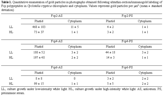
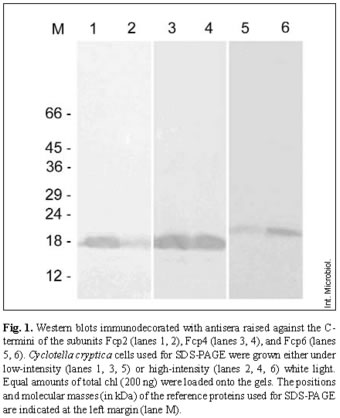
<body><![CDATA[<p><font face="Verdana, Arial, Helvetica, sans-serif"><b><font size="2">Gel electrophoresis and Western immunoblotting. </font></b></font><font size="2" face="Verdana, Arial, Helvetica, sans-serif">Total cell protein of <i>C. cryptica</i> was analyzed by SDS-PAGE, according to the method of Laemmli &#091;15&#093;, using 17.5% separating gels and 5% stacking gels. Equal amounts of total chl (chl <i>a</i> + chl <i>c</i>) were loaded onto the gels. Samples were prepared by resuspending the pelleted cells in small amounts of TE buffer (10 mM Tris/HCl, pH 7, 1 mM EDTA). Defined amounts, generally 10 or 20 &#181;l, were used to estimate the chl (<i>a</i> + <i>c</i>) contents. Appropriate amounts of sample buffer were then added to yield a final concentration of 4 &#181;g chl/ml. The protein-containing samples were boiled for 2-4 min at 100oC, and 50 &#181;l, equivalent to 200 ng chl, were loaded onto the gel. Western blotting was carried out in a Biorad Trans-blot SD semidry transfer cell according to the method of Towbin et al. &#091;28&#093;. The proteins were transferred onto Protran nitrocellulose transfer membrane (Schleicher &amp; Schuell, Dassel, Germany) at a constant voltage of 20 V for 1.5 h. For immunodecoration, the blots were preincubated for 2 h in TBS-buffer (10 mM Tris-HCl, pH 7.5, 50 mM NaCl) saturated with skim-milk powder and washed in TBS, followed by decorating with the first antibodies &#091;serum-diluted 1:1000 in TBS with 1% (w/v) bovine serum albumin (BSA)&#093; for 2-16 h. Antibodies raised against the C-termini of distinct Fcps of <i>Cyclotella cryptica</i> were described in Westermann and Rhiel &#091;31&#093;. After several washings with TBS, the nitrocellulose membranes were incubated for 2 h with peroxidase-conjugated goat anti-rabbit IgG (diluted 1:1000 in TBS with 1% BSA), provided by Sigma (Munich, Germany). The membranes were washed again and stained with 4-chloro-1-naphthol and H<sub>2</sub>O<sub>2</sub>. Images of the blots were recorded with a Herolab EASY CCD camera (type 429K) in an illumination chamber (Herolab RH-3) and stored as digitized files. The results were documented using the enhanced analysis system EASY (Herolab, Wiesloch, Germany). For quantitative investigations of the immunodecorated protein bands, the EASY Image Plus software package was used.</font></p>     <p><font face="Verdana, Arial, Helvetica, sans-serif"><b><font size="2">Spectroscopy, pigment extraction, and pigment estimation. </font></b></font><font size="2" face="Verdana, Arial, Helvetica, sans-serif">Pigments were extracted with 90% (v/v) acetone and the concentrations of chl <i>a</i> and chl <i>c</i> concentrations were estimated in a U-3000 spectrophotometer (Hitachi, Tokyo, Japan) according to the equations of Jeffrey and Humphrey &#091;13&#093;.</font></p>     <p><font face="Verdana, Arial, Helvetica, sans-serif"><b><font size="2">Ultrathin section/immunogold-labeling electron microscopy. </font></b></font><font size="2" face="Verdana, Arial, Helvetica, sans-serif">For ultrathin sectioning and immunogold-labeling experiments, the pelleted cells were fixed at 4&deg;C for 2 h in 0.1 M Na-K-phosphate buffer, pH 7, containing 4% glutardialdehyde. Excess glutardialdehyde was removed by washing the cells with 0.1 M Na-K-phosphate buffer, pH 7. The cells were then dehydrated in ethanol and embedded in Lowicryl K4M at 4&deg;C, according to the manufacturer's specifications (Chemische Werke Lowi, Waldkreuzburg, Germany). The samples were polymerized by UV radiation for 24 h at 4&deg;C and for an additional 48 h at room temperature. Ultrathin sections were cut with a Reichard-Jung Ultracut ultramicrotome. The sections were immunogold-labeled for 1 h using preimmune sera and antisera in dilutions of 1:100, according to the procedure of Rhiel et al. &#091;23&#093;, and a 1:100 diluted solution of the second gold-conjugated antibody (goat anti-rabbit IgG with 10 nm gold, British Biocell International, Cardiff, UK). Finally, the sections were examined in a Zeiss EM 109 operated at 50 kV or in a Zeiss EM 902A electron microscope operated at 50 kV. A post-staining step was omitted. Photographs were taken on negative films (Zeiss EM 109) or as digitized images (Zeiss EM 902A) using a digital camera (Proscan High Speed SSCCD camera, 1kx1k, Proscan elektronische Systeme GmbH, Lagerlechfeld, Germany). The digital camera was operated by the SIS Esivision, Version 3.1 (Soft Imaging System GmbH, M&uuml;nster, Germany). The images were stored as digitized files (tiff-formatted). Measurements of the gold-particle density were made on positive prints enlarged 58,500&times;. The number of gold particles was counted within defined areas, selected arbitrarily in the region of the plastids or in the cytoplasm. A minimum of ten areas from ten different positive prints was counted.</font></p>     <p>&nbsp;</p>     <p><font face="Verdana, Arial, Helvetica, sans-serif"><b><font size="3">Results</font></b></font></p>     <p><b><font size="2" face="Verdana, Arial, Helvetica, sans-serif">Western immunoblotting. </font></b><font size="2" face="Verdana, Arial, Helvetica, sans-serif"><i>C. cryptica</i> was grown for 2 weeks under low- or high-intensity white light, harvested by centrifugation, and subjected to SDS-PAGE. Equal amounts of total chl were loaded onto the gels, which allowed estimation of the relative abundances of the distinct Fcp subunits under the two light regimes. The three antisera specifically immunodecorated protein bands with relative molecular mass (Mr) of 18,000 (Fcp2), 18,000 (Fcp4), and 22,000 (Fcp6) in both low- and high-intensity white-light-treated cells (<a href="#f1">Fig. 1</a>).</font></p>     <p align="center"><a name="f1"><img src="/img/revistas/im/v9n1/04fig1becker.jpg" width="340" height="418"></a></p>     <p>&nbsp;</p>     <p><font size="2" face="Verdana, Arial, Helvetica, sans-serif">Quantification of the immunodecorated bands revealed that the amount of Fcp2 was approximately twice as high in low-light-grown cells than in cells grown in high-intensity white light. The amounts of Fcp4 were almost the same under the two light regimes, whereas for Fcp6 an approximately four- to five-fold higher signal was registered for cells grown under high-intensity white light.</font></p>     <p><b><font size="2" face="Verdana, Arial, Helvetica, sans-serif">Ultrathin sectioning and immunogold labeling. </font></b><font size="2" face="Verdana, Arial, Helvetica, sans-serif">Ultrathin sections of chloroplasts of <i>C. cryptica</i> immunogold-labeled with the three antisera and preimmune sera are shown in <a target="_blank" href="/img/revistas/im/v9n1/04fig2becker.jpg">Figs. 2</a> and  <a target="_blank" href="/img/revistas/im/v9n1/04fig3becker.jpg">3</a>, and the quantitative data are compiled in <a href="#t1">Table 1</a>.</font></p>     ]]></body>
<body><![CDATA[<p align="center"><a name="t1"><img src="/img/revistas/im/v9n1/04tab1becker.gif" width="560" height="332"></a></p>     <p>&nbsp;</p>     <p><font size="2" face="Verdana, Arial, Helvetica, sans-serif">The highest counts of gold particles per &#181;m<sup>2</sup> plastid area were obtained with antiserum against Fcp2, followed by antiserum against Fcp4. Antiserum raised against Fcp6 yielded the lowest densities of gold particles (Table 1). For all three antisera, no differences concerning the allocation of gold particles could be detected. In addition, the particles were not restricted to thylakoids of the plastid center or periphery (girdle lamellas), but were instead distributed randomly within the plastids. Gold labeling of cells grown under low light and then probed with antiserum against Fcp2 was approximately six-fold higher than that of high-light-grown cells (<a href="#t1">Table 1</a>). Antiserum directed against the C-terminus of Fcp4 gave rise to similar gold-labeling densities; 180 &plusmn; 52 and 197 &plusmn; 48 gold particles per &#181;m<sup>2</sup> plastid area, for cells grown under low- and high-intensity white light, respectively (<a href="#t1">Table 1</a>). When cells were probed with antiserum against Fcp6, the density of gold particles per &#181;m<sup>2</sup> plastid area was approximately 10- to 12-fold higher in cells grown under high-intensity white light than in those grown under low-light conditions (<a target="_blank" href="/img/revistas/im/v9n1/04fig2becker.jpg">Fig. 2E, F</a>, <a href="#t1">Table 1</a>).</font></p>     <p><font size="2" face="Verdana, Arial, Helvetica, sans-serif">All values measured within the chloroplasts significantly exceeded those measured within the cytoplasm. The number of gold particles per &#181;m<sup>2</sup> in cytoplasm ranged from 11 &plusmn; 5 to 0 (<a href="#t1">Table 1</a>), which was similar to the number of particles counted on immunogold-labeled ultrathin sections treated with preimmune sera (<a target="_blank" href="/img/revistas/im/v9n1/04fig3becker.jpg">Fig. 3</a>, <a href="#t1">Table 1</a>). Generally, rather low amounts of gold particles were counted in both plastids and cytoplasm when the preimmune sera were used. The exception was the preimmune serum of Fcp4, which resulted in a significantly higher amount of labeling within the chloroplasts than in the cytoplasm (<a target="_blank" href="/img/revistas/im/v9n1/04fig3becker.jpg">Fig. 3C, D</a>, <a href="#t1">Table 1</a>).</font></p>     <p>&nbsp;</p>     <p><b><font size="3" face="Verdana, Arial, Helvetica, sans-serif">Discussion</font></b></p>     <p><b><font size="2" face="Verdana, Arial, Helvetica, sans-serif">Western immunoblotting.</font></b> <font size="2" face="Verdana, Arial, Helvetica, sans-serif">The estimated relative molecular weights are in line with those described by Westermann and Rhiel &#091;31&#093; and confirm the specificity of the antisera. The latter were raised against synthetic polypeptides whose amino acid sequences were deduced from the corresponding <i>fcp</i> gene sequences, and which represent the 16 amino acids at the very C-terminal ends of the three Fcps of <i>C. cryptica</i>. In a previous report, Western blots of total protein extracts of <i>C. cryptica</i> were challenged with antisera directed against either the <i>Cyclotella</i> LHC fraction, composed of several Fcp subunits (cc-antiserum), or the LHC of the cryptophyte <i>Cryptomonas maculata</i> (cm-antiserum) &#091;10&#093;. The cc-antiserum decorated two bands, with Mr 18,000 and 22,000, under both light regimes and the signal intensities of the bands decreased simultaneously under high-light conditions. Quantitative estimations revealed that high-light-grown cells of <i>C. cryptica</i> showed only 67.2% (&plusmn; 6.1) of the signal intensities for Fcp that were measured for cells cultivated under low-intensity white light. When Western blots were challenged with cm-antiserum, only two polypeptides, of approximately Mr 24,000 and 23,000, were immunodecorated in <i>C. cryptica</i>. The band with a Mr of 24,000 was found in cells grown under low-intensity white light, while the band with a Mr of 23,000 was decorated in protein extracts of cells grown under high-intensity white light. It was therefore concluded that specific Fcp subunits are present in <i>C. cryptica</i>, and they are expressed in response to high-intensity white light &#091;10&#093;</font></p>     <p><font size="2" face="Verdana, Arial, Helvetica, sans-serif">The results obtained in the current work using the three monospecific antisera confirm this assumption and show that Fcp2 expression is higher in response to moderate light, whereas Fcp6 is specifically induced in response to high-intensity white light. The results for Fcp2 and Fcp6 are in line with data on the steady-state mRNA abundances measured for <i>fcp2</i> and <i>fcp6</i> &#091;20,21&#093;. Gene <i>fcp2</i> was expressed at a higher level under low-light conditions, whereas the steady-state mRNA concentration of <i>fcp6</i> increased under high-intensity white light. Interestingly, the steady-state mRNA concentration measured for <i>fcp4</i> was higher under low light and decreased under high light. This difference does not become manifested with respect to the abundance of the protein, Fcp4, and can be interpreted as demonstrating that the turnover rate of <i>fcp4</i> mRNA exceeds the translation rate. The specific induction of Fcp6 in response to high-intensity white light supports a role for Fcp6 in photoprotection, as also suggested by phylogenetic studies, in which <i>fcp6</i>, <i>fcp7</i>, and <i>fcp12</i> of <i>C. cryptica</i> showed homology to Fcp of the haptophyte <i>Isochrysis galbana</i> and to the light-inducible proteins LI818r3 and LI818 of <i>Chlamydomonas reinhardtii</i> and <i><i>Chlamydomonas</i> eugametos</i>, respectively &#091;2&#093;.</font></p>     <p><b><font size="2" face="Verdana, Arial, Helvetica, sans-serif">Ultrathin sectioning and immunogold labeling. </font></b><font size="2" face="Verdana, Arial, Helvetica, sans-serif">The overall ultrastructure of low- and high-light grown cells of <i>C. cryptica</i> was previously described in detail by Janssen et al. &#091;10&#093;, and confirmed in the current study. Several small chloroplasts, comprising approximately 35% of the sectioned area of cells, were the dominating organelles in low-light-grown cells. The chloroplasts were densely packed with thylakoid membranes, which, as known for diatoms, were predominantly arranged in groups of three. When cultured under high-intensity white light, the area covered by vacuoles increased whereas the chloroplast area was reduced to 12-18% of the total cell area sectioned. The thylakoid membranes remained arranged in groups of three, but the overall amount of thylakoids was reduced significantly. Similar results were described by Rosen and Lowe in the diatom <i>Cyclotella meneghiniana</i> &#091;25&#093;, in which the chloroplast volume and thylakoid surface density decreased in response to simultaneous light and nutrient stress. In the current study, nutrient stress was excluded as the cause of the observed morphological changes, as the medium was changed after one week of growth under low- and high-intensity white light.</font></p>     <p><font size="2" face="Verdana, Arial, Helvetica, sans-serif">Similarly, Pyszniak and Gibbs &#091;22&#093; used immunogold labeling to localize the fucoxanthin-chlorophyll <i>a/c</i> light-harvesting complex in the diatom <i>Phaeodactylum tricornutum</i>. They found an equal distribution of the complex in appressed and non-appressed thylakoid membranes.</font></p>     ]]></body>
<body><![CDATA[<p><b><font size="2" face="Verdana, Arial, Helvetica, sans-serif">Comparison of Western immunoblotting with ultrathin sectioning and immunogold labeling. </font></b><font size="2" face="Verdana, Arial, Helvetica, sans-serif">While the same tendencies were observed in Western immunoblotting and immunogold-labeling experiments, quantitative measurements of the signal intensities on immunodecorated blots and of gold particles per &#181;m<sup>2</sup> deviated by a factor of 2-2.5. This might have been due to the different experimental approaches. Hust et al. &#091;9&#093; attempted to quantify Western immunoblots of sediment samples that had been extracted with SDS loading buffer. The authors stepwise diluted the extracts with the buffer and obtained fractions with decreasing chl <i>a</i> and protein concentrations. SDS-PAGE followed by Western immunoblotting gave rise to blots in which the signal intensities were almost linear in the chl <i>a</i> concentration range of 100-20 ng, but the curve flattened somewhat at higher chl <i>a</i> loads. An additional explanation may be found in the Western immunoblotting protocol, since prolonged development of the blots with 4-chloro-1-naphthol and H<sub>2</sub>O<sub>2</sub> might lead to excessive staining. As a result, the signal intensity would saturate with increasing time of immunodecoration.</font></p>     <p><font size="2" face="Verdana, Arial, Helvetica, sans-serif">Incubation with Fcp4 preimmune serum resulted in significantly higher labeling than obtained with the other preimmune sera. Although the amount was still four-fold lower than that registered for Fcp4 antiserum, it indicates that the rabbit must have raised immunoglobulins against a distinct plastidal protein prior to immunization with the oligopeptide corresponding to the very C-terminal amino acid sequence of Fcp4. Westermann and Rhiel &#091;31&#093; showed that none of the three preimmune sera taken prior to the first immunization gave rise to a signal on nitrocellulose membrane blotted with total cell protein extracts of <i>C. cryptica</i>. Plastidal labeling with the Fcp4 preimmune serum therefore might have been caused by a protein present in minor amounts within the chloroplasts of <i>C. cryptica</i>.</font></p>     <p><font size="2" face="Verdana, Arial, Helvetica, sans-serif">Westermann and Rhiel &#091;31&#093; tested the three antisera in immunogold-electron microscopy of both ultrathin-sectioned cells and freeze-fracture replicas of thylakoid membranes of <i>C. cryptica</i> grown under low-intensity white light. The antisera densely labeled the chloroplasts in a manner that was specific for and restricted to the thylakoid membranes. Thylakoid membranes that had been cut superficially rather than at a right angle to their planes showed a rather patchy distribution of gold particles, with areas of higher or less-dense labeling. For each of the three antisera, approximately ten-fold fewer gold particles were visible outside the chloroplast in the cytoplasm, indicating either unspecific background labeling or confirming the observation that Fcps are nuclear-encoded &#091;4&#093;. Estimations of the label density per plastid area indicated nearly equal amounts of gold particles per &#181;m<sup>2</sup> plastid area for Fcp2 and Fcp4, but an eight- to ten-fold reduction for Fcp6 &#091;31&#093;. In a previous report &#091;31&#093;, enumeration of gold particles observed in photographs yielded values of 1681 &plusmn; 784, 1995 &plusmn; 422, and 208 &plusmn; 151 gold particles per &#181;m<sup>2</sup> for Fcp2, Fcp4, and Fcp6, respectively. These values are higher than those measured in the current work and are most probably due to the fact that: (i) fresh cells of <i>C. cryptica</i> were embedded, (ii) different batches of preimmune sera, antisera, and gold-conjugated second antibodies were used, and (iii) different persons participated in the previous and recent experiments.</font></p>     <p><b><font size="2" face="Verdana, Arial, Helvetica, sans-serif">Concluding remarks. </font></b><font size="2" face="Verdana, Arial, Helvetica, sans-serif">Western immunoblotting and ultrathin sectioning/immunogold labeling together provide strong evidence for the light-regulated expression of distinct Fcp subunits: <i>fcp6</i> mRNA transcription is induced and its gene product, Fcp6, was synthesized in high amounts when <i>C. cryptica</i> was cultured in high-intensity white light. By contrast, transcription and translation of <i>fcp2</i> were enhanced when <i>C. cryptica</i> was grown in low-intensity white light. Therefore, it is expected that Fcp6 is involved in photoprotection, whereas Fcp2 most likely plays a role in harvesting light energy under light-limiting conditions. Currently, experiments are in progress to isolate Fcp2 and Fcp6 in their native states. It will be interesting to determine which pigments, especially carotenoids, are attached to the protein backbones and how they function in photoprotection/light-harvesting. Additional experiments will be aimed at identifying the photosystem (PSI, PSII, or both) to which Fcp6 and Fcp2 are bound, and whether this association occurs in addition to the attachment of the existing antenna or whether the latter are able to replace distinct Fcps.</font></p>     <p>&nbsp;</p>     <p><b><font size="3" face="Verdana, Arial, Helvetica, sans-serif">Acknowledgements</font></b></p>     <p><font size="2" face="Verdana, Arial, Helvetica, sans-serif">The authors express their gratitude to Ms. Renate Kort, Ms. Heike Oetting, and Mr. Michael Pilzen for technical assistance. The financial support of the Deutsche Forschungsgemeinschaft (Rh 20/3) is gratefully acknowledged. We hereby declare that the experiments comply with the current laws of Germany.</font></p>     <p>&nbsp;</p>     <p><b><font size="3" face="Verdana, Arial, Helvetica, sans-serif">References</font></b></p>     <!-- ref --><p><font size="2" face="Verdana, Arial, Helvetica, sans-serif">1. Eppard M, Rhiel E (1998) The genes encoding light-harvesting subunits of <i>Cyclotella cryptica</i> (Bacillariophyceae) constitute a complex and heterogeneous family. Mol Gen Genet 260:335-345</font>&nbsp;&nbsp;&nbsp;&nbsp;&nbsp;&nbsp;&nbsp;&nbsp;[&#160;<a href="javascript:void(0);" onclick="javascript: window.open('/scielo.php?script=sci_nlinks&ref=2803785&pid=S1139-6709200600010000400001&lng=','','width=640,height=500,resizable=yes,scrollbars=1,menubar=yes,');">Links</a>&#160;]<!-- end-ref --><!-- ref --><p><font size="2" face="Verdana, Arial, Helvetica, sans-serif">2. Eppard M, Krumbein WE, von Haeseler A, Rhiel E (2000) Characterization of <i>fcp</i>4 and <i>fcp</i>12, two additional genes encoding light-harvesting proteins of <i>Cyclotella cryptica</i> (Bacillariophyceae) and phylogenetic analysis of this complex gene family. Plant Biology 2:283-289</font>&nbsp;&nbsp;&nbsp;&nbsp;&nbsp;&nbsp;&nbsp;&nbsp;[&#160;<a href="javascript:void(0);" onclick="javascript: window.open('/scielo.php?script=sci_nlinks&ref=2803786&pid=S1139-6709200600010000400002&lng=','','width=640,height=500,resizable=yes,scrollbars=1,menubar=yes,');">Links</a>&#160;]<!-- end-ref --><!-- ref --><p><font size="2" face="Verdana, Arial, Helvetica, sans-serif">3. Eppard M, Rhiel E (2000) Investigations on gene copy number, introns and chromosomal arrangement of genes encoding the fucoxanthin chlorophyll <i>a/c</i>-binding proteins of the centric diatom <i>Cyclotella cryptica</i>. Protist 151:27-39</font>&nbsp;&nbsp;&nbsp;&nbsp;&nbsp;&nbsp;&nbsp;&nbsp;[&#160;<a href="javascript:void(0);" onclick="javascript: window.open('/scielo.php?script=sci_nlinks&ref=2803787&pid=S1139-6709200600010000400003&lng=','','width=640,height=500,resizable=yes,scrollbars=1,menubar=yes,');">Links</a>&#160;]<!-- end-ref --><!-- ref --><p><font size="2" face="Verdana, Arial, Helvetica, sans-serif">4. Fawley MW, Grossman AR (1986) Polypeptides of a light-harvesting complex of the diatom <i>Phaeodactylum tricornutum</i> are synthesized in the cytoplasm of the cell as precursors. Plant Physiol 81:149-155</font>&nbsp;&nbsp;&nbsp;&nbsp;&nbsp;&nbsp;&nbsp;&nbsp;[&#160;<a href="javascript:void(0);" onclick="javascript: window.open('/scielo.php?script=sci_nlinks&ref=2803788&pid=S1139-6709200600010000400004&lng=','','width=640,height=500,resizable=yes,scrollbars=1,menubar=yes,');">Links</a>&#160;]<!-- end-ref --><!-- ref --><p><font size="2" face="Verdana, Arial, Helvetica, sans-serif">5. Gagne G, Guertin M (1992) The early genetic response to light in the green unicellular alga <i><i>Chlamydomonas</i> eugametos</i> grown under light/dark cycles involves genes that represent direct responses to light and photosynthesis. Plant Mol Biol 18:429-445</font>&nbsp;&nbsp;&nbsp;&nbsp;&nbsp;&nbsp;&nbsp;&nbsp;[&#160;<a href="javascript:void(0);" onclick="javascript: window.open('/scielo.php?script=sci_nlinks&ref=2803789&pid=S1139-6709200600010000400005&lng=','','width=640,height=500,resizable=yes,scrollbars=1,menubar=yes,');">Links</a>&#160;]<!-- end-ref --><!-- ref --><p><font size="2" face="Verdana, Arial, Helvetica, sans-serif">6. Green BR, Durnford DG (1996) The chlorophyll-carotenoid proteins of oxygenic photosynthesis. Annu Rev Plant Physiol Plant Mol Biol 47:685-714</font>&nbsp;&nbsp;&nbsp;&nbsp;&nbsp;&nbsp;&nbsp;&nbsp;[&#160;<a href="javascript:void(0);" onclick="javascript: window.open('/scielo.php?script=sci_nlinks&ref=2803790&pid=S1139-6709200600010000400006&lng=','','width=640,height=500,resizable=yes,scrollbars=1,menubar=yes,');">Links</a>&#160;]<!-- end-ref --><!-- ref --><p><font size="2" face="Verdana, Arial, Helvetica, sans-serif">7. Grossman AR, Bhaya D, Apt KE, Kehoe DM (1995) Light-harvesting complexes in oxygenic photosynthesis: diversity, control, and evolution. Annu Rev Genetics 29:231-288</font>&nbsp;&nbsp;&nbsp;&nbsp;&nbsp;&nbsp;&nbsp;&nbsp;[&#160;<a href="javascript:void(0);" onclick="javascript: window.open('/scielo.php?script=sci_nlinks&ref=2803791&pid=S1139-6709200600010000400007&lng=','','width=640,height=500,resizable=yes,scrollbars=1,menubar=yes,');">Links</a>&#160;]<!-- end-ref --><!-- ref --><p><font size="2" face="Verdana, Arial, Helvetica, sans-serif">8. Hager A, Stransky H (1970) Das Carotinoidmuster und die Verbreitung des lichtinduzierten Xanthophyllcyclus in verschiedenen Algenklassen. V. Einzelne Vertreter der Cryptophyceae, Euglenophyceae, Bacillariophyceae, Chrysophyceae und Phaeophyceae. Arch Mikrobiol 73:77-89</font>&nbsp;&nbsp;&nbsp;&nbsp;&nbsp;&nbsp;&nbsp;&nbsp;[&#160;<a href="javascript:void(0);" onclick="javascript: window.open('/scielo.php?script=sci_nlinks&ref=2803792&pid=S1139-6709200600010000400008&lng=','','width=640,height=500,resizable=yes,scrollbars=1,menubar=yes,');">Links</a>&#160;]<!-- end-ref --><!-- ref --><p><font size="2" face="Verdana, Arial, Helvetica, sans-serif">9. Hust M, Krumbein WE, Rhiel E (1999) An immunochemical in situ approach to detect adaptation processes in the photosynthetic apparatus of diatoms of the Wadden Sea sediment surface layers. J Microbiol Methods 38:69-80</font>&nbsp;&nbsp;&nbsp;&nbsp;&nbsp;&nbsp;&nbsp;&nbsp;[&#160;<a href="javascript:void(0);" onclick="javascript: window.open('/scielo.php?script=sci_nlinks&ref=2803793&pid=S1139-6709200600010000400009&lng=','','width=640,height=500,resizable=yes,scrollbars=1,menubar=yes,');">Links</a>&#160;]<!-- end-ref --><!-- ref --><p><font size="2" face="Verdana, Arial, Helvetica, sans-serif">10. Janssen M, Bathke L, Marquardt J, Krumbein WE, Rhiel E (2001) Changes in the photosynthetic apparatus of diatoms in response to low and high light intensities. Int Microbiol 4:27-33</font>&nbsp;&nbsp;&nbsp;&nbsp;&nbsp;&nbsp;&nbsp;&nbsp;[&#160;<a href="javascript:void(0);" onclick="javascript: window.open('/scielo.php?script=sci_nlinks&ref=2803794&pid=S1139-6709200600010000400010&lng=','','width=640,height=500,resizable=yes,scrollbars=1,menubar=yes,');">Links</a>&#160;]<!-- end-ref --><!-- ref --><p><font size="2" face="Verdana, Arial, Helvetica, sans-serif">11. Jansson S, Pichersky E, Bassi R, Green BR, Ikeuchi M, Melis A, Simpson DJ, Spangfort M, Staehelin LA, Thornber JP (1992) A nomenclature for the genes encoding the chlorophyll <i>a/b</i>-binding proteins of higher plants. Plant Mol Biol Rep 10:242-253</font>&nbsp;&nbsp;&nbsp;&nbsp;&nbsp;&nbsp;&nbsp;&nbsp;[&#160;<a href="javascript:void(0);" onclick="javascript: window.open('/scielo.php?script=sci_nlinks&ref=2803795&pid=S1139-6709200600010000400011&lng=','','width=640,height=500,resizable=yes,scrollbars=1,menubar=yes,');">Links</a>&#160;]<!-- end-ref --><!-- ref --><p><font size="2" face="Verdana, Arial, Helvetica, sans-serif">12. Jansson S (1994) The light-harvesting chlorophyll <i>a/b</i>-binding proteins. Biochim Biophys Acta 1184: 1-19</font>&nbsp;&nbsp;&nbsp;&nbsp;&nbsp;&nbsp;&nbsp;&nbsp;[&#160;<a href="javascript:void(0);" onclick="javascript: window.open('/scielo.php?script=sci_nlinks&ref=2803796&pid=S1139-6709200600010000400012&lng=','','width=640,height=500,resizable=yes,scrollbars=1,menubar=yes,');">Links</a>&#160;]<!-- end-ref --><!-- ref --><p><font size="2" face="Verdana, Arial, Helvetica, sans-serif">13. Jeffrey SW, Humphrey GF (1975) New spectrophotometric equations for determining chlorophylls <i>a</i>, <i>b</i>, <i>c<sub>1</sub></i> and <i>c<sub>2</sub></i> in higher plants, algae and natural phytoplankton. Biochem Physiol Pflanzen 167:191-194</font>&nbsp;&nbsp;&nbsp;&nbsp;&nbsp;&nbsp;&nbsp;&nbsp;[&#160;<a href="javascript:void(0);" onclick="javascript: window.open('/scielo.php?script=sci_nlinks&ref=2803797&pid=S1139-6709200600010000400013&lng=','','width=640,height=500,resizable=yes,scrollbars=1,menubar=yes,');">Links</a>&#160;]<!-- end-ref --><!-- ref --><p><font size="2" face="Verdana, Arial, Helvetica, sans-serif">14. Kruse O (2001) Light-induced short-term adaptation mechanisms under redox control in the PSII-LHCII supercomplex: LHC II state transitions and PS II repair cycle. Naturwissenschaften 88:284-292</font>&nbsp;&nbsp;&nbsp;&nbsp;&nbsp;&nbsp;&nbsp;&nbsp;[&#160;<a href="javascript:void(0);" onclick="javascript: window.open('/scielo.php?script=sci_nlinks&ref=2803798&pid=S1139-6709200600010000400014&lng=','','width=640,height=500,resizable=yes,scrollbars=1,menubar=yes,');">Links</a>&#160;]<!-- end-ref --><!-- ref --><p><font size="2" face="Verdana, Arial, Helvetica, sans-serif">15. Laemmli UK (1970) Cleavage of structural proteins during the assembly of the head of bacteriophage T4. Nature 227:680-685</font>&nbsp;&nbsp;&nbsp;&nbsp;&nbsp;&nbsp;&nbsp;&nbsp;[&#160;<a href="javascript:void(0);" onclick="javascript: window.open('/scielo.php?script=sci_nlinks&ref=2803799&pid=S1139-6709200600010000400015&lng=','','width=640,height=500,resizable=yes,scrollbars=1,menubar=yes,');">Links</a>&#160;]<!-- end-ref --><!-- ref --><p><font size="2" face="Verdana, Arial, Helvetica, sans-serif">16. Lavaud J, Rousseau B, Etienne, A-L (2003) Enrichment of the light-harvesting complex in diadinoxanthin and implications for the nonphotochemical fluorescence quenching in diatoms. Biochemistry 42:5802-5808</font>&nbsp;&nbsp;&nbsp;&nbsp;&nbsp;&nbsp;&nbsp;&nbsp;[&#160;<a href="javascript:void(0);" onclick="javascript: window.open('/scielo.php?script=sci_nlinks&ref=2803800&pid=S1139-6709200600010000400016&lng=','','width=640,height=500,resizable=yes,scrollbars=1,menubar=yes,');">Links</a>&#160;]<!-- end-ref --><!-- ref --><p><font size="2" face="Verdana, Arial, Helvetica, sans-serif">17. Li XP, Bjorkman O, Shih C, Grossman AR, Rosenquist M, Jansson S, Niyogi KK (2000) A pigment-binding protein essential for regulation of photosynthetic light harvesting. Nature 403:391-395</font>&nbsp;&nbsp;&nbsp;&nbsp;&nbsp;&nbsp;&nbsp;&nbsp;[&#160;<a href="javascript:void(0);" onclick="javascript: window.open('/scielo.php?script=sci_nlinks&ref=2803801&pid=S1139-6709200600010000400017&lng=','','width=640,height=500,resizable=yes,scrollbars=1,menubar=yes,');">Links</a>&#160;]<!-- end-ref --><!-- ref --><p><font size="2" face="Verdana, Arial, Helvetica, sans-serif">18. Montane MH, Kloppstech K (2000) The family of light-harvesting-related proteins (LHCs, ELIPs, HLIPs): was the harvesting of light their primary function? Gene 258:1-8</font>&nbsp;&nbsp;&nbsp;&nbsp;&nbsp;&nbsp;&nbsp;&nbsp;[&#160;<a href="javascript:void(0);" onclick="javascript: window.open('/scielo.php?script=sci_nlinks&ref=2803802&pid=S1139-6709200600010000400018&lng=','','width=640,height=500,resizable=yes,scrollbars=1,menubar=yes,');">Links</a>&#160;]<!-- end-ref --><!-- ref --><p><font size="2" face="Verdana, Arial, Helvetica, sans-serif">19. Morosinotto T, Caffarri S, Dall'Osto L, Bassi R (2003) Mechanistic aspects of the xanthophyll dynamics in higher plant thylakoids. Physiol Plant 119:347-354</font>&nbsp;&nbsp;&nbsp;&nbsp;&nbsp;&nbsp;&nbsp;&nbsp;[&#160;<a href="javascript:void(0);" onclick="javascript: window.open('/scielo.php?script=sci_nlinks&ref=2803803&pid=S1139-6709200600010000400019&lng=','','width=640,height=500,resizable=yes,scrollbars=1,menubar=yes,');">Links</a>&#160;]<!-- end-ref --><!-- ref --><p><font size="2" face="Verdana, Arial, Helvetica, sans-serif">20. Oeltjen A, Krumbein WE, Rhiel E (2002) Investigations on transcript sizes, steady-state mRNA concentrations and diurnal expression of genes encoding fucoxanthin chlorophyll <i>a/c</i> light harvesting polypeptides in the centric diatom <i>Cyclotella cryptica</i>. Plant Biol (Stuttg) 4:250-257</font>&nbsp;&nbsp;&nbsp;&nbsp;&nbsp;&nbsp;&nbsp;&nbsp;[&#160;<a href="javascript:void(0);" onclick="javascript: window.open('/scielo.php?script=sci_nlinks&ref=2803804&pid=S1139-6709200600010000400020&lng=','','width=640,height=500,resizable=yes,scrollbars=1,menubar=yes,');">Links</a>&#160;]<!-- end-ref --><!-- ref --><p><font size="2" face="Verdana, Arial, Helvetica, sans-serif">21. Oeltjen A, Marquardt J, Rhiel E (2004) Differential circadian expression of genes <i>fcp2</i> and <i>fcp6</i> in <i>Cyclotella cryptica</i>. Int Microbiol 7:127-131</font>&nbsp;&nbsp;&nbsp;&nbsp;&nbsp;&nbsp;&nbsp;&nbsp;[&#160;<a href="javascript:void(0);" onclick="javascript: window.open('/scielo.php?script=sci_nlinks&ref=2803805&pid=S1139-6709200600010000400021&lng=','','width=640,height=500,resizable=yes,scrollbars=1,menubar=yes,');">Links</a>&#160;]<!-- end-ref --><!-- ref --><p><font size="2" face="Verdana, Arial, Helvetica, sans-serif">22. Pyszniak AM, Gibbs SP (1992) Immunocytochemical localization of photosystem I and the fucoxanthin-chlorophyll <i>a/c</i> light-harvesting complex in the diatom <i>Phaeodactylum tricornutum</i>. Protoplasma 166:208-217</font>&nbsp;&nbsp;&nbsp;&nbsp;&nbsp;&nbsp;&nbsp;&nbsp;[&#160;<a href="javascript:void(0);" onclick="javascript: window.open('/scielo.php?script=sci_nlinks&ref=2803806&pid=S1139-6709200600010000400022&lng=','','width=640,height=500,resizable=yes,scrollbars=1,menubar=yes,');">Links</a>&#160;]<!-- end-ref --><!-- ref --><p><font size="2" face="Verdana, Arial, Helvetica, sans-serif">23. Rhiel E, Kunz J, Wehrmeyer W (1989) Immunocytochemical localization of phycoerythrin-545 and of a chlorophyll <i>a/c</i> light harvesting complex in <i>Cryptomonas maculata</i> (Cryptophyceae). Bot Acta 102:46-53</font>&nbsp;&nbsp;&nbsp;&nbsp;&nbsp;&nbsp;&nbsp;&nbsp;[&#160;<a href="javascript:void(0);" onclick="javascript: window.open('/scielo.php?script=sci_nlinks&ref=2803807&pid=S1139-6709200600010000400023&lng=','','width=640,height=500,resizable=yes,scrollbars=1,menubar=yes,');">Links</a>&#160;]<!-- end-ref --><!-- ref --><p><font size="2" face="Verdana, Arial, Helvetica, sans-serif">24. Rhiel E, Marquardt J, Eppard M, M&ouml;rschel E, Krumbein WE (1997) The light-harvesting system of the diatom <i>Cyclotella cryptica</i>. Isolation and characterization of the main light-harvesting complex and evidence for the existence of minor pigment proteins. Bot Acta 110:109-117</font>&nbsp;&nbsp;&nbsp;&nbsp;&nbsp;&nbsp;&nbsp;&nbsp;[&#160;<a href="javascript:void(0);" onclick="javascript: window.open('/scielo.php?script=sci_nlinks&ref=2803808&pid=S1139-6709200600010000400024&lng=','','width=640,height=500,resizable=yes,scrollbars=1,menubar=yes,');">Links</a>&#160;]<!-- end-ref --><!-- ref --><p><font size="2" face="Verdana, Arial, Helvetica, sans-serif">25. Rosen BH, Lowe RL (1984) Physiological and ultrastructural responses of <i>Cyclotella meneghiniana</i> (Bacillariophyta) to light intensity and nutrient limitation. J Phycol 20:173-183</font>&nbsp;&nbsp;&nbsp;&nbsp;&nbsp;&nbsp;&nbsp;&nbsp;[&#160;<a href="javascript:void(0);" onclick="javascript: window.open('/scielo.php?script=sci_nlinks&ref=2803809&pid=S1139-6709200600010000400025&lng=','','width=640,height=500,resizable=yes,scrollbars=1,menubar=yes,');">Links</a>&#160;]<!-- end-ref --><!-- ref --><p><font size="2" face="Verdana, Arial, Helvetica, sans-serif">26. Savard F, Richard C, Guertin M (1996) The <i>Chlamydomonas reinhardtii</i> LI818 gene represents a distant relative of the <i>cabI</i>/<i>II</i> genes that is regulated during the cell cycle and in response to illumination. Plant Mol Biol 32:461-47</font>&nbsp;&nbsp;&nbsp;&nbsp;&nbsp;&nbsp;&nbsp;&nbsp;[&#160;<a href="javascript:void(0);" onclick="javascript: window.open('/scielo.php?script=sci_nlinks&ref=2803810&pid=S1139-6709200600010000400026&lng=','','width=640,height=500,resizable=yes,scrollbars=1,menubar=yes,');">Links</a>&#160;]<!-- end-ref --><!-- ref --><p><font size="2" face="Verdana, Arial, Helvetica, sans-serif">27. Schl&ouml;sser UG (1994) SAG-Sammlung von Algenkulturen at the University of G&ouml;ttingen. Catalogue of strains. Bot Acta 107:111-186</font>&nbsp;&nbsp;&nbsp;&nbsp;&nbsp;&nbsp;&nbsp;&nbsp;[&#160;<a href="javascript:void(0);" onclick="javascript: window.open('/scielo.php?script=sci_nlinks&ref=2803811&pid=S1139-6709200600010000400027&lng=','','width=640,height=500,resizable=yes,scrollbars=1,menubar=yes,');">Links</a>&#160;]<!-- end-ref --><!-- ref --><p><font size="2" face="Verdana, Arial, Helvetica, sans-serif">28. Towbin H, Staehelin T, Gordon J (1979) Electrophoretic transfer of proteins from polyacrylamide gels to nitrocellulose sheets: Procedure and some applications. Proc Natl Acad Sci USA 76:4350-4354</font>&nbsp;&nbsp;&nbsp;&nbsp;&nbsp;&nbsp;&nbsp;&nbsp;[&#160;<a href="javascript:void(0);" onclick="javascript: window.open('/scielo.php?script=sci_nlinks&ref=2803812&pid=S1139-6709200600010000400028&lng=','','width=640,height=500,resizable=yes,scrollbars=1,menubar=yes,');">Links</a>&#160;]<!-- end-ref --><!-- ref --><p><font size="2" face="Verdana, Arial, Helvetica, sans-serif">29. Uhrmacher S, Hanelt D, Nultsch W (1995) Zeaxanthin content and the degree of photoinhibition are linearly correlated in the brown alga <i>Dictyota dichotoma</i>. Mar Biol 123:159-165</font>&nbsp;&nbsp;&nbsp;&nbsp;&nbsp;&nbsp;&nbsp;&nbsp;[&#160;<a href="javascript:void(0);" onclick="javascript: window.open('/scielo.php?script=sci_nlinks&ref=2803813&pid=S1139-6709200600010000400029&lng=','','width=640,height=500,resizable=yes,scrollbars=1,menubar=yes,');">Links</a>&#160;]<!-- end-ref --><!-- ref --><p><font size="2" face="Verdana, Arial, Helvetica, sans-serif">30. Werner D (1969) Silicoborate als erste nicht C-haltige Wachstumsfaktoren. Arch Mikrobiol 65:258-274</font>&nbsp;&nbsp;&nbsp;&nbsp;&nbsp;&nbsp;&nbsp;&nbsp;[&#160;<a href="javascript:void(0);" onclick="javascript: window.open('/scielo.php?script=sci_nlinks&ref=2803814&pid=S1139-6709200600010000400030&lng=','','width=640,height=500,resizable=yes,scrollbars=1,menubar=yes,');">Links</a>&#160;]<!-- end-ref --><!-- ref --><p><font size="2" face="Verdana, Arial, Helvetica, sans-serif">31. Westermann M, Rhiel E (2005) Localisation of fucoxanthin chlorophyll <i>a/c</i>-binding polypeptides of the centric diatom <i>Cyclotella cryptica</i> by immuno-electron microscopy. Protoplasma 225:217-223</font>&nbsp;&nbsp;&nbsp;&nbsp;&nbsp;&nbsp;&nbsp;&nbsp;[&#160;<a href="javascript:void(0);" onclick="javascript: window.open('/scielo.php?script=sci_nlinks&ref=2803815&pid=S1139-6709200600010000400031&lng=','','width=640,height=500,resizable=yes,scrollbars=1,menubar=yes,');">Links</a>&#160;]<!-- end-ref --><p>&nbsp;</p>     <p>&nbsp;</p>     <p><font face="Verdana, Arial, Helvetica, sans-serif" size="2"><a href="#top"><img border="0" src="img/revistas/im/v9n1/seta.gif" width="15" height="17"></a><b><a name="back"></a>Corresponding author </b>    <br> E. Rhiel    <br> Geomikrobiologie, ICBM    <br> Carl-von-Ossietzky-Universit&auml;t Oldenburg    <br> POB 2503    <br> D-26129 Oldenburg, Germany    <br> Tel. +49-4417983389. Fax +49-4417983384    <br> E-mail: <a href="mailto:erhard.rhiel@uni-oldenburg.de">erhard.rhiel@uni-oldenburg.de</a></font></p>     ]]></body>
<body><![CDATA[<p><font size="2" face="Verdana, Arial, Helvetica, sans-serif">Received 5 September 2005    <br> Accepted 12 December 2005</font></p>       ]]></body><back>
<ref-list>
<ref id="B1">
<label>1</label><nlm-citation citation-type="journal">
<person-group person-group-type="author">
<name>
<surname><![CDATA[Eppard]]></surname>
<given-names><![CDATA[M]]></given-names>
</name>
<name>
<surname><![CDATA[Rhiel]]></surname>
<given-names><![CDATA[E]]></given-names>
</name>
</person-group>
<article-title xml:lang="en"><![CDATA[The genes encoding light-harvesting subunits of Cyclotella cryptica (Bacillariophyceae) constitute a complex and heterogeneous family]]></article-title>
<source><![CDATA[Mol Gen Genet]]></source>
<year>1998</year>
<volume>260</volume>
<page-range>335-345</page-range></nlm-citation>
</ref>
<ref id="B2">
<label>2</label><nlm-citation citation-type="journal">
<person-group person-group-type="author">
<name>
<surname><![CDATA[Eppard]]></surname>
<given-names><![CDATA[M]]></given-names>
</name>
<name>
<surname><![CDATA[Krumbein]]></surname>
<given-names><![CDATA[WE]]></given-names>
</name>
<name>
<surname><![CDATA[von Haeseler]]></surname>
<given-names><![CDATA[A]]></given-names>
</name>
<name>
<surname><![CDATA[Rhiel]]></surname>
<given-names><![CDATA[E]]></given-names>
</name>
</person-group>
<article-title xml:lang="en"><![CDATA[Characterization of fcp4 and fcp12, two additional genes encoding light-harvesting proteins of Cyclotella cryptica (Bacillariophyceae) and phylogenetic analysis of this complex gene family]]></article-title>
<source><![CDATA[Plant Biology]]></source>
<year>2000</year>
<volume>2</volume>
<page-range>283-289</page-range></nlm-citation>
</ref>
<ref id="B3">
<label>3</label><nlm-citation citation-type="journal">
<person-group person-group-type="author">
<name>
<surname><![CDATA[Eppard]]></surname>
<given-names><![CDATA[M]]></given-names>
</name>
<name>
<surname><![CDATA[Rhiel]]></surname>
<given-names><![CDATA[E]]></given-names>
</name>
</person-group>
<article-title xml:lang="en"><![CDATA[Investigations on gene copy number, introns and chromosomal arrangement of genes encoding the fucoxanthin chlorophyll a/c-binding proteins of the centric diatom Cyclotella cryptica]]></article-title>
<source><![CDATA[Protist]]></source>
<year>2000</year>
<volume>151</volume>
<page-range>27-39</page-range></nlm-citation>
</ref>
<ref id="B4">
<label>4</label><nlm-citation citation-type="journal">
<person-group person-group-type="author">
<name>
<surname><![CDATA[Fawley]]></surname>
<given-names><![CDATA[MW]]></given-names>
</name>
<name>
<surname><![CDATA[Grossman]]></surname>
<given-names><![CDATA[AR]]></given-names>
</name>
</person-group>
<article-title xml:lang="en"><![CDATA[Polypeptides of a light-harvesting complex of the diatom Phaeodactylum tricornutum are synthesized in the cytoplasm of the cell as precursors]]></article-title>
<source><![CDATA[Plant Physiol]]></source>
<year>1986</year>
<volume>81</volume>
<page-range>149-155</page-range></nlm-citation>
</ref>
<ref id="B5">
<label>5</label><nlm-citation citation-type="journal">
<person-group person-group-type="author">
<name>
<surname><![CDATA[Gagne]]></surname>
<given-names><![CDATA[G]]></given-names>
</name>
<name>
<surname><![CDATA[Guertin]]></surname>
<given-names><![CDATA[M]]></given-names>
</name>
</person-group>
<article-title xml:lang="en"><![CDATA[The early genetic response to light in the green unicellular alga Chlamydomonas eugametos grown under light/dark cycles involves genes that represent direct responses to light and photosynthesis]]></article-title>
<source><![CDATA[Plant Mol Biol]]></source>
<year>1992</year>
<volume>18</volume>
<page-range>429-445</page-range></nlm-citation>
</ref>
<ref id="B6">
<label>6</label><nlm-citation citation-type="journal">
<person-group person-group-type="author">
<name>
<surname><![CDATA[Green]]></surname>
<given-names><![CDATA[BR]]></given-names>
</name>
<name>
<surname><![CDATA[Durnford]]></surname>
<given-names><![CDATA[DG]]></given-names>
</name>
</person-group>
<article-title xml:lang="en"><![CDATA[The chlorophyll-carotenoid proteins of oxygenic photosynthesis]]></article-title>
<source><![CDATA[Annu Rev Plant Physiol Plant Mol Biol]]></source>
<year>1996</year>
<volume>47</volume>
<page-range>685-714</page-range></nlm-citation>
</ref>
<ref id="B7">
<label>7</label><nlm-citation citation-type="journal">
<person-group person-group-type="author">
<name>
<surname><![CDATA[Grossman]]></surname>
<given-names><![CDATA[AR]]></given-names>
</name>
<name>
<surname><![CDATA[Bhaya]]></surname>
<given-names><![CDATA[D]]></given-names>
</name>
<name>
<surname><![CDATA[Apt]]></surname>
<given-names><![CDATA[KE]]></given-names>
</name>
<name>
<surname><![CDATA[Kehoe]]></surname>
<given-names><![CDATA[DM]]></given-names>
</name>
</person-group>
<article-title xml:lang="en"><![CDATA[Light-harvesting complexes in oxygenic photosynthesis: diversity, control, and evolution]]></article-title>
<source><![CDATA[Annu Rev Genetics]]></source>
<year>1995</year>
<volume>29</volume>
<page-range>231-288</page-range></nlm-citation>
</ref>
<ref id="B8">
<label>8</label><nlm-citation citation-type="journal">
<person-group person-group-type="author">
<name>
<surname><![CDATA[Hager]]></surname>
<given-names><![CDATA[A]]></given-names>
</name>
<name>
<surname><![CDATA[Stransky]]></surname>
<given-names><![CDATA[H]]></given-names>
</name>
</person-group>
<article-title xml:lang="de"><![CDATA[Das Carotinoidmuster und die Verbreitung des lichtinduzierten Xanthophyllcyclus in verschiedenen Algenklassen: V. Einzelne Vertreter der Cryptophyceae, Euglenophyceae, Bacillariophyceae, Chrysophyceae und Phaeophyceae]]></article-title>
<source><![CDATA[Arch Mikrobiol]]></source>
<year>1970</year>
<volume>73</volume>
<page-range>77-89</page-range></nlm-citation>
</ref>
<ref id="B9">
<label>9</label><nlm-citation citation-type="journal">
<person-group person-group-type="author">
<name>
<surname><![CDATA[Hust]]></surname>
<given-names><![CDATA[M]]></given-names>
</name>
<name>
<surname><![CDATA[Krumbein]]></surname>
<given-names><![CDATA[WE]]></given-names>
</name>
<name>
<surname><![CDATA[Rhiel]]></surname>
<given-names><![CDATA[E]]></given-names>
</name>
</person-group>
<article-title xml:lang="en"><![CDATA[An immunochemical in situ approach to detect adaptation processes in the photosynthetic apparatus of diatoms of the Wadden Sea sediment surface layers]]></article-title>
<source><![CDATA[J Microbiol Methods]]></source>
<year>1999</year>
<volume>38</volume>
<page-range>69-80</page-range></nlm-citation>
</ref>
<ref id="B10">
<label>10</label><nlm-citation citation-type="journal">
<person-group person-group-type="author">
<name>
<surname><![CDATA[Janssen]]></surname>
<given-names><![CDATA[M]]></given-names>
</name>
<name>
<surname><![CDATA[Bathke]]></surname>
<given-names><![CDATA[L]]></given-names>
</name>
<name>
<surname><![CDATA[Marquardt]]></surname>
<given-names><![CDATA[J]]></given-names>
</name>
<name>
<surname><![CDATA[Krumbein]]></surname>
<given-names><![CDATA[WE]]></given-names>
</name>
<name>
<surname><![CDATA[Rhiel]]></surname>
<given-names><![CDATA[E]]></given-names>
</name>
</person-group>
<article-title xml:lang="en"><![CDATA[Changes in the photosynthetic apparatus of diatoms in response to low and high light intensities]]></article-title>
<source><![CDATA[Int Microbiol]]></source>
<year>2001</year>
<volume>4</volume>
<page-range>27-33</page-range></nlm-citation>
</ref>
<ref id="B11">
<label>11</label><nlm-citation citation-type="journal">
<person-group person-group-type="author">
<name>
<surname><![CDATA[Jansson]]></surname>
<given-names><![CDATA[S]]></given-names>
</name>
<name>
<surname><![CDATA[Pichersky]]></surname>
<given-names><![CDATA[E]]></given-names>
</name>
<name>
<surname><![CDATA[Bassi]]></surname>
<given-names><![CDATA[R]]></given-names>
</name>
<name>
<surname><![CDATA[Green]]></surname>
<given-names><![CDATA[BR]]></given-names>
</name>
<name>
<surname><![CDATA[Ikeuchi]]></surname>
<given-names><![CDATA[M]]></given-names>
</name>
<name>
<surname><![CDATA[Melis]]></surname>
<given-names><![CDATA[A]]></given-names>
</name>
<name>
<surname><![CDATA[Simpson]]></surname>
<given-names><![CDATA[DJ]]></given-names>
</name>
<name>
<surname><![CDATA[Spangfort]]></surname>
<given-names><![CDATA[M]]></given-names>
</name>
<name>
<surname><![CDATA[Staehelin]]></surname>
<given-names><![CDATA[LA]]></given-names>
</name>
<name>
<surname><![CDATA[Thornber]]></surname>
<given-names><![CDATA[JP]]></given-names>
</name>
</person-group>
<article-title xml:lang="en"><![CDATA[A nomenclature for the genes encoding the chlorophyll a/b-binding proteins of higher plants]]></article-title>
<source><![CDATA[Plant Mol Biol Rep]]></source>
<year>1992</year>
<volume>10</volume>
<page-range>242-253</page-range></nlm-citation>
</ref>
<ref id="B12">
<label>12</label><nlm-citation citation-type="journal">
<person-group person-group-type="author">
<name>
<surname><![CDATA[Jansson]]></surname>
<given-names><![CDATA[S]]></given-names>
</name>
</person-group>
<article-title xml:lang="en"><![CDATA[The light-harvesting chlorophyll a/b-binding proteins]]></article-title>
<source><![CDATA[Biochim Biophys Acta]]></source>
<year>1994</year>
<volume>1184</volume>
<page-range>1-19</page-range></nlm-citation>
</ref>
<ref id="B13">
<label>13</label><nlm-citation citation-type="journal">
<person-group person-group-type="author">
<name>
<surname><![CDATA[Jeffrey]]></surname>
<given-names><![CDATA[SW]]></given-names>
</name>
<name>
<surname><![CDATA[Humphrey]]></surname>
<given-names><![CDATA[GF]]></given-names>
</name>
</person-group>
<article-title xml:lang="en"><![CDATA[New spectrophotometric equations for determining chlorophylls a, b, c1 and c2 in higher plants, algae and natural phytoplankton]]></article-title>
<source><![CDATA[Biochem Physiol Pflanzen]]></source>
<year>1975</year>
<volume>167</volume>
<page-range>191-194</page-range></nlm-citation>
</ref>
<ref id="B14">
<label>14</label><nlm-citation citation-type="journal">
<person-group person-group-type="author">
<name>
<surname><![CDATA[Kruse]]></surname>
<given-names><![CDATA[O]]></given-names>
</name>
</person-group>
<article-title xml:lang="en"><![CDATA[Light-induced short-term adaptation mechanisms under redox control in the PSII-LHCII supercomplex: LHC II state transitions and PS II repair cycle]]></article-title>
<source><![CDATA[Naturwissenschaften]]></source>
<year>2001</year>
<volume>88</volume>
<page-range>284-292</page-range></nlm-citation>
</ref>
<ref id="B15">
<label>15</label><nlm-citation citation-type="journal">
<person-group person-group-type="author">
<name>
<surname><![CDATA[Laemmli]]></surname>
<given-names><![CDATA[UK]]></given-names>
</name>
</person-group>
<article-title xml:lang="en"><![CDATA[Cleavage of structural proteins during the assembly of the head of bacteriophage T4]]></article-title>
<source><![CDATA[Nature]]></source>
<year>1970</year>
<volume>227</volume>
<page-range>680-685</page-range></nlm-citation>
</ref>
<ref id="B16">
<label>16</label><nlm-citation citation-type="journal">
<person-group person-group-type="author">
<name>
<surname><![CDATA[Lavaud]]></surname>
<given-names><![CDATA[J]]></given-names>
</name>
<name>
<surname><![CDATA[Rousseau]]></surname>
<given-names><![CDATA[B]]></given-names>
</name>
<name>
<surname><![CDATA[Etienne]]></surname>
<given-names><![CDATA[A-L]]></given-names>
</name>
</person-group>
<article-title xml:lang="en"><![CDATA[Enrichment of the light-harvesting complex in diadinoxanthin and implications for the nonphotochemical fluorescence quenching in diatoms]]></article-title>
<source><![CDATA[Biochemistry]]></source>
<year>2003</year>
<volume>42</volume>
<page-range>5802-5808</page-range></nlm-citation>
</ref>
<ref id="B17">
<label>17</label><nlm-citation citation-type="journal">
<person-group person-group-type="author">
<name>
<surname><![CDATA[Li]]></surname>
<given-names><![CDATA[XP]]></given-names>
</name>
<name>
<surname><![CDATA[Bjorkman]]></surname>
<given-names><![CDATA[O]]></given-names>
</name>
<name>
<surname><![CDATA[Shih]]></surname>
<given-names><![CDATA[C]]></given-names>
</name>
<name>
<surname><![CDATA[Grossman]]></surname>
<given-names><![CDATA[AR]]></given-names>
</name>
<name>
<surname><![CDATA[Rosenquist]]></surname>
<given-names><![CDATA[M]]></given-names>
</name>
<name>
<surname><![CDATA[Jansson]]></surname>
<given-names><![CDATA[S]]></given-names>
</name>
<name>
<surname><![CDATA[Niyogi]]></surname>
<given-names><![CDATA[KK]]></given-names>
</name>
</person-group>
<article-title xml:lang="en"><![CDATA[A pigment-binding protein essential for regulation of photosynthetic light harvesting]]></article-title>
<source><![CDATA[Nature]]></source>
<year>2000</year>
<volume>403</volume>
<page-range>391-395</page-range></nlm-citation>
</ref>
<ref id="B18">
<label>18</label><nlm-citation citation-type="journal">
<person-group person-group-type="author">
<name>
<surname><![CDATA[Montane]]></surname>
<given-names><![CDATA[MH]]></given-names>
</name>
<name>
<surname><![CDATA[Kloppstech]]></surname>
<given-names><![CDATA[K]]></given-names>
</name>
</person-group>
<article-title xml:lang="en"><![CDATA[The family of light-harvesting-related proteins (LHCs, ELIPs, HLIPs): was the harvesting of light their primary function]]></article-title>
<source><![CDATA[Gene]]></source>
<year>2000</year>
<volume>258</volume>
<page-range>1-8</page-range></nlm-citation>
</ref>
<ref id="B19">
<label>19</label><nlm-citation citation-type="journal">
<person-group person-group-type="author">
<name>
<surname><![CDATA[Morosinotto]]></surname>
<given-names><![CDATA[T]]></given-names>
</name>
<name>
<surname><![CDATA[Caffarri]]></surname>
<given-names><![CDATA[S]]></given-names>
</name>
<name>
<surname><![CDATA[Dall'Osto]]></surname>
<given-names><![CDATA[L]]></given-names>
</name>
<name>
<surname><![CDATA[Bassi]]></surname>
<given-names><![CDATA[R]]></given-names>
</name>
</person-group>
<article-title xml:lang="en"><![CDATA[Mechanistic aspects of the xanthophyll dynamics in higher plant thylakoids]]></article-title>
<source><![CDATA[Physiol Plant]]></source>
<year>2003</year>
<volume>119</volume>
<page-range>347-354</page-range></nlm-citation>
</ref>
<ref id="B20">
<label>20</label><nlm-citation citation-type="journal">
<person-group person-group-type="author">
<name>
<surname><![CDATA[Oeltjen]]></surname>
<given-names><![CDATA[A]]></given-names>
</name>
<name>
<surname><![CDATA[Krumbein]]></surname>
<given-names><![CDATA[WE]]></given-names>
</name>
<name>
<surname><![CDATA[Rhiel]]></surname>
<given-names><![CDATA[E]]></given-names>
</name>
</person-group>
<article-title xml:lang="en"><![CDATA[Investigations on transcript sizes, steady-state mRNA concentrations and diurnal expression of genes encoding fucoxanthin chlorophyll a/c light harvesting polypeptides in the centric diatom Cyclotella cryptica]]></article-title>
<source><![CDATA[Plant Biol (Stuttg)]]></source>
<year>2002</year>
<volume>4</volume>
<page-range>250-257</page-range></nlm-citation>
</ref>
<ref id="B21">
<label>21</label><nlm-citation citation-type="journal">
<person-group person-group-type="author">
<name>
<surname><![CDATA[Oeltjen]]></surname>
<given-names><![CDATA[A]]></given-names>
</name>
<name>
<surname><![CDATA[Marquardt]]></surname>
<given-names><![CDATA[J]]></given-names>
</name>
<name>
<surname><![CDATA[Rhiel]]></surname>
<given-names><![CDATA[E]]></given-names>
</name>
</person-group>
<article-title xml:lang="en"><![CDATA[Differential circadian expression of genes fcp2 and fcp6 in Cyclotella cryptica]]></article-title>
<source><![CDATA[Int Microbiol]]></source>
<year>2004</year>
<volume>7</volume>
<page-range>127-131</page-range></nlm-citation>
</ref>
<ref id="B22">
<label>22</label><nlm-citation citation-type="journal">
<person-group person-group-type="author">
<name>
<surname><![CDATA[Pyszniak]]></surname>
<given-names><![CDATA[AM]]></given-names>
</name>
<name>
<surname><![CDATA[Gibbs]]></surname>
<given-names><![CDATA[SP]]></given-names>
</name>
</person-group>
<article-title xml:lang="en"><![CDATA[Immunocytochemical localization of photosystem I and the fucoxanthin-chlorophyll a/c light-harvesting complex in the diatom Phaeodactylum tricornutum]]></article-title>
<source><![CDATA[Protoplasma]]></source>
<year>1992</year>
<volume>166</volume>
<page-range>208-217</page-range></nlm-citation>
</ref>
<ref id="B23">
<label>23</label><nlm-citation citation-type="journal">
<person-group person-group-type="author">
<name>
<surname><![CDATA[Rhiel]]></surname>
<given-names><![CDATA[E]]></given-names>
</name>
<name>
<surname><![CDATA[Kunz]]></surname>
<given-names><![CDATA[J]]></given-names>
</name>
<name>
<surname><![CDATA[Wehrmeyer]]></surname>
<given-names><![CDATA[W]]></given-names>
</name>
</person-group>
<article-title xml:lang="en"><![CDATA[Immunocytochemical localization of phycoerythrin-545 and of a chlorophyll a/c light harvesting complex in Cryptomonas maculata (Cryptophyceae)]]></article-title>
<source><![CDATA[Bot Acta]]></source>
<year>1989</year>
<volume>102</volume>
<page-range>46-53</page-range></nlm-citation>
</ref>
<ref id="B24">
<label>24</label><nlm-citation citation-type="journal">
<person-group person-group-type="author">
<name>
<surname><![CDATA[Rhiel]]></surname>
<given-names><![CDATA[E]]></given-names>
</name>
<name>
<surname><![CDATA[Marquardt]]></surname>
<given-names><![CDATA[J]]></given-names>
</name>
<name>
<surname><![CDATA[Eppard]]></surname>
<given-names><![CDATA[M]]></given-names>
</name>
<name>
<surname><![CDATA[Mörschel]]></surname>
<given-names><![CDATA[E]]></given-names>
</name>
<name>
<surname><![CDATA[Krumbein]]></surname>
<given-names><![CDATA[WE]]></given-names>
</name>
</person-group>
<article-title xml:lang="en"><![CDATA[The light-harvesting system of the diatom Cyclotella cryptica: Isolation and characterization of the main light-harvesting complex and evidence for the existence of minor pigment proteins]]></article-title>
<source><![CDATA[Bot Acta]]></source>
<year>1997</year>
<volume>110</volume>
<page-range>109-117</page-range></nlm-citation>
</ref>
<ref id="B25">
<label>25</label><nlm-citation citation-type="journal">
<person-group person-group-type="author">
<name>
<surname><![CDATA[Rosen]]></surname>
<given-names><![CDATA[BH]]></given-names>
</name>
<name>
<surname><![CDATA[Lowe]]></surname>
<given-names><![CDATA[RL]]></given-names>
</name>
</person-group>
<article-title xml:lang="en"><![CDATA[Physiological and ultrastructural responses of Cyclotella meneghiniana (Bacillariophyta) to light intensity and nutrient limitation]]></article-title>
<source><![CDATA[J Phycol]]></source>
<year>1984</year>
<volume>20</volume>
<page-range>173-183</page-range></nlm-citation>
</ref>
<ref id="B26">
<label>26</label><nlm-citation citation-type="journal">
<person-group person-group-type="author">
<name>
<surname><![CDATA[Savard]]></surname>
<given-names><![CDATA[F]]></given-names>
</name>
<name>
<surname><![CDATA[Richard]]></surname>
<given-names><![CDATA[C]]></given-names>
</name>
<name>
<surname><![CDATA[Guertin]]></surname>
<given-names><![CDATA[M]]></given-names>
</name>
</person-group>
<article-title xml:lang="en"><![CDATA[The Chlamydomonas reinhardtii LI818 gene represents a distant relative of the cabI/II genes that is regulated during the cell cycle and in response to illumination]]></article-title>
<source><![CDATA[Plant Mol Biol]]></source>
<year>1996</year>
<volume>32</volume>
<page-range>461-47</page-range></nlm-citation>
</ref>
<ref id="B27">
<label>27</label><nlm-citation citation-type="journal">
<person-group person-group-type="author">
<name>
<surname><![CDATA[Schlösser]]></surname>
<given-names><![CDATA[UG]]></given-names>
</name>
</person-group>
<article-title xml:lang="en"><![CDATA[SAG-Sammlung von Algenkulturen at the University of Göttingen: Catalogue of strains]]></article-title>
<source><![CDATA[Bot Acta]]></source>
<year>1994</year>
<volume>107</volume>
<page-range>111-186</page-range></nlm-citation>
</ref>
<ref id="B28">
<label>28</label><nlm-citation citation-type="journal">
<person-group person-group-type="author">
<name>
<surname><![CDATA[Towbin]]></surname>
<given-names><![CDATA[H]]></given-names>
</name>
<name>
<surname><![CDATA[Staehelin]]></surname>
<given-names><![CDATA[T]]></given-names>
</name>
<name>
<surname><![CDATA[Gordon]]></surname>
<given-names><![CDATA[J]]></given-names>
</name>
</person-group>
<article-title xml:lang="en"><![CDATA[Electrophoretic transfer of proteins from polyacrylamide gels to nitrocellulose sheets: Procedure and some applications]]></article-title>
<source><![CDATA[Proc Natl Acad Sci USA]]></source>
<year>1979</year>
<volume>76</volume>
<page-range>4350-4354</page-range></nlm-citation>
</ref>
<ref id="B29">
<label>29</label><nlm-citation citation-type="journal">
<person-group person-group-type="author">
<name>
<surname><![CDATA[Uhrmacher]]></surname>
<given-names><![CDATA[S]]></given-names>
</name>
<name>
<surname><![CDATA[Hanelt]]></surname>
<given-names><![CDATA[D]]></given-names>
</name>
<name>
<surname><![CDATA[Nultsch]]></surname>
<given-names><![CDATA[W]]></given-names>
</name>
</person-group>
<article-title xml:lang="en"><![CDATA[Zeaxanthin content and the degree of photoinhibition are linearly correlated in the brown alga Dictyota dichotoma]]></article-title>
<source><![CDATA[Mar Biol]]></source>
<year>1995</year>
<volume>123</volume>
<page-range>159-165</page-range></nlm-citation>
</ref>
<ref id="B30">
<label>30</label><nlm-citation citation-type="journal">
<person-group person-group-type="author">
<name>
<surname><![CDATA[Werner]]></surname>
<given-names><![CDATA[D]]></given-names>
</name>
</person-group>
<article-title xml:lang="de"><![CDATA[Silicoborate als erste nicht C-haltige Wachstumsfaktoren]]></article-title>
<source><![CDATA[Arch Mikrobiol]]></source>
<year>1969</year>
<volume>65</volume>
<page-range>258-274</page-range></nlm-citation>
</ref>
<ref id="B31">
<label>31</label><nlm-citation citation-type="journal">
<person-group person-group-type="author">
<name>
<surname><![CDATA[Westermann]]></surname>
<given-names><![CDATA[M]]></given-names>
</name>
<name>
<surname><![CDATA[Rhiel]]></surname>
<given-names><![CDATA[E]]></given-names>
</name>
</person-group>
<article-title xml:lang="en"><![CDATA[Localisation of fucoxanthin chlorophyll a/c-binding polypeptides of the centric diatom Cyclotella cryptica by immuno-electron microscopy]]></article-title>
<source><![CDATA[Protoplasma]]></source>
<year>2005</year>
<volume>225</volume>
<page-range>217-223</page-range></nlm-citation>
</ref>
</ref-list>
</back>
</article>