<?xml version="1.0" encoding="ISO-8859-1"?><article xmlns:mml="http://www.w3.org/1998/Math/MathML" xmlns:xlink="http://www.w3.org/1999/xlink" xmlns:xsi="http://www.w3.org/2001/XMLSchema-instance">
<front>
<journal-meta>
<journal-id>1698-6946</journal-id>
<journal-title><![CDATA[Medicina Oral, Patología Oral y Cirugía Bucal (Internet)]]></journal-title>
<abbrev-journal-title><![CDATA[Med. oral patol. oral cir.bucal (Internet)]]></abbrev-journal-title>
<issn>1698-6946</issn>
<publisher>
<publisher-name><![CDATA[Sociedad Española de Medicina Oral]]></publisher-name>
</publisher>
</journal-meta>
<article-meta>
<article-id>S1698-69462006000200003</article-id>
<title-group>
<article-title xml:lang="en"><![CDATA[Progressive hemifacial atrophy: Case report]]></article-title>
</title-group>
<contrib-group>
<contrib contrib-type="author">
<name>
<surname><![CDATA[da Silva Pinheiro]]></surname>
<given-names><![CDATA[Thiago Pastor]]></given-names>
</name>
<xref ref-type="aff" rid="A01"/>
</contrib>
<contrib contrib-type="author">
<name>
<surname><![CDATA[Camarinha da Silva]]></surname>
<given-names><![CDATA[Camila]]></given-names>
</name>
<xref ref-type="aff" rid="A02"/>
</contrib>
<contrib contrib-type="author">
<name>
<surname><![CDATA[Limeira da Silveira]]></surname>
<given-names><![CDATA[Carolina Souza]]></given-names>
</name>
<xref ref-type="aff" rid="A02"/>
</contrib>
<contrib contrib-type="author">
<name>
<surname><![CDATA[Ereno Botelho]]></surname>
<given-names><![CDATA[Patrícia Cristina]]></given-names>
</name>
</contrib>
<contrib contrib-type="author">
<name>
<surname><![CDATA[Rodrigues Pinheiro]]></surname>
<given-names><![CDATA[Maria das Graças]]></given-names>
</name>
<xref ref-type="aff" rid="A01"/>
</contrib>
<contrib contrib-type="author">
<name>
<surname><![CDATA[Viana Pinheiro]]></surname>
<given-names><![CDATA[João de Jesus]]></given-names>
</name>
<xref ref-type="aff" rid="A02"/>
<xref ref-type="aff" rid="A03"/>
</contrib>
</contrib-group>
<aff id="A01">
<institution><![CDATA[,University Center of Pará School of Dentistry Department of Estomatology]]></institution>
<addr-line><![CDATA[ ]]></addr-line>
</aff>
<aff id="A02">
<institution><![CDATA[,Federal University of Pará School of Dentistry Department of Oral Pathology]]></institution>
<addr-line><![CDATA[ ]]></addr-line>
</aff>
<aff id="A03">
<institution><![CDATA[,Universitary Hospital João de Barros Barreto  ]]></institution>
<addr-line><![CDATA[ ]]></addr-line>
</aff>
<pub-date pub-type="pub">
<day>00</day>
<month>04</month>
<year>2006</year>
</pub-date>
<pub-date pub-type="epub">
<day>00</day>
<month>04</month>
<year>2006</year>
</pub-date>
<volume>11</volume>
<numero>2</numero>
<fpage>112</fpage>
<lpage>114</lpage>
<copyright-statement/>
<copyright-year/>
<self-uri xlink:href="http://scielo.isciii.es/scielo.php?script=sci_arttext&amp;pid=S1698-69462006000200003&amp;lng=en&amp;nrm=iso"></self-uri><self-uri xlink:href="http://scielo.isciii.es/scielo.php?script=sci_abstract&amp;pid=S1698-69462006000200003&amp;lng=en&amp;nrm=iso"></self-uri><self-uri xlink:href="http://scielo.isciii.es/scielo.php?script=sci_pdf&amp;pid=S1698-69462006000200003&amp;lng=en&amp;nrm=iso"></self-uri><abstract abstract-type="short" xml:lang="en"><p><![CDATA[Progressive Hemifacial Atrophy, also known as Parry-Romberg Syndrome, is an uncommon degenerative and poorly understood condition. It is characterized by a slow and progressive atrophy affecting one side of the face. The incidence and the cause of this alteration is unknown. A cerebral disturbance of fat metabolism has been proposed as a primary cause. This can be the result of a trophic malformation of Cervical Sympathetic Nervous System. Possible factors that are involved in the pathogenesis include trauma, viral infections, heredity, endocrine disturbances and auto-immunity, among others. The most common complications that appear in association to this health disorder are: trigeminal neuritis, facial paresthesia, severe headache and epilepsy, being this last one the most frequent complication of the Central Nervous System. Characteristically, the atrophy progresses slowly for several years and, soon after, it become stable. Now, plastic surgery with graft of autogenous fat can be performed, after stabilization of the disease, to correct the deformity. Orthodontic treatment can help in the correction of any associated malformation. The objective of this work is, through the presentation of a clinical case, to accomplish a literature review concerning general characteristics, etiology, physiopathology, differential diagnosis and treatment of progressive hemifacial atrophy.]]></p></abstract>
<kwd-group>
<kwd lng="en"><![CDATA[Hemifacial atrophy]]></kwd>
<kwd lng="en"><![CDATA[Parry-Romberg syndrome]]></kwd>
<kwd lng="en"><![CDATA[physiopathology]]></kwd>
<kwd lng="en"><![CDATA[treatment]]></kwd>
</kwd-group>
</article-meta>
</front><body><![CDATA[ <p align="right"><a name="top"><font face="Verdana" size="2"></font></a><font face="Verdana" size="2"><b>MEDICINA Y PATOLOGÍA ORAL</b></font></font>     <p align="right">&nbsp;</p>     <p align="left"><b><font face="Verdana" size="4">Progressive hemifacial atrophy – Case report</font></b></p>     <p align="left">&nbsp;</p>     <p align="left">&nbsp;</p>     <p align="left"><b><font face="Verdana" size="2">Thiago Pastor da Silva Pinheiro <sup>1</sup>, Camila  Camarinha da Silva <sup>2</sup>, Carolina Souza Limeira da Silveira <sup>2</sup>,&nbsp;    <br>Patrícia Cristina Ereno Botelho <sup>3</sup>, Maria das Graças Rodrigues Pinheiro <sup>4</sup>, João de Jesus Viana Pinheiro <sup>5</sup></font></p> </b>     <p><font face="Verdana" size="2"> (1) DDS, Department of Estomatology, School of Dentistry, University Center of Pará    <br> (2) Student of Department of Oral Pathology, School of Dentistry of Federal University of Pará    <br> (3) DDS, Orthodontist    ]]></body>
<body><![CDATA[<br> (4) MS, Department of Estomatology, School of Dentistry, University Center of Pará    <br> (5) PhD, Universitary Hospital João de Barros Barreto, Department of Oral Pathology,    <br>  School of Dentistry of Federal University of Pará</font></p>     <p><font face="Verdana" size="2"><a href="#down">Correspondence</a></font></p>     <p>&nbsp;</p>     <p>&nbsp;</p> <hr size="1">     <p><b><font size="2" face="Verdana">ABSTRACT</font></p> </b>     <p><font size="2" face="Verdana"> Progressive Hemifacial Atrophy, also known as Parry-Romberg  Syndrome, is an uncommon degenerative and poorly understood condition. It is  characterized by a slow and progressive atrophy affecting one side of the face.  The incidence and the cause of this alteration is unknown. A cerebral  disturbance of fat metabolism has been proposed as a primary cause. This can be  the result of a trophic malformation of Cervical Sympathetic Nervous System.  Possible factors that are involved in the pathogenesis include trauma, viral  infections, heredity, endocrine disturbances and auto-immunity, among others.  The most common complications that appear in association to this health disorder  are: trigeminal neuritis, facial paresthesia, severe headache and epilepsy,  being this last one the most frequent complication of the Central Nervous System.  Characteristically, the atrophy progresses slowly for several years and, soon  after, it become stable. Now, plastic surgery with graft of autogenous fat can  be performed, after stabilization of the disease, to correct the deformity.  Orthodontic treatment can help in the correction of any associated malformation.  The objective of this work is, through the presentation of a clinical case, to  accomplish a literature review concerning general characteristics, etiology,  physiopathology, differential diagnosis and treatment of progressive hemifacial atrophy.</font></p>     <p><b><font size="2" face="Verdana">Key words:</font></b> <font size="2" face="Verdana"> Hemifacial atrophy, Parry-Romberg syndrome, physiopathology, treatment.</font></p> <hr size="1"> <b>     <p>&nbsp;</p>     ]]></body>
<body><![CDATA[<p><font face="Verdana">Introduction</font></p> </b>     <p><font size="2" face="Verdana"> Progressive Hemifacial Atrophy, also known as Parry-Romberg  Syndrome, is an uncommon degenerative condition characterized by a slow and  progressive atrophy, generally unilateral, of facial tissues, including muscles,  bones and skin (1, 2). More than an aesthetic trouble, this illness bring  several functional and psychological problems, when a &quot;symmetric&quot; face lose its  identity. The incidence and cause of this alteration is unknown. A cerebral  disturbance on fat metabolism has been proposed as a primary cause (3, 4).  Trauma, viral infections, endocrine disturbances, auto-immunity and heredity are  believed to be associated to the pathogenesis of the disease (5-9). </font></p>     <p><font size="2" face="Verdana"> Frequently, the onset of this syndrome occur along first and  second decades of life. Characteristically, the atrophy progress slowly during  many years and, then, it become stable (5, 10-12). Alterations concerning  involvement, duration and deformity can stabilize in any stage of growing and  development (2, 5). Patients who manifest atrophy in early ages, have bigger  repercussions (11). </font></p>     <p><font size="2" face="Verdana"> This syndrome seems to have higher incidence in women (1, 3,  7). The extension of the atrophy is frequently limited to on one side of the  face, and the ipsilateral involvement of body is rare (2). 5% to 10% of cases  were described as being bilateral (6). </font> </p>     <p><font size="2" face="Verdana"> The most important features of this pathology are the  enophthalmy, the deviation of mouth and nose to the affected side, and  unilateral exposition of teeth (when lips are involved) (7), among others. </font></p>     <p><font size="2" face="Verdana"> Parry-Romberg Syndrome is an auto-limitable condition and  there is no cure. Affected patients should have multidisciplinary attendance of  physicians, dentists, phonoaudiologists and psychologists. Treatment were  developed trying to give a better appearance to carriers of this syndrome.  Nowadays, cosmetic surgeries with autogenous fat graft, injections of silicone  or bovine collagen, and inorganic implants are some alternatives to correct  deformities (13). Besides esthetic improvement, symptomatic treatment for  neurological disorders is indicated (9). </font></p>     <p><font size="2" face="Verdana"> The objective of this work is, through the presentation of a  clinical case, to accomplish a literature review concerning general  characteristics, etiology, physiopathology, differential diagnosis and treatment  of progressive hemifacial atrophy.</font></p>     <p>&nbsp;</p>     <p><b><font face="Verdana">Case Report</font></p> </b>     <p><font size="2" face="Verdana"> 
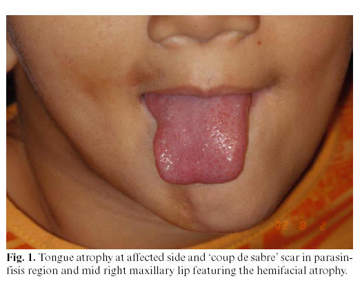
In 2001, an 8-year-old female patient, resident in Belém,  State of Pará, Brazil, was conducted do the radiological clinic for orthodontic  documentation. During the physical examination, it was noted that this patient  presented facial asymmetry with a marked hypoplasia of the right side of the  face, with deviation of lips and nose, with a big linear dark scar (<i>coup de  sabre</i>) in the right side of mandibular mentum region, enophthalmy in right  eye region and areas of hyperpigmentation on the affected skin. Intraorally, the  most relevant alteration was the unilateral atrophy of tongue (<a href="#f1">Figure 1</a>).  Radiographically, it was observed an atrophic root development or a pathologic  reabsorption of permanent teeth numbers 34, 33, 41, 42, 43, 44, 45, 46 and 47 (<a href="#f2">Figure 2</a>).</font> </p>     ]]></body>
<body><![CDATA[<p align="center"><font size="2" face="Verdana"> <a name="f1"><img border="0" src="/img/revistas/medicorpa/v11n2/03i.ht1.jpg" width="525" height="424"></a> </font></p>     <p align="center"><font size="2" face="Verdana"> <a name="f2"><img border="0" src="/img/revistas/medicorpa/v11n2/03i.ht2.gif" width="525" height="291"></a> </font></p>     <p align="center">&nbsp;</p>     <p><font size="2" face="Verdana"> 
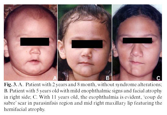
The patient presented good health condition, without any  sickness that would explain the cause of that facial asymmetry. Photos of  previous years were requested to the responsible for the patient, who provided  them from the 1st until the 8th year of age. Clinical signs of the disturb were  just noticed from the 6th year onwards (<a href="#f3">Figure 3 – A, B, C</a>). </font></p>     <p align="center"><font size="2" face="Verdana"> <a name="f3"><img border="0" src="/img/revistas/medicorpa/v11n2/03i.ht3.jpg" width="531" height="367"></a> </font></p>     <p><font size="2" face="Verdana"> With these clinical and radiologic findings, the diagnosis  was of progressive hemifacial atrophy. At present, the patient is being  periodically reviewed until atrophic manifestation stops and specific  intervention could be accomplished. </font></p>     <p><font size="2" face="Verdana"> In 2003, at age of 10, a new photographic and radiographic  documentation was done in order to analyze the evolution of the case.  Extraorally, there was an aggravation of clinical signs observed two years ago,  such as enophthalmy, hypoplasia of right hemiface, with deviation of lips and  nose, and also cutaneous hyperpigmentation. During an intraoral exam, it was  observed the persistence of the canine and the first molar deciduous teeth, as  well as dental crowding. Radiographically, it was noticed an image suggestive of  pathologic root reabsorption of teeth number 14, 15, 16, 17, 41, 42, 43, 44, 45,  46 and 47, as well as impactation of element 45 by 84 and of element 47 by 48 (developing tooth).</font></p>     <p>&nbsp;</p>     <p><b><font face="Verdana">Discussion</font></p> </b>     <p><font size="2" face="Verdana"> Progressive hemifacial atrophy, as described in the case  above, is a rare pathology, of unknown cause, whose degenerative condition  affect not only the esthetic, but also the functionality of the attained hemiface. </font></p>     ]]></body>
<body><![CDATA[<p><font size="2" face="Verdana"> Clinically, the skin can be dry and with a dark pigmentation.  Some patients present a demarcation line between normal and abnormal skin,  reminding a big linear scar, known as <i>coup de sabre</i>, as we could notice  in the case reported. Ocular involvement is common, and the most frequent  manifestation is the enophthalmy, due to fat loss around the orbit, as was  observed in our patient. The eye usually works normally. Even not noticed in the  present case, the ear can be smaller than normal ones due to the atrophy (5).  There may be localized areas of alopecia. Occasionally, there may be some  neurological complications, such as trigeminal neuralgia, facial paresthesia,  severe headache and contralateral epilepsy (1, 11, 14-16), this last being the  most common complication (17). Mouth and nose are deviated to the affected side,  deviating also facial and dental midlines. Atrophy of superior lip led the  anterior teeth to be exposed, and there may be also unilateral atrophy of tongue.  Our case showed clearly those features of facial asymmetry and tongue atrophy.  Radiographically, teeth of affected side can present some deficiency in root  development and, consequently, delayed eruption. However, the affected teeth are  normal and vital clinically. This situation occurred with our patient, as most  of her teeth of the affected side presented a reabsorption/malformation of their  roots, causing delayed eruption and dental crowding. Very often, there is  unilateral posterior crossbite, as a result of jaw hypoplasia and delayed teeth  eruption (4, 10). The intraoral soft tissue and chewing muscles are, sometimes,  normal, without any movement, speeching or deglutition implications (5, 7).  Histologically, atrophy of epidermis, dermis and subcutaneous tissue are  observed (7). Variable infiltrate of lymphocytes and monocytes at dermis (11, 7)  and lack of subcutaneous fat in the affected tissue are also characteristic (7).  Besides, it can be identified degenerative alterations on vascular endothelia in  electron microscopy (7). </font> </p>     <p><font size="2" face="Verdana"> The treatment is usually based on reposition of adipose  tissue that was lost due to atrophy (12). Autogenous fat grafts, cartilage  grafts, silicon injections and prostheses, bovine collagen and inorganic  implants are some alternatives to esthetic correction of the atrophy (12, 13).  However, these treatment modalities resolve just momentarily the good appearance,  whereas all the structure projected in the cosmetic surgery is lost with time,  due to gravity action, and the patient needs a new intervention. Moreover, a  backing treatment is indicated to functional and neurological troubles. The  cosmetic treatment is just recommended when the illness stops its evolution, so  this is the reason why our patient hasn’t been submitted to any surgical  intervention yet. </font> </p>     <p><font size="2" face="Verdana"> Precocious diagnostic is relevant, mainly in cases of  systemic sicknesses with idiopathic origin, in which a simple anamnesis and a  conventional clinical exam do not give enough data for a precise diagnosis and a  suitable treatment.</font> </p>     <p>&nbsp; </p>     <p><b><font face="Verdana">References</font> </p></b>     <!-- ref --><p><font size="2" face="Verdana"> 1. Jurkiewicz MJ, Nahai F. The use of free revascularized  grafts in the amelioration of hemifacial atrophy. Plast Reconstr Surg  1985;76:44-55.</font>&nbsp;&nbsp;&nbsp;&nbsp;&nbsp;&nbsp;&nbsp;&nbsp;[&#160;<a href="javascript:void(0);" onclick="javascript: window.open('/scielo.php?script=sci_nlinks&ref=2945543&pid=S1698-6946200600020000300001&lng=','','width=640,height=500,resizable=yes,scrollbars=1,menubar=yes,');">Links</a>&#160;]<!-- end-ref --><!-- ref --><p><font face="Verdana" size="2"> 2. Lakhani PK, David TJ. Progressive hemifacial atrophy with  scleroderma and ipsilateral limb wasting (Parry Romberg Syndrome). J R Soc Med  1984;77:138-9.</font>&nbsp;&nbsp;&nbsp;&nbsp;&nbsp;&nbsp;&nbsp;&nbsp;[&#160;<a href="javascript:void(0);" onclick="javascript: window.open('/scielo.php?script=sci_nlinks&ref=2945544&pid=S1698-6946200600020000300002&lng=','','width=640,height=500,resizable=yes,scrollbars=1,menubar=yes,');">Links</a>&#160;]<!-- end-ref --><!-- ref --><p><font face="Verdana" size="2"> 3. Finesilver B, Rosow HN. Total hemiatrophy. JAMA  1938;5:366-8.</font>&nbsp;&nbsp;&nbsp;&nbsp;&nbsp;&nbsp;&nbsp;&nbsp;[&#160;<a href="javascript:void(0);" onclick="javascript: window.open('/scielo.php?script=sci_nlinks&ref=2945545&pid=S1698-6946200600020000300003&lng=','','width=640,height=500,resizable=yes,scrollbars=1,menubar=yes,');">Links</a>&#160;]<!-- end-ref --><!-- ref --><p><font face="Verdana" size="2"> 4. Foster TD. The effects of hemifacial atrophy of dental  growth. Br Dent J 1979;146:148-50.</font>&nbsp;&nbsp;&nbsp;&nbsp;&nbsp;&nbsp;&nbsp;&nbsp;[&#160;<a href="javascript:void(0);" onclick="javascript: window.open('/scielo.php?script=sci_nlinks&ref=2945546&pid=S1698-6946200600020000300004&lng=','','width=640,height=500,resizable=yes,scrollbars=1,menubar=yes,');">Links</a>&#160;]<!-- end-ref --><!-- ref --><p><font face="Verdana" size="2"> 5. Mazzeo N, Fisher JG, Mayer MH, Mathieu GP, Mcade FGG.  Progressive hemifacial atrophy (Parry Romberg Syndrome). Oral Surg Oral Med Oral  Pathol Oral Radiol Endod 1995;79:30-5.</font>&nbsp;&nbsp;&nbsp;&nbsp;&nbsp;&nbsp;&nbsp;&nbsp;[&#160;<a href="javascript:void(0);" onclick="javascript: window.open('/scielo.php?script=sci_nlinks&ref=2945547&pid=S1698-6946200600020000300005&lng=','','width=640,height=500,resizable=yes,scrollbars=1,menubar=yes,');">Links</a>&#160;]<!-- end-ref --><!-- ref --><p><font face="Verdana" size="2"> 6. Miller MT, Sloane H, Goldberg MF, Grisolano J, Frenkel M,  Mafee MF. Progressive hemifacial atrophy (Parry Romberg Disease). J Pediatr  Oftamol Strabismus 1987;24:27-36.</font>&nbsp;&nbsp;&nbsp;&nbsp;&nbsp;&nbsp;&nbsp;&nbsp;[&#160;<a href="javascript:void(0);" onclick="javascript: window.open('/scielo.php?script=sci_nlinks&ref=2945548&pid=S1698-6946200600020000300006&lng=','','width=640,height=500,resizable=yes,scrollbars=1,menubar=yes,');">Links</a>&#160;]<!-- end-ref --><!-- ref --><p><font face="Verdana" size="2"> 7. Pensler JM, Murphy GF, Muliken JB. Clinical and ultra-structural  studies of Romberg´s hemifacial atrophy. Plast Reconstr Surg 1990;85:669-76.</font>&nbsp;&nbsp;&nbsp;&nbsp;&nbsp;&nbsp;&nbsp;&nbsp;[&#160;<a href="javascript:void(0);" onclick="javascript: window.open('/scielo.php?script=sci_nlinks&ref=2945549&pid=S1698-6946200600020000300007&lng=','','width=640,height=500,resizable=yes,scrollbars=1,menubar=yes,');">Links</a>&#160;]<!-- end-ref --><!-- ref --><p><font face="Verdana" size="2"> 8. Shaffer WG, Hine MK, Levy BM, eds. Textbook of oral  pathology. Philadelphia: WB Saunders; 1983. p. 9-10.</font>&nbsp;&nbsp;&nbsp;&nbsp;&nbsp;&nbsp;&nbsp;&nbsp;[&#160;<a href="javascript:void(0);" onclick="javascript: window.open('/scielo.php?script=sci_nlinks&ref=2945550&pid=S1698-6946200600020000300008&lng=','','width=640,height=500,resizable=yes,scrollbars=1,menubar=yes,');">Links</a>&#160;]<!-- end-ref --><!-- ref --><p><font face="Verdana" size="2"> 9. Zafarulla MY. Progressive hemifacial atrophy: a case  report. Br J Ophthalmol 1985;69:545-7.</font>&nbsp;&nbsp;&nbsp;&nbsp;&nbsp;&nbsp;&nbsp;&nbsp;[&#160;<a href="javascript:void(0);" onclick="javascript: window.open('/scielo.php?script=sci_nlinks&ref=2945551&pid=S1698-6946200600020000300009&lng=','','width=640,height=500,resizable=yes,scrollbars=1,menubar=yes,');">Links</a>&#160;]<!-- end-ref --><!-- ref --><p><font face="Verdana" size="2"> 10. Moore MH, Wong KS, Proudman TW, David DJ. Progressive  hemifacial atrophy (Romberg´s Disease): skeletal involvement and treatment. Br J  Plast Surg 1993; 46: 39-44.</font>&nbsp;&nbsp;&nbsp;&nbsp;&nbsp;&nbsp;&nbsp;&nbsp;[&#160;<a href="javascript:void(0);" onclick="javascript: window.open('/scielo.php?script=sci_nlinks&ref=2945552&pid=S1698-6946200600020000300010&lng=','','width=640,height=500,resizable=yes,scrollbars=1,menubar=yes,');">Links</a>&#160;]<!-- end-ref --><!-- ref --><p><font face="Verdana" size="2"> 11. Neville BW, Damm DD, Allen CN, Bouquout JE, eds.  Patologia oral e Maxilofacial. Rio de Janeiro: Guanabara Koogan; 1998. p. 35.</font>&nbsp;&nbsp;&nbsp;&nbsp;&nbsp;&nbsp;&nbsp;&nbsp;[&#160;<a href="javascript:void(0);" onclick="javascript: window.open('/scielo.php?script=sci_nlinks&ref=2945553&pid=S1698-6946200600020000300011&lng=','','width=640,height=500,resizable=yes,scrollbars=1,menubar=yes,');">Links</a>&#160;]<!-- end-ref --><!-- ref --><p><font face="Verdana" size="2"> 12. Roddi R, Riggio E, Gilbert PM, Houvius SER, Vaandrager  JM, van der Meulen JCH. Clinical evaluation of techiniques used in the surgical  treatment of progressive hemifacial atrophy. J Craniomaxilofac Surg  1994;22:23-32.</font>&nbsp;&nbsp;&nbsp;&nbsp;&nbsp;&nbsp;&nbsp;&nbsp;[&#160;<a href="javascript:void(0);" onclick="javascript: window.open('/scielo.php?script=sci_nlinks&ref=2945554&pid=S1698-6946200600020000300012&lng=','','width=640,height=500,resizable=yes,scrollbars=1,menubar=yes,');">Links</a>&#160;]<!-- end-ref --><!-- ref --><p><font face="Verdana" size="2"> 13. de la Fuente A, Jimenez A. Latissimus dorsi free flap for  restauration of facial contour defects. Ann Plast Surg 1989;22:1-8.</font>&nbsp;&nbsp;&nbsp;&nbsp;&nbsp;&nbsp;&nbsp;&nbsp;[&#160;<a href="javascript:void(0);" onclick="javascript: window.open('/scielo.php?script=sci_nlinks&ref=2945555&pid=S1698-6946200600020000300013&lng=','','width=640,height=500,resizable=yes,scrollbars=1,menubar=yes,');">Links</a>&#160;]<!-- end-ref --><!-- ref --><p><font face="Verdana" size="2"> 14. Gorlin RJ, Pinborg JJ, eds. Syndromes of the head and  neck. New York: McGraw Hill; 1964. p. 475 – 7.</font>&nbsp;&nbsp;&nbsp;&nbsp;&nbsp;&nbsp;&nbsp;&nbsp;[&#160;<a href="javascript:void(0);" onclick="javascript: window.open('/scielo.php?script=sci_nlinks&ref=2945556&pid=S1698-6946200600020000300014&lng=','','width=640,height=500,resizable=yes,scrollbars=1,menubar=yes,');">Links</a>&#160;]<!-- end-ref --><!-- ref --><p><font face="Verdana" size="2"> 15. Roed-Petersen B. Hemifacial lipodystrophy – report of a  case. Oral Surg Oral Med Oral Pathol 1979;43:230-2.</font>&nbsp;&nbsp;&nbsp;&nbsp;&nbsp;&nbsp;&nbsp;&nbsp;[&#160;<a href="javascript:void(0);" onclick="javascript: window.open('/scielo.php?script=sci_nlinks&ref=2945557&pid=S1698-6946200600020000300015&lng=','','width=640,height=500,resizable=yes,scrollbars=1,menubar=yes,');">Links</a>&#160;]<!-- end-ref --><!-- ref --><p><font face="Verdana" size="2"> 16. Sagild JC, Alving J. Hemiplegic migraine and progressive  hemifacial atrophy. Ann Neurol 1985;17:620.</font>&nbsp;&nbsp;&nbsp;&nbsp;&nbsp;&nbsp;&nbsp;&nbsp;[&#160;<a href="javascript:void(0);" onclick="javascript: window.open('/scielo.php?script=sci_nlinks&ref=2945558&pid=S1698-6946200600020000300016&lng=','','width=640,height=500,resizable=yes,scrollbars=1,menubar=yes,');">Links</a>&#160;]<!-- end-ref --><!-- ref --><p><font face="Verdana" size="2"> 17. Chbicheb M, Gelot A, Rivier F, Roubertie A, Humbert  Claude V, Coubes P. et al. Parry-Romberg´s syndrome and epilepsy. Rev Neurol  2005;161:92-7.</font>&nbsp;&nbsp;&nbsp;&nbsp;&nbsp;&nbsp;&nbsp;&nbsp;[&#160;<a href="javascript:void(0);" onclick="javascript: window.open('/scielo.php?script=sci_nlinks&ref=2945559&pid=S1698-6946200600020000300017&lng=','','width=640,height=500,resizable=yes,scrollbars=1,menubar=yes,');">Links</a>&#160;]<!-- end-ref --><p>&nbsp;</p>      <p>&nbsp;</p>      <p><b><a href="#top"><font face="Verdana" size="2"><img border="0" src="/img/revistas/medicorpa/v11n2/seta.gif" width="15" height="17"></font></a> <font face="Verdana" size="2"> <a name="down">Correspondence</a></font></font></b><font face="Verdana" size="2">    <br> Dr. João de Jesus Viana Pinheiro    <br> Av. Almirante Wandenkolk, 1243,1006    <br> Umarizal. Belém-Pará-Brazil –</font><font face="Verdana" size="2">    <br> ZIP Code: 66050-030    <br> E-mail- <a href="mailto:radface@hotmail.com">radface@hotmail.com</a></font></p>      ]]></body>
<body><![CDATA[<p><font face="Verdana" size="2"> Received: 10-03-2005    <br> Accepted: 14-08-2005</font> </p>      ]]></body><back>
<ref-list>
<ref id="B1">
<label>1</label><nlm-citation citation-type="journal">
<person-group person-group-type="author">
<name>
<surname><![CDATA[Jurkiewicz]]></surname>
<given-names><![CDATA[MJ]]></given-names>
</name>
<name>
<surname><![CDATA[Nahai]]></surname>
<given-names><![CDATA[F]]></given-names>
</name>
</person-group>
<article-title xml:lang="en"><![CDATA[The use of free revascularized grafts in the amelioration of hemifacial atrophy]]></article-title>
<source><![CDATA[Plast Reconstr Surg]]></source>
<year>1985</year>
<volume>76</volume>
<page-range>44-55</page-range></nlm-citation>
</ref>
<ref id="B2">
<label>2</label><nlm-citation citation-type="journal">
<person-group person-group-type="author">
<name>
<surname><![CDATA[Lakhani]]></surname>
<given-names><![CDATA[PK]]></given-names>
</name>
<name>
<surname><![CDATA[David]]></surname>
<given-names><![CDATA[TJ]]></given-names>
</name>
</person-group>
<article-title xml:lang="en"><![CDATA[Progressive hemifacial atrophy with scleroderma and ipsilateral limb wasting (Parry Romberg Syndrome)]]></article-title>
<source><![CDATA[J R Soc Med]]></source>
<year>1984</year>
<volume>77</volume>
<page-range>138-9</page-range></nlm-citation>
</ref>
<ref id="B3">
<label>3</label><nlm-citation citation-type="journal">
<person-group person-group-type="author">
<name>
<surname><![CDATA[Finesilver]]></surname>
<given-names><![CDATA[B]]></given-names>
</name>
<name>
<surname><![CDATA[Rosow]]></surname>
<given-names><![CDATA[HN]]></given-names>
</name>
</person-group>
<article-title xml:lang="en"><![CDATA[Total hemiatrophy]]></article-title>
<source><![CDATA[JAMA]]></source>
<year>1938</year>
<volume>5</volume>
<page-range>366-8</page-range></nlm-citation>
</ref>
<ref id="B4">
<label>4</label><nlm-citation citation-type="journal">
<person-group person-group-type="author">
<name>
<surname><![CDATA[Foster]]></surname>
<given-names><![CDATA[TD]]></given-names>
</name>
</person-group>
<article-title xml:lang="en"><![CDATA[The effects of hemifacial atrophy of dental growth]]></article-title>
<source><![CDATA[Br Dent J]]></source>
<year>1979</year>
<volume>146</volume>
<page-range>148-50</page-range></nlm-citation>
</ref>
<ref id="B5">
<label>5</label><nlm-citation citation-type="journal">
<person-group person-group-type="author">
<name>
<surname><![CDATA[Mazzeo]]></surname>
<given-names><![CDATA[N]]></given-names>
</name>
<name>
<surname><![CDATA[Fisher]]></surname>
<given-names><![CDATA[JG]]></given-names>
</name>
<name>
<surname><![CDATA[Mayer]]></surname>
<given-names><![CDATA[MH]]></given-names>
</name>
<name>
<surname><![CDATA[Mathieu]]></surname>
<given-names><![CDATA[GP]]></given-names>
</name>
<name>
<surname><![CDATA[Mcade]]></surname>
<given-names><![CDATA[FGG]]></given-names>
</name>
</person-group>
<article-title xml:lang="en"><![CDATA[Progressive hemifacial atrophy (Parry Romberg Syndrome)]]></article-title>
<source><![CDATA[Oral Surg Oral Med Oral Pathol Oral Radiol Endod]]></source>
<year>1995</year>
<volume>79</volume>
<page-range>30-5</page-range></nlm-citation>
</ref>
<ref id="B6">
<label>6</label><nlm-citation citation-type="journal">
<person-group person-group-type="author">
<name>
<surname><![CDATA[Miller]]></surname>
<given-names><![CDATA[MT]]></given-names>
</name>
<name>
<surname><![CDATA[Sloane]]></surname>
<given-names><![CDATA[H]]></given-names>
</name>
<name>
<surname><![CDATA[Goldberg]]></surname>
<given-names><![CDATA[MF]]></given-names>
</name>
<name>
<surname><![CDATA[Grisolano]]></surname>
<given-names><![CDATA[J]]></given-names>
</name>
<name>
<surname><![CDATA[Frenkel]]></surname>
<given-names><![CDATA[M]]></given-names>
</name>
<name>
<surname><![CDATA[Mafee]]></surname>
<given-names><![CDATA[MF]]></given-names>
</name>
</person-group>
<article-title xml:lang="en"><![CDATA[Progressive hemifacial atrophy (Parry Romberg Disease)]]></article-title>
<source><![CDATA[J Pediatr Oftamol Strabismus]]></source>
<year>1987</year>
<volume>24</volume>
<page-range>27-36</page-range></nlm-citation>
</ref>
<ref id="B7">
<label>7</label><nlm-citation citation-type="journal">
<person-group person-group-type="author">
<name>
<surname><![CDATA[Pensler]]></surname>
<given-names><![CDATA[JM]]></given-names>
</name>
<name>
<surname><![CDATA[Murphy]]></surname>
<given-names><![CDATA[GF]]></given-names>
</name>
<name>
<surname><![CDATA[Muliken]]></surname>
<given-names><![CDATA[JB]]></given-names>
</name>
</person-group>
<article-title xml:lang="en"><![CDATA[Clinical and ultra-structural studies of Romberg´s hemifacial atrophy]]></article-title>
<source><![CDATA[Plast Reconstr Surg]]></source>
<year>1990</year>
<volume>85</volume>
<page-range>669-76</page-range></nlm-citation>
</ref>
<ref id="B8">
<label>8</label><nlm-citation citation-type="book">
<person-group person-group-type="author">
<name>
<surname><![CDATA[Shaffer]]></surname>
<given-names><![CDATA[WG]]></given-names>
</name>
<name>
<surname><![CDATA[Hine]]></surname>
<given-names><![CDATA[MK]]></given-names>
</name>
<name>
<surname><![CDATA[Levy]]></surname>
<given-names><![CDATA[BM]]></given-names>
</name>
</person-group>
<source><![CDATA[Textbook of oral pathology]]></source>
<year>1983</year>
<page-range>9-10</page-range><publisher-loc><![CDATA[Philadelphia ]]></publisher-loc>
<publisher-name><![CDATA[WB Saunders]]></publisher-name>
</nlm-citation>
</ref>
<ref id="B9">
<label>9</label><nlm-citation citation-type="journal">
<person-group person-group-type="author">
<name>
<surname><![CDATA[Zafarulla]]></surname>
<given-names><![CDATA[MY]]></given-names>
</name>
</person-group>
<article-title xml:lang="en"><![CDATA[Progressive hemifacial atrophy: a case report]]></article-title>
<source><![CDATA[Br J Ophthalmol]]></source>
<year>1985</year>
<volume>69</volume>
<page-range>545-7</page-range></nlm-citation>
</ref>
<ref id="B10">
<label>10</label><nlm-citation citation-type="journal">
<person-group person-group-type="author">
<name>
<surname><![CDATA[Moore]]></surname>
<given-names><![CDATA[MH]]></given-names>
</name>
<name>
<surname><![CDATA[Wong]]></surname>
<given-names><![CDATA[KS]]></given-names>
</name>
<name>
<surname><![CDATA[Proudman]]></surname>
<given-names><![CDATA[TW]]></given-names>
</name>
<name>
<surname><![CDATA[David]]></surname>
<given-names><![CDATA[DJ]]></given-names>
</name>
</person-group>
<article-title xml:lang="en"><![CDATA[Progressive hemifacial atrophy (Romberg´s Disease): skeletal involvement and treatment]]></article-title>
<source><![CDATA[Br J Plast Surg]]></source>
<year>1993</year>
<volume>46</volume>
<page-range>39-44</page-range></nlm-citation>
</ref>
<ref id="B11">
<label>11</label><nlm-citation citation-type="book">
<person-group person-group-type="author">
<name>
<surname><![CDATA[Neville]]></surname>
<given-names><![CDATA[BW]]></given-names>
</name>
<name>
<surname><![CDATA[Damm]]></surname>
<given-names><![CDATA[DD]]></given-names>
</name>
<name>
<surname><![CDATA[Allen]]></surname>
<given-names><![CDATA[CN]]></given-names>
</name>
<name>
<surname><![CDATA[Bouquout]]></surname>
<given-names><![CDATA[JE]]></given-names>
</name>
</person-group>
<source><![CDATA[Patologia oral e Maxilofacial]]></source>
<year>1998</year>
<page-range>35</page-range><publisher-loc><![CDATA[Rio de Janeiro ]]></publisher-loc>
<publisher-name><![CDATA[Guanabara Koogan]]></publisher-name>
</nlm-citation>
</ref>
<ref id="B12">
<label>12</label><nlm-citation citation-type="journal">
<person-group person-group-type="author">
<name>
<surname><![CDATA[Roddi]]></surname>
<given-names><![CDATA[R]]></given-names>
</name>
<name>
<surname><![CDATA[Riggio]]></surname>
<given-names><![CDATA[E]]></given-names>
</name>
<name>
<surname><![CDATA[Gilbert]]></surname>
<given-names><![CDATA[PM]]></given-names>
</name>
<name>
<surname><![CDATA[Houvius]]></surname>
<given-names><![CDATA[SER]]></given-names>
</name>
<name>
<surname><![CDATA[Vaandrager]]></surname>
<given-names><![CDATA[JM]]></given-names>
</name>
<name>
<surname><![CDATA[van der Meulen]]></surname>
<given-names><![CDATA[JCH]]></given-names>
</name>
</person-group>
<article-title xml:lang="en"><![CDATA[Clinical evaluation of techiniques used in the surgical treatment of progressive hemifacial atrophy]]></article-title>
<source><![CDATA[J Craniomaxilofac Surg]]></source>
<year>1994</year>
<volume>22</volume>
<page-range>23-32</page-range></nlm-citation>
</ref>
<ref id="B13">
<label>13</label><nlm-citation citation-type="journal">
<person-group person-group-type="author">
<name>
<surname><![CDATA[de la Fuente]]></surname>
<given-names><![CDATA[A]]></given-names>
</name>
<name>
<surname><![CDATA[Jimenez]]></surname>
<given-names><![CDATA[A]]></given-names>
</name>
</person-group>
<article-title xml:lang="en"><![CDATA[Latissimus dorsi free flap for restauration of facial contour defects]]></article-title>
<source><![CDATA[Ann Plast Surg]]></source>
<year>1989</year>
<volume>22</volume>
<page-range>1-8</page-range></nlm-citation>
</ref>
<ref id="B14">
<label>14</label><nlm-citation citation-type="book">
<person-group person-group-type="author">
<name>
<surname><![CDATA[Gorlin]]></surname>
<given-names><![CDATA[RJ]]></given-names>
</name>
<name>
<surname><![CDATA[Pinborg]]></surname>
<given-names><![CDATA[JJ]]></given-names>
</name>
</person-group>
<source><![CDATA[Syndromes of the head and neck]]></source>
<year>1964</year>
<page-range>475 - 7</page-range><publisher-loc><![CDATA[New York ]]></publisher-loc>
<publisher-name><![CDATA[McGraw Hill]]></publisher-name>
</nlm-citation>
</ref>
<ref id="B15">
<label>15</label><nlm-citation citation-type="journal">
<person-group person-group-type="author">
<name>
<surname><![CDATA[Roed-Petersen]]></surname>
<given-names><![CDATA[B]]></given-names>
</name>
</person-group>
<article-title xml:lang="en"><![CDATA[Hemifacial lipodystrophy: report of a case]]></article-title>
<source><![CDATA[Oral Surg Oral Med Oral Pathol]]></source>
<year>1979</year>
<volume>43</volume>
<page-range>230-2</page-range></nlm-citation>
</ref>
<ref id="B16">
<label>16</label><nlm-citation citation-type="journal">
<person-group person-group-type="author">
<name>
<surname><![CDATA[Sagild]]></surname>
<given-names><![CDATA[JC]]></given-names>
</name>
<name>
<surname><![CDATA[Alving]]></surname>
<given-names><![CDATA[J]]></given-names>
</name>
</person-group>
<article-title xml:lang="en"><![CDATA[Hemiplegic migraine and progressive hemifacial atrophy]]></article-title>
<source><![CDATA[Ann Neurol]]></source>
<year>1985</year>
<volume>17</volume>
<page-range>620</page-range></nlm-citation>
</ref>
<ref id="B17">
<label>17</label><nlm-citation citation-type="journal">
<person-group person-group-type="author">
<name>
<surname><![CDATA[Chbicheb]]></surname>
<given-names><![CDATA[M]]></given-names>
</name>
<name>
<surname><![CDATA[Gelot]]></surname>
<given-names><![CDATA[A]]></given-names>
</name>
<name>
<surname><![CDATA[Rivier]]></surname>
<given-names><![CDATA[F]]></given-names>
</name>
<name>
<surname><![CDATA[Roubertie]]></surname>
<given-names><![CDATA[A]]></given-names>
</name>
<name>
<surname><![CDATA[Humbert Claude]]></surname>
<given-names><![CDATA[V]]></given-names>
</name>
<name>
<surname><![CDATA[Coubes]]></surname>
<given-names><![CDATA[P]]></given-names>
</name>
</person-group>
<article-title xml:lang="en"><![CDATA[Parry-Romberg´s syndrome and epilepsy]]></article-title>
<source><![CDATA[Rev Neurol]]></source>
<year>2005</year>
<volume>161</volume>
<page-range>92-7</page-range></nlm-citation>
</ref>
</ref-list>
</back>
</article>
