<?xml version="1.0" encoding="ISO-8859-1"?><article xmlns:mml="http://www.w3.org/1998/Math/MathML" xmlns:xlink="http://www.w3.org/1999/xlink" xmlns:xsi="http://www.w3.org/2001/XMLSchema-instance">
<front>
<journal-meta>
<journal-id>1698-6946</journal-id>
<journal-title><![CDATA[Medicina Oral, Patología Oral y Cirugía Bucal (Internet)]]></journal-title>
<abbrev-journal-title><![CDATA[Med. oral patol. oral cir.bucal (Internet)]]></abbrev-journal-title>
<issn>1698-6946</issn>
<publisher>
<publisher-name><![CDATA[Sociedad Española de Medicina Oral]]></publisher-name>
</publisher>
</journal-meta>
<article-meta>
<article-id>S1698-69462006000200019</article-id>
<title-group>
<article-title xml:lang="en"><![CDATA[An unusual clinicoradiographic presentation of a lateral periodontal cyst: report of two cases]]></article-title>
</title-group>
<contrib-group>
<contrib contrib-type="author">
<name>
<surname><![CDATA[Amaral Mendes]]></surname>
<given-names><![CDATA[Rui]]></given-names>
</name>
<xref ref-type="aff" rid="A01"/>
</contrib>
<contrib contrib-type="author">
<name>
<surname><![CDATA[van der Waal]]></surname>
<given-names><![CDATA[Isaac]]></given-names>
</name>
<xref ref-type="aff" rid="A02"/>
</contrib>
</contrib-group>
<aff id="A01">
<institution><![CDATA[,University of Porto Faculty of Dental Medicine Department of Oral Surgery]]></institution>
<addr-line><![CDATA[Porto ]]></addr-line>
<country>Portugal</country>
</aff>
<aff id="A02">
<institution><![CDATA[,VU University Medical Center Department of Oral and Maxillofacial Surgery/Oral Pathology ]]></institution>
<addr-line><![CDATA[Amsterdam ]]></addr-line>
<country>Netherlands</country>
</aff>
<pub-date pub-type="pub">
<day>00</day>
<month>04</month>
<year>2006</year>
</pub-date>
<pub-date pub-type="epub">
<day>00</day>
<month>04</month>
<year>2006</year>
</pub-date>
<volume>11</volume>
<numero>2</numero>
<fpage>185</fpage>
<lpage>187</lpage>
<copyright-statement/>
<copyright-year/>
<self-uri xlink:href="http://scielo.isciii.es/scielo.php?script=sci_arttext&amp;pid=S1698-69462006000200019&amp;lng=en&amp;nrm=iso"></self-uri><self-uri xlink:href="http://scielo.isciii.es/scielo.php?script=sci_abstract&amp;pid=S1698-69462006000200019&amp;lng=en&amp;nrm=iso"></self-uri><self-uri xlink:href="http://scielo.isciii.es/scielo.php?script=sci_pdf&amp;pid=S1698-69462006000200019&amp;lng=en&amp;nrm=iso"></self-uri><abstract abstract-type="short" xml:lang="en"><p><![CDATA[The lateral periodontal cyst and the botryoid odontogenic cyst are two rare nosological entities, who, despite their radiological and clinical presentation can only be diagnosed by their rather typical histopathological characteristics. The purpose of this article is to report two cases of radiolucent cystic lesions of the mandible, located in the premolar area, with a clinical and radiographic diagnosis of residual cyst, which showed histological features of a lateral periodontal cyst. Histopathologically, the lateral periodontal cyst lining is characterized by a thin cuboidal to stratified squamous non-keratinizing epithelium, ranging from one to five cell layers and presence of one or more epithelial thickenings or plaques. Furthermore, glycogen-rich clear cells encountered either in the epithelial plaques or in the superficial layer of the lining epithelium.]]></p></abstract>
<kwd-group>
<kwd lng="en"><![CDATA[Odontogenic cysts]]></kwd>
<kwd lng="en"><![CDATA[lateral periodontal cyst]]></kwd>
<kwd lng="en"><![CDATA[residual cyst]]></kwd>
</kwd-group>
</article-meta>
</front><body><![CDATA[ <p align="right"><font face="Verdana" size="2"><a name="top"></a><b>CIRUGÍA BUCAL</b></font>     <p>&nbsp;</p>     <p><b><font size="4" face="Verdana">An unusual clinicoradiographic presentation of a lateral periodontal cyst –&nbsp;    <br> report of two cases</font></b></p>     <p>&nbsp;</p>     <p>&nbsp;</p>     <p><b><font face="Verdana" size="2">Rui Amaral Mendes <sup>1</sup>, Isaac van der Waal <sup>2</sup></font> </font></b></p>     <p><font face="Verdana" size="2">(1) DMD. Clinical Instructor of Oral and Maxillofacial  Surgery. Doctoral Student of Oral Surgery and Pathology.&nbsp;    <br>  Department of Oral  Surgery. Faculty of Dental Medicine, University of Porto, Portugal    <br> (2) DDS, PhD. Head, Department of Oral and Maxillofacial  Surgery/Oral Pathology. Department of Oral and&nbsp;    ]]></body>
<body><![CDATA[<br>  Maxillofacial Surgery/Oral  Pathology. VU University Medical Center/ACTA, Amsterdam, the Netherlands</font></p>     <p><font face="Verdana" size="2"><a href="#down">Correspondence</a></font></p>     <p>&nbsp;</p>     <p>&nbsp;</p> <hr size="1">     <p><b><font face="Verdana" size="2">ABSTRACT</font></b></p>      <p><font face="Verdana" size="2">The lateral periodontal cyst and the botryoid odontogenic  cyst are two rare nosological entities, who, despite their radiological and  clinical presentation can only be diagnosed by their rather typical  histopathological characteristics.    <br> The purpose of this article is to report two cases of  radiolucent cystic lesions of the mandible, located in the premolar area, with a  clinical and radiographic diagnosis of residual cyst, which showed histological  features of a lateral periodontal cyst. Histopathologically, the lateral  periodontal cyst lining is characterized by a thin cuboidal to stratified  squamous non-keratinizing epithelium, ranging from one to five cell layers and  presence of one or more epithelial thickenings or plaques. Furthermore, glycogen-rich  clear cells encountered either in the epithelial plaques or in the superficial  layer of the lining epithelium.</font></p>     <p><b><font face="Verdana" size="2">Key words:</font></b><font face="Verdana" size="2"> Odontogenic cysts, lateral periodontal cyst, residual cyst.</font></p> <hr size="1"> <b>     <p>&nbsp;</p>     <p><font face="Verdana">Introduction</font></p> </b>     ]]></body>
<body><![CDATA[<p><font face="Verdana" size="2">The epithelial lining of the odontogenic cysts is derived  from the epithelial remains of the tooth-forming organ, such as 1) the  epithelial rests of Malassez, left in the periodontal ligament by the breakdown  of the epithelial root sheath of Hertwig; 2) the reduced enamel epithelium,  which covers the completely formed crown of an unerupted tooth and is derived  from the enamel organ; and 3) the epithelial rests or glands of Serres, which  persist after the dissolution of the dental lamina (1).</font></p>     <p><font face="Verdana" size="2">The lateral periodontal lateral cyst is a developmental odontogenic cyst  defined as a radiolucent lesion which develops along the lateral aspect of an  erupted vital</font> </p>     <p><font face="Verdana" size="2">tooth, in which an inflammatory etiology and a diagnosis of  collateral keratocyst have been excluded based on clinical and histological  grounds (2).</font></p>     <p><font face="Verdana" size="2">The pathogenesis of the lateral periodontal cyst is a matter  of controversy dealt with in numerous articles, with opposing views regarding  the origin, being either from the reduced enamel epithelium or the dental lamina  or even the epithelial rest of Malassez (1-5).</font></p>     <p><font face="Verdana" size="2">In the past, the lateral periodontal cyst was largely</font> <font face="Verdana" size="2">defined as a cyst located between the roots of two vital teeth,</font> <font face="Verdana" size="2">usually in the bicuspid or cuspid area (1,2,5). However in  1992, in the WHO’s monograph on the &quot;Histological typing of odontogenic tumours&quot;,  rather characteristic histopathological criteria were added to the  clinicoradiographic ones (<a href="#t1">Table 1</a>) (6).</font> </p>     <p align="center"><font face="Verdana" size="2"><a name="t1"><img border="0" src="/img/revistas/medicorpa/v11n2/19i.ht9.gif" width="675" height="306"></a></font></p>     <p>&nbsp;</p>     <p><b><font face="Verdana">Case Report</font></p> </b><i>     <p><font face="Verdana" size="2">Case 1</font></p> </i>     <p><font face="Verdana" size="2">A 54 year-old was treated for a residual cyst at the site of  a 36, that has been extracted twelve years before. The lesion was discovered as  an incidental finding by his dentist (<a href="#f1">Fig. 1</a>). Treatment consisted of enucleation.</font> </p>      ]]></body>
<body><![CDATA[<p align="center"><font face="Verdana" size="2"><a name="f1"><img border="0" src="/img/revistas/medicorpa/v11n2/19i.ht10.gif" width="313" height="251"></a></font></p>      <p><font face="Verdana" size="2">Histopathological examination showed the typical features of  a lateral periodontal cyst (<a href="#f2">Fig. 2</a>). Healing was uneventful. The patient has  been lost to follow-up.</font></p>      <p align="center"><font face="Verdana" size="2"><a name="f2"><img border="0" src="/img/revistas/medicorpa/v11n2/19i.ht11.jpg" width="309" height="444"></a></font></p>      <p align="center">&nbsp;</p>      <p align="left"><i><font face="Verdana" size="2">Case 2</font></p>  </i>     <p><font face="Verdana" size="2">In May 2004, a 65-year-old man was referred because of a  cystic swelling distally of 33 in an edentulous part of the mandible. The 34 had been extracted four years before. At that time there were no distinct  radiographic abnormalities. At oral examination a bluish, cyst-like swelling was  seen distally of 33 and measuring approximately 1.5 cm (<a href="#f3">Fig. 3</a>).</font></p>      <p>&nbsp;</p>      <p align="center"><font face="Verdana" size="2"><a name="f3"><img border="0" src="/img/revistas/medicorpa/v11n2/19i.ht12.jpg" width="602" height="203"></a></font></p>      <p></p>     <p><font face="Verdana" size="2">
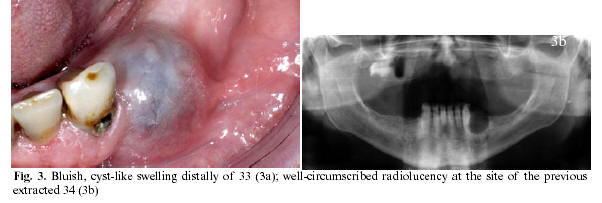
On the panoramic view a well-circumscribed radiolucency was  observed at the site of the previously extracted 34. A tentative  clinicoradiographic diagnosis of residual cyst was made.</font></p>     ]]></body>
<body><![CDATA[<p><font face="Verdana" size="2">Treatment consisted of enucleation. Histopathological  examination showed the typical features of lateral periodontal cyst. Healing was  uneventful. The patient will be scheduled for regular follow-up visits.</font></p>     <p>&nbsp;</p>     <p><b><font face="Verdana">Discussion</font></p> </b>     <p><font face="Verdana" size="2">Clinical information about the lateral periodontal cyst is  sparse since there have only been a few analyses of large series of cases  (1,4,5).</font></p>     <p><font face="Verdana" size="2">Epidemiologically, the lateral periodontal cyst presents at a  low frequency, without a distinct gender predilection (1,2,4,6,7).  Radiologically, the lateral periodontal cyst appears as a round, oval or  teardrop-like well-circumscribed interradicular radiolucent area, usually with a  sclerotic margin, lying somewhere between the apex and the cervical margin of  the teeth (1,6). Although uncommon, resorption of adjacent teeth has been  reported (8). Loss of lamina dura and periodontal ligament space may be present  (8). A swelling may occur on the buccal aspect, in which case the differential  diagnosis might include a gingival cyst (2).</font></p>     <p><font face="Verdana" size="2">Histologically, the lateral periodontal cyst is lined by a  thin non-proliferating cuboidal to stratified squamous non-keratinizing  epithelium, ranging from 1 to 5 cell layers, and thus resembling the reduced  enamel epithelium(1,6). The cyst wall and the lining are usually  free of inflammation(4).The lateral periodontal cyst presents two main  characteristic features, being (1,2,4,6):</font></p>     <blockquote>     <p><font face="Verdana" size="2">a) the presence of epithelial thickenings or &quot;plaques&quot;,  which, according to Shear and Pindborg are an ontogenic recapitulation by  odontogenic epithelium under pathological conditions (1,3);</font></p>     <p><font face="Verdana" size="2">b) the presence of glycogen-rich clear cells either in  &quot;plaques&quot; or in the superficial layers of the lining epithelium.</font></p> </blockquote>     <p><font face="Verdana" size="2">The clinical and radiographic characteristics per se are not  distinctive as diagnostic criteria for a diagnosis of lateral periodontal cyst. Actually, the WHO’s &quot;Histological typing of odontogenic tumours&quot; has more or  less changed the lateral periodontal cyst from a clinicoradiological entity into  a histopathological one. In fact, today, the diagnosis of lateral periodontal  cyst seems to be primarily based on histopathologic features. Therefore, when  dealing with a radiographic and clinical diagnosis of residual cyst, the  pathologist report may occasionally be that of a lateral periodontal cyst, as  was the case in the two presently reported patients.</font></p>     ]]></body>
<body><![CDATA[<p><font face="Verdana" size="2">The lateral periodontal cyst has characteristic histological  features which separate it from other odontogenic cysts, and for that reason it  seems preferable to define this cyst primarily based on the histopathologic  features and to give less weight to its exact location in the jaw or its  relation to the teeth.</font></p>     <p>&nbsp;</p>     <p><b><font face="Verdana">References</font></p> </b>     <!-- ref --><p><font face="Verdana" size="2">1. Shear M. Cyst of the Oral Regions (ed 3). Oxford: Wright;  1992. p. 51-71.</font>&nbsp;&nbsp;&nbsp;&nbsp;&nbsp;&nbsp;&nbsp;&nbsp;[&#160;<a href="javascript:void(0);" onclick="javascript: window.open('/scielo.php?script=sci_nlinks&ref=2955360&pid=S1698-6946200600020001900001&lng=','','width=640,height=500,resizable=yes,scrollbars=1,menubar=yes,');">Links</a>&#160;]<!-- end-ref --><!-- ref --><p><font face="Verdana" size="2">2. Altini M, Shear M: The lateral periodontal cyst: an  update. J Oral Pathol Med 1992;21:245-50.</font>&nbsp;&nbsp;&nbsp;&nbsp;&nbsp;&nbsp;&nbsp;&nbsp;[&#160;<a href="javascript:void(0);" onclick="javascript: window.open('/scielo.php?script=sci_nlinks&ref=2955361&pid=S1698-6946200600020001900002&lng=','','width=640,height=500,resizable=yes,scrollbars=1,menubar=yes,');">Links</a>&#160;]<!-- end-ref --><!-- ref --><p><font face="Verdana" size="2">3. Shear M: Developmental odontogenic cysts. An update. J  Oral Pathol Med 1994;23:1-11.</font>&nbsp;&nbsp;&nbsp;&nbsp;&nbsp;&nbsp;&nbsp;&nbsp;[&#160;<a href="javascript:void(0);" onclick="javascript: window.open('/scielo.php?script=sci_nlinks&ref=2955362&pid=S1698-6946200600020001900003&lng=','','width=640,height=500,resizable=yes,scrollbars=1,menubar=yes,');">Links</a>&#160;]<!-- end-ref --><!-- ref --><p><font face="Verdana" size="2">4. Rasmusson LG, Magnusson BC, Borrman H: The lateral  periodontal cyst. A histopathological and radiographic study of 32 cases. Br J  Oral Maxillofac Surg 1991;29:54-7.</font>&nbsp;&nbsp;&nbsp;&nbsp;&nbsp;&nbsp;&nbsp;&nbsp;[&#160;<a href="javascript:void(0);" onclick="javascript: window.open('/scielo.php?script=sci_nlinks&ref=2955363&pid=S1698-6946200600020001900004&lng=','','width=640,height=500,resizable=yes,scrollbars=1,menubar=yes,');">Links</a>&#160;]<!-- end-ref --><!-- ref --><p><font face="Verdana" size="2">5. Fantasia JE. Lateral periodontal cyst. An analysis of  forty-six cases. Oral Surg Oral Med Oral Pathol 1979;48:237-43.</font>&nbsp;&nbsp;&nbsp;&nbsp;&nbsp;&nbsp;&nbsp;&nbsp;[&#160;<a href="javascript:void(0);" onclick="javascript: window.open('/scielo.php?script=sci_nlinks&ref=2955364&pid=S1698-6946200600020001900005&lng=','','width=640,height=500,resizable=yes,scrollbars=1,menubar=yes,');">Links</a>&#160;]<!-- end-ref --><!-- ref --><p><font face="Verdana" size="2">6. Kramer IRH, Pindborg JJ, Shear M. World Health  Organization. International Classification of Tumours. Histological Typing of  Odontogenic Tumours (ed 2). Berlin-Budapest: Springer-Verlag; 1992. p. 37.</font>&nbsp;&nbsp;&nbsp;&nbsp;&nbsp;&nbsp;&nbsp;&nbsp;[&#160;<a href="javascript:void(0);" onclick="javascript: window.open('/scielo.php?script=sci_nlinks&ref=2955365&pid=S1698-6946200600020001900006&lng=','','width=640,height=500,resizable=yes,scrollbars=1,menubar=yes,');">Links</a>&#160;]<!-- end-ref --><!-- ref --><p><font face="Verdana" size="2">7. Mosqueda Taylor A, Irigoyen Camacho ME, Diaz Franco MA,  Torres Tejero MA. Odontogenic cysts. Analysis of 856 cases. Medicina Oral  2002;7:89-96.</font>&nbsp;&nbsp;&nbsp;&nbsp;&nbsp;&nbsp;&nbsp;&nbsp;[&#160;<a href="javascript:void(0);" onclick="javascript: window.open('/scielo.php?script=sci_nlinks&ref=2955366&pid=S1698-6946200600020001900007&lng=','','width=640,height=500,resizable=yes,scrollbars=1,menubar=yes,');">Links</a>&#160;]<!-- end-ref --><!-- ref --><p><font face="Verdana" size="2">8. Suljak JP, Bohay RN, Wysocki GP. Lateral periodontal cyst:  A case report and review of the literature. J Can Dent Assoc 1998;64:48-51.</font>&nbsp;&nbsp;&nbsp;&nbsp;&nbsp;&nbsp;&nbsp;&nbsp;[&#160;<a href="javascript:void(0);" onclick="javascript: window.open('/scielo.php?script=sci_nlinks&ref=2955367&pid=S1698-6946200600020001900008&lng=','','width=640,height=500,resizable=yes,scrollbars=1,menubar=yes,');">Links</a>&#160;]<!-- end-ref --><p>&nbsp;</p>     <p>&nbsp;</p>     <p><b><font face="Verdana" size="2"><a href="#top"><img border="0" src="/img/revistas/medicorpa/v11n2/seta.gif" width="15" height="17"></a>  <a name="down">Correspondence    <br></a></font></b><font face="Verdana" size="2">Dr. Isaac van der Waal    <br> Vrije Universiteit medical center    <br> P.O.Box 7057    <br> 1107 MB, Amsterdam, the Netherlands    <br> E-mail: <a href="mailto:i.vanderwaal@vumc.nl">i.vanderwaal@vumc.nl</a></font></p>     <p><font face="Verdana" size="2">Received: 15-08-2005    ]]></body>
<body><![CDATA[<br> Accepted: 2-10-2005</font></p>      ]]></body><back>
<ref-list>
<ref id="B1">
<label>1</label><nlm-citation citation-type="book">
<person-group person-group-type="author">
<name>
<surname><![CDATA[Shear]]></surname>
<given-names><![CDATA[M]]></given-names>
</name>
</person-group>
<source><![CDATA[Cyst of the Oral Regions]]></source>
<year>1992</year>
<edition>3</edition>
<page-range>51-71</page-range><publisher-loc><![CDATA[Oxford ]]></publisher-loc>
<publisher-name><![CDATA[Wright]]></publisher-name>
</nlm-citation>
</ref>
<ref id="B2">
<label>2</label><nlm-citation citation-type="journal">
<person-group person-group-type="author">
<name>
<surname><![CDATA[Altini]]></surname>
<given-names><![CDATA[M]]></given-names>
</name>
<name>
<surname><![CDATA[Shear]]></surname>
<given-names><![CDATA[M]]></given-names>
</name>
</person-group>
<article-title xml:lang="en"><![CDATA[The lateral periodontal cyst: an update]]></article-title>
<source><![CDATA[J Oral Pathol Med]]></source>
<year>1992</year>
<volume>21</volume>
<page-range>245-50</page-range></nlm-citation>
</ref>
<ref id="B3">
<label>3</label><nlm-citation citation-type="journal">
<person-group person-group-type="author">
<name>
<surname><![CDATA[Shear]]></surname>
<given-names><![CDATA[M]]></given-names>
</name>
</person-group>
<article-title xml:lang="en"><![CDATA[Developmental odontogenic cysts: An update]]></article-title>
<source><![CDATA[J Oral Pathol Med]]></source>
<year>1994</year>
<volume>23</volume>
<page-range>1-11</page-range></nlm-citation>
</ref>
<ref id="B4">
<label>4</label><nlm-citation citation-type="journal">
<person-group person-group-type="author">
<name>
<surname><![CDATA[Rasmusson]]></surname>
<given-names><![CDATA[LG]]></given-names>
</name>
<name>
<surname><![CDATA[Magnusson]]></surname>
<given-names><![CDATA[BC]]></given-names>
</name>
<name>
<surname><![CDATA[Borrman]]></surname>
<given-names><![CDATA[H]]></given-names>
</name>
</person-group>
<article-title xml:lang="en"><![CDATA[The lateral periodontal cyst: A histopathological and radiographic study of 32 cases]]></article-title>
<source><![CDATA[Br J Oral Maxillofac Surg]]></source>
<year>1991</year>
<volume>29</volume>
<page-range>54-7</page-range></nlm-citation>
</ref>
<ref id="B5">
<label>5</label><nlm-citation citation-type="journal">
<person-group person-group-type="author">
<name>
<surname><![CDATA[Fantasia]]></surname>
<given-names><![CDATA[JE]]></given-names>
</name>
</person-group>
<article-title xml:lang="en"><![CDATA[Lateral periodontal cyst: An analysis of forty-six cases]]></article-title>
<source><![CDATA[Oral Surg Oral Med Oral Pathol]]></source>
<year>1979</year>
<volume>48</volume>
<page-range>237-43</page-range></nlm-citation>
</ref>
<ref id="B6">
<label>6</label><nlm-citation citation-type="book">
<person-group person-group-type="author">
<name>
<surname><![CDATA[Kramer]]></surname>
<given-names><![CDATA[IRH]]></given-names>
</name>
<name>
<surname><![CDATA[Pindborg]]></surname>
<given-names><![CDATA[JJ]]></given-names>
</name>
<name>
<surname><![CDATA[Shear]]></surname>
<given-names><![CDATA[M]]></given-names>
</name>
</person-group>
<collab>World Health Organization</collab>
<source><![CDATA[International Classification of Tumours: Histological Typing of Odontogenic Tumours]]></source>
<year>1992</year>
<edition>2</edition>
<page-range>37</page-range><publisher-loc><![CDATA[BerlinBudapest ]]></publisher-loc>
<publisher-name><![CDATA[Springer-Verlag]]></publisher-name>
</nlm-citation>
</ref>
<ref id="B7">
<label>7</label><nlm-citation citation-type="journal">
<person-group person-group-type="author">
<name>
<surname><![CDATA[Mosqueda Taylor]]></surname>
<given-names><![CDATA[A]]></given-names>
</name>
<name>
<surname><![CDATA[Irigoyen Camacho]]></surname>
<given-names><![CDATA[ME]]></given-names>
</name>
<name>
<surname><![CDATA[Diaz Franco]]></surname>
<given-names><![CDATA[MA]]></given-names>
</name>
<name>
<surname><![CDATA[Torres Tejero]]></surname>
<given-names><![CDATA[MA]]></given-names>
</name>
</person-group>
<article-title xml:lang="en"><![CDATA[Odontogenic cysts: Analysis of 856 cases]]></article-title>
<source><![CDATA[Medicina Oral]]></source>
<year>2002</year>
<volume>7</volume>
<page-range>89-96</page-range></nlm-citation>
</ref>
<ref id="B8">
<label>8</label><nlm-citation citation-type="journal">
<person-group person-group-type="author">
<name>
<surname><![CDATA[Suljak]]></surname>
<given-names><![CDATA[JP]]></given-names>
</name>
<name>
<surname><![CDATA[Bohay]]></surname>
<given-names><![CDATA[RN]]></given-names>
</name>
<name>
<surname><![CDATA[Wysocki]]></surname>
<given-names><![CDATA[GP]]></given-names>
</name>
</person-group>
<article-title xml:lang="en"><![CDATA[Lateral periodontal cyst: A case report and review of the literature]]></article-title>
<source><![CDATA[J Can Dent Assoc]]></source>
<year>1998</year>
<volume>64</volume>
<page-range>48-51</page-range></nlm-citation>
</ref>
</ref-list>
</back>
</article>
