<?xml version="1.0" encoding="ISO-8859-1"?><article xmlns:mml="http://www.w3.org/1998/Math/MathML" xmlns:xlink="http://www.w3.org/1999/xlink" xmlns:xsi="http://www.w3.org/2001/XMLSchema-instance">
<front>
<journal-meta>
<journal-id>1698-6946</journal-id>
<journal-title><![CDATA[Medicina Oral, Patología Oral y Cirugía Bucal (Internet)]]></journal-title>
<abbrev-journal-title><![CDATA[Med. oral patol. oral cir.bucal (Internet)]]></abbrev-journal-title>
<issn>1698-6946</issn>
<publisher>
<publisher-name><![CDATA[Sociedad Española de Medicina Oral]]></publisher-name>
</publisher>
</journal-meta>
<article-meta>
<article-id>S1698-69462006000600006</article-id>
<title-group>
<article-title xml:lang="en"><![CDATA[Benign cementoblastoma: A case report]]></article-title>
</title-group>
<contrib-group>
<contrib contrib-type="author">
<name>
<surname><![CDATA[Sumer]]></surname>
<given-names><![CDATA[Mahmut]]></given-names>
</name>
</contrib>
<contrib contrib-type="author">
<name>
<surname><![CDATA[Gunduz]]></surname>
<given-names><![CDATA[Kaan]]></given-names>
</name>
</contrib>
<contrib contrib-type="author">
<name>
<surname><![CDATA[Sumer]]></surname>
<given-names><![CDATA[A. Pinar]]></given-names>
</name>
</contrib>
<contrib contrib-type="author">
<name>
<surname><![CDATA[Gunhan]]></surname>
<given-names><![CDATA[Omer]]></given-names>
</name>
</contrib>
</contrib-group>
<aff id="A01">
<institution><![CDATA[,Ondokuz Mayis University Faculty of Dentistry Department of Oral and Maxillofacial Surgery]]></institution>
<addr-line><![CDATA[Samsun ]]></addr-line>
<country>Turkey</country>
</aff>
<aff id="A02">
<institution><![CDATA[,Ondokuz Mayis University Faculty of Dentistry Department of Oral Diagnosis and Radiology]]></institution>
<addr-line><![CDATA[Samsun ]]></addr-line>
<country>Turkey</country>
</aff>
<aff id="A03">
<institution><![CDATA[,GATA Department of Pathology ]]></institution>
<addr-line><![CDATA[Ankara ]]></addr-line>
<country>Turkey</country>
</aff>
<pub-date pub-type="pub">
<day>00</day>
<month>12</month>
<year>2006</year>
</pub-date>
<pub-date pub-type="epub">
<day>00</day>
<month>12</month>
<year>2006</year>
</pub-date>
<volume>11</volume>
<numero>6</numero>
<fpage>483</fpage>
<lpage>485</lpage>
<copyright-statement/>
<copyright-year/>
<self-uri xlink:href="http://scielo.isciii.es/scielo.php?script=sci_arttext&amp;pid=S1698-69462006000600006&amp;lng=en&amp;nrm=iso"></self-uri><self-uri xlink:href="http://scielo.isciii.es/scielo.php?script=sci_abstract&amp;pid=S1698-69462006000600006&amp;lng=en&amp;nrm=iso"></self-uri><self-uri xlink:href="http://scielo.isciii.es/scielo.php?script=sci_pdf&amp;pid=S1698-69462006000600006&amp;lng=en&amp;nrm=iso"></self-uri><abstract abstract-type="short" xml:lang="en"><p><![CDATA[In the past the benign cementoblastoma was recognized in the World Health Organization’s classification of odontogenic tumours as one of the cementoma neoplasias. Recently the benign cementoblastoma is included into ‘Mesenchyme and/or odontogenic ectomesenchyme, with or without odontogenic epithelium’ odontogenic tumours. Benign cementoblastoma has characteristic radiologic and microscopic features and it appears to be fused to the tooth roots. Symptoms may be totally absent, and when they do occur, pain and swelling are frequent findings. The final diagnosis is usually made histopathologically, but the clinical diagnosis is comparatively easy if it is examined radiographically. The tumour has unlimited growth potential. Most frequently tends to be associated with an erupted permanent tooth, most often the first molar: rarely has an association with an impacted or partial impacted tooth been reported. This case represents a case of benign cementoblastoma associated with a partially impacted mandibular third molar.]]></p></abstract>
<kwd-group>
<kwd lng="en"><![CDATA[Benign cementoblastoma]]></kwd>
<kwd lng="en"><![CDATA[odontogenic tumour]]></kwd>
<kwd lng="en"><![CDATA[partially impacted]]></kwd>
</kwd-group>
</article-meta>
</front><body><![CDATA[ <p>&nbsp;</p>     <p>&nbsp;</p>     <p><B><font face="Verdana" size="2"><a name="top"></a></font> <font face="Verdana" size="4">Benign cementoblastoma: A case report</font></B></p>     <p>&nbsp;</p>     <p>&nbsp;</p>     <p><font face="Verdana" size="4"><b>Mahmut Sumer <sup>1</sup>, Kaan Gunduz <sup>2</sup>, A. Pinar. Sumer <sup>2</sup>, Omer Gunhan <sup>3</sup></b></font></p>      <p><font face="Verdana" size="2">(1) Ondokuz Mayis University, Faculty of Dentistry, Department of Oral and Maxillofacial Surgery, Samsun, Turkey    <BR> (2) Ondokuz Mayis University, Faculty of Dentistry, Department of Oral Diagnosis and Radiology, Samsun, Turkey    <BR> (3) Department of Pathology, GATA, Ankara, Turkey</font></p>     <p><font face="Verdana" size="2"><a href="#bajo">Correspondence</a></font></p>     ]]></body>
<body><![CDATA[<p>&nbsp;</p>     <p>&nbsp;</p><hr size="1">      <p><font face="Verdana" size="2"><B>ABSTRACT</B></font></p>      <p><font face="Verdana" size="2">In the past the benign cementoblastoma was recognized in the World Health Organization’s classification of odontogenic tumours as one of the cementoma neoplasias. Recently the benign cementoblastoma is included into ‘Mesenchyme and/or odontogenic ectomesenchyme, with or without odontogenic epithelium’ odontogenic tumours. Benign cementoblastoma has characteristic radiologic and microscopic features and it appears to be fused to the tooth roots. Symptoms may be totally absent, and when they do occur, pain and swelling are frequent findings. The final diagnosis is usually made histopathologically, but the clinical diagnosis is comparatively easy if it is examined radiographically. The tumour has unlimited growth potential. Most frequently tends to be associated with an erupted permanent tooth, most often the first molar: rarely has an association with an impacted or partial impacted tooth been reported. This case represents a case of benign cementoblastoma associated with a partially impacted mandibular third molar.</font></p>       <p><font face="Verdana" size="2"><B>Key words:</B> Benign cementoblastoma, odontogenic tumour, partially impacted.</font></p> <hr size="1">     <p>&nbsp;</p>     <p><font face="Verdana"><B>Introduction</B></font></p>     <p><font face="Verdana" size="2">The benign cementoblastoma is a relatively rare odontogenic neoplasm of the jaws and was first described by Dewey (1) in 1927. The lesion is considered as the only true neoplasm of cementum origin (2). The benign cementoblastoma, which generally occurs in young persons, comprises less than 1% to 6.2% of all odontogenic tumours (2) and is characterized as being attached to the roots, most frequently tends to be associated with an erupted permanent tooth, most often the first molar or second premolar in the lower jaw: only rarely has an association with an impacted or partially impacted tooth been reported (3).</font></p>     <p><font face="Verdana" size="2">Symptoms may be totally absent, and when they do occur, pain and swelling are frequent findings (3-5). The final diagnosis is usually made histopathologically, but the clinical diagnosis is comparatively easy if it is examined radiographically. The tumour has unlimited growth potential (3-6). The recommended treatment is complete enuclation of the tumor mass with extraction of the involved tooth (2-6).</font></p>     <p><font face="Verdana" size="2">This case report describes a benign cementoblastoma that attached to the lateral portion of the root of the partially impacted mandibular third molar; treatment included surgical excision of the lesion.</font></p>     ]]></body>
<body><![CDATA[<p>&nbsp;</p>     <p><font face="Verdana"><B>Case report</B></font></p>      <p><font face="Verdana" size="2">A 46-year-old male presented complaining of pain, trismus and swelling in the left third molar area for a period of 2 months. His medical and family history was noncontribibutory. There was no reported history of orafacial trauma. The clinical examination revealed acute pericoronitis involved with a partially impacted left mandibular third molar with swelling of the overlying mucosa. Also there was a slight expansion of bone on the buccal side of the mandible. A panoramic radiography is taken for radiological examination. The panoramic radiograph revealed a 20 mm, radiopaque mass attached to the lateral portion of the root of the lower left third molar that was surrounded by a radiolucent periphery (<a href="#f1">Figure 1</a>). </font> </p>     <p align="center">&nbsp;</p>     <p align="center"><font face="Verdana" size="2"><a name="f1"><img border="0" src="/img/revistas/medicorpa/v11n6/06i.ht26.jpg" width="380" height="210"></a></font></p>      <p>&nbsp;</p>     <p><font face="Verdana" size="2">After the symptoms improved with treatment with antibiotic and anti-inflammatory agents, enucleation of the calcified mass with the extraction of the tooth involved was performed under local anesthesia. The wound was irrigated and closed. Soft tissue healing was complete by 3 weeks. Eleven-month follow up shows no evidence of recurrence.</font></p>     <p><font face="Verdana" size="2">
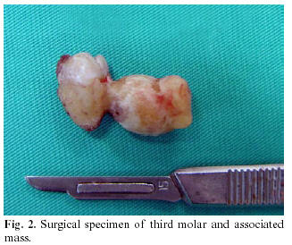
Macroscopically, a hard ovoid mass of calcified material measuring 20 x 10 x 10 mm, attached to the lateral portion of the root of the mandibular third molar (<a href="#f2">Figure 2</a>). Radiographs and macroscopic appearance led to diagnose the lesion as a cementoblastoma. The surgical specimen was sent for pathologic examination.</font></p>     <p align="center"><font face="Verdana" size="2"><a name="f2"><img border="0" src="/img/revistas/medicorpa/v11n6/06i.ht27.jpg" width="327" height="274"></a></font></p>      <p>&nbsp;</p>     ]]></body>
<body><![CDATA[<p><font face="Verdana" size="2">Histopathologic examination showed dens, irregularly  lamellated, osteocementum-like material with lack of interstitial tissue. A diagnosis of benign cementoblastoma was made (<a href="#f3">Figure 3</a>).</font></p>     <p align="center"><font face="Verdana" size="2"><a name="f3"><img border="0" src="/img/revistas/medicorpa/v11n6/06i.ht28.jpg" width="300" height="273"></a></font></p>      <p>&nbsp;</p>     <p><font face="Verdana"><B>Discussion</B></font></p>     <p><font face="Verdana" size="2">In the past the benign cementoblastoma was recognized in the  World Health Organization’s classification of odontogenic tumours as one of the cementoma neoplasias (3). Recently the benign cementoblastoma is included into ‘Mesenchyme and/or odontogenic ectomesenchyme, with or without odontogenic epithelium’ odontogenic tumours (7). The lesion derives from mesenchymal tissue, although its aetiology is unknown (4,5).</font></p>     <p><font face="Verdana" size="2">Benign cementoblastomas are predominantly seen in young persons. Ulmansky et al. (3), has reviewed literature and reported that close to three quarters of the patients (73%) are under the age of 30. Although it has been reported that there is no significant sex predilection between two sexes (8, 9) some authors (10, 11) indicates that males are affected more frequently than females. The mandible is involved more often than the maxilla (2-6, 8-11). The tumour usually involves an erupted permanent tooth. The most commonly affected tooth is the first permanent molar (2). The benign cementoblastoma rarely has an association with a partially impacted third molar tooth like in this case.</font></p>     <p><font face="Verdana" size="2">The lesion is slow growing and usually asymptomatic; however, pain and swelling have been reported in a number of cases including the one presented in this article. Cortical expansion and facial asymmetry are common (4-6, 8, 9).</font></p>     <p><font face="Verdana" size="2">Radiographically, the lesion usually shows a radiopaque mass often fused with a root or roots of a tooth and surrounded and limited peripherally by a radiolucent halo. When the intimate relation with roots is present, the radiographic appearance is nearly pathognomonic (3). The present case providing this parameter had this characteristic. Few authors have reported a more radiolucent form of the lesion and considered it to represent an early uncalcified matrix stage (12).</font></p>     <p><font face="Verdana" size="2">There are a few lesions which should be distinguished from this lesion such as osteoma, benign osteoblastoma, chronic focal sclerosing osteotitis, osteomyelitis and osteosarcoma, etc (3-5, 6, 9, 14). Clinical and especially radiological findings are helpful for differential diagnosis. Sometimes the general appearance of the lesion is very similar with osteoma or osteoblastoma. Microscopic differential diagnosis between cementoblastoma and osteoblastoma, the direct connection with the radicular surface is the most significant finding (13).</font></p>     <p><font face="Verdana" size="2">Because benign cementoblastoma has unlimited growth potential, the usual treatment is complete surgical excision with extraction of the associated teeth. In literature, there are 13 recurrent cases associated with cemetoblastomas. Extraction of the involved tooth or teeth along with removal of the lesion was performed in 9 of the 13 recurrent cases. Four cases were treated with curettage only without extraction of the involved tooth. Recommended treatment of cementoblastoma should consist of removal of the lesion along with the affected tooth or teeth, followed by thorough curettage or peripheral ostectomy (8). In our case, there has been no recurrence more than 11 months after the surgical procedure.</font></p>     ]]></body>
<body><![CDATA[<p><font face="Verdana" size="2">The benign cementoblastomas usually involve an erupted permanent tooth and most commonly found in the patients younger than 30 year old. In this case report; a case of benign cementoblastoma of a 46-year-old male is presented involving a mandibular semi-impacted third molar and arising from the lateral portion of the root of the involved tooth which had a very characteristic macroscopic appearance.</font></p>     <p>&nbsp;</p>     <p><font face="Verdana"><B>References</B></font></p>     <!-- ref --><p><font face="Verdana" size="2">1. Dewey KW. Osteoma of a molar. Dent Cosmos 1927; 69:1143-9.</font>&nbsp;&nbsp;&nbsp;&nbsp;&nbsp;&nbsp;&nbsp;&nbsp;[&#160;<a href="javascript:void(0);" onclick="javascript: window.open('/scielo.php?script=sci_nlinks&ref=2957617&pid=S1698-6946200600060000600001&lng=','','width=640,height=500,resizable=yes,scrollbars=1,menubar=yes,');">Links</a>&#160;]<!-- end-ref --><!-- ref --><p><font face="Verdana" size="2">2. Lu Y, Xaun M, Takata T, Wang C, He Z, Zhou Z, et al. Odontogenic tumours. A demographic study of 759 cases in a Chinese population. Oral Surg Oral Med Oral Pathol Oral Radiol Endod 1998;86:707-14.</font>&nbsp;&nbsp;&nbsp;&nbsp;&nbsp;&nbsp;&nbsp;&nbsp;[&#160;<a href="javascript:void(0);" onclick="javascript: window.open('/scielo.php?script=sci_nlinks&ref=2957618&pid=S1698-6946200600060000600002&lng=','','width=640,height=500,resizable=yes,scrollbars=1,menubar=yes,');">Links</a>&#160;]<!-- end-ref --><!-- ref --><p><font face="Verdana" size="2">3. Ulmansky M, Hjorting-Hansen E, Praetorius F, Haque MF. Benign cementoblastoma; a review and five new cases. Oral Surg Oral Med Oral Pathol 1994;77:48-55.</font>&nbsp;&nbsp;&nbsp;&nbsp;&nbsp;&nbsp;&nbsp;&nbsp;[&#160;<a href="javascript:void(0);" onclick="javascript: window.open('/scielo.php?script=sci_nlinks&ref=2957619&pid=S1698-6946200600060000600003&lng=','','width=640,height=500,resizable=yes,scrollbars=1,menubar=yes,');">Links</a>&#160;]<!-- end-ref --><!-- ref --><p><font face="Verdana" size="2">4. Piattelli A, Di Alberti L, Scarano A, Piattelli M. Benign cementoblastoma associated with an unerupted third molar. Oral Oncol 1998;34:229-31.</font>&nbsp;&nbsp;&nbsp;&nbsp;&nbsp;&nbsp;&nbsp;&nbsp;[&#160;<a href="javascript:void(0);" onclick="javascript: window.open('/scielo.php?script=sci_nlinks&ref=2957620&pid=S1698-6946200600060000600004&lng=','','width=640,height=500,resizable=yes,scrollbars=1,menubar=yes,');">Links</a>&#160;]<!-- end-ref --><!-- ref --><p><font face="Verdana" size="2">5. Piatelli A, D’addona A, Piatelli M. Benign cemento-blastoma: Review of the literature and report of a case at unusual location. Acta Stomatol Belg 1990;87:209-15.</font>&nbsp;&nbsp;&nbsp;&nbsp;&nbsp;&nbsp;&nbsp;&nbsp;[&#160;<a href="javascript:void(0);" onclick="javascript: window.open('/scielo.php?script=sci_nlinks&ref=2957621&pid=S1698-6946200600060000600005&lng=','','width=640,height=500,resizable=yes,scrollbars=1,menubar=yes,');">Links</a>&#160;]<!-- end-ref --><!-- ref --><p><font face="Verdana" size="2">6. Cundiff J. Developing cementoblastoma: Case report and update of differential diagnosis. Quintessence Int 2000; 31:191-5.</font>&nbsp;&nbsp;&nbsp;&nbsp;&nbsp;&nbsp;&nbsp;&nbsp;[&#160;<a href="javascript:void(0);" onclick="javascript: window.open('/scielo.php?script=sci_nlinks&ref=2957622&pid=S1698-6946200600060000600006&lng=','','width=640,height=500,resizable=yes,scrollbars=1,menubar=yes,');">Links</a>&#160;]<!-- end-ref --><!-- ref --><p><font face="Verdana" size="2">7. Barnes L, Eveson JW, Reichart P, Sidransky D, eds. Pathology &amp; Genetics: Head and Neck Tumours. Geneva: WHO; 2005.</font>&nbsp;&nbsp;&nbsp;&nbsp;&nbsp;&nbsp;&nbsp;&nbsp;[&#160;<a href="javascript:void(0);" onclick="javascript: window.open('/scielo.php?script=sci_nlinks&ref=2957623&pid=S1698-6946200600060000600007&lng=','','width=640,height=500,resizable=yes,scrollbars=1,menubar=yes,');">Links</a>&#160;]<!-- end-ref --><!-- ref --><p><font face="Verdana" size="2">8. Brannon RB, Fowler CB, Carpenter WM, Corio RL. Cementoblastomas: an innocuous neoplasm.? A clinicopathologic study of 44 cases and review of the literature with special emphasis on recurrence. Oral Surg Oral Med Oral Pathol Oral Radiol Endod 2002;93:311-20.</font>&nbsp;&nbsp;&nbsp;&nbsp;&nbsp;&nbsp;&nbsp;&nbsp;[&#160;<a href="javascript:void(0);" onclick="javascript: window.open('/scielo.php?script=sci_nlinks&ref=2957624&pid=S1698-6946200600060000600008&lng=','','width=640,height=500,resizable=yes,scrollbars=1,menubar=yes,');">Links</a>&#160;]<!-- end-ref --><!-- ref --><p><font face="Verdana" size="2">9. Regezi JA, Sciubba JJ, eds. Oral pathology: Clinical-pathological correlations. Philadelphia: Saunders; 1989. p. 359-61.</font>&nbsp;&nbsp;&nbsp;&nbsp;&nbsp;&nbsp;&nbsp;&nbsp;[&#160;<a href="javascript:void(0);" onclick="javascript: window.open('/scielo.php?script=sci_nlinks&ref=2957625&pid=S1698-6946200600060000600009&lng=','','width=640,height=500,resizable=yes,scrollbars=1,menubar=yes,');">Links</a>&#160;]<!-- end-ref --><!-- ref --><p><font face="Verdana" size="2">10. Ohki K, Kumamoto H, Nitta Y, Nagasaka H, Kawamura H, Ooya K. Benign cementoblastoma involving multiple maxillary teeth: report of a case with a review of the literature. Oral Surg Oral Med Oral Pathol Oral Radiol Endod 2004;97:53-8.</font>&nbsp;&nbsp;&nbsp;&nbsp;&nbsp;&nbsp;&nbsp;&nbsp;[&#160;<a href="javascript:void(0);" onclick="javascript: window.open('/scielo.php?script=sci_nlinks&ref=2957626&pid=S1698-6946200600060000600010&lng=','','width=640,height=500,resizable=yes,scrollbars=1,menubar=yes,');">Links</a>&#160;]<!-- end-ref --><!-- ref --><p><font face="Verdana" size="2">11. Monks FT, Bradley JC, Turner EP. Central osteoblastoma or cementoblastoma? A case report and 12-year review. Br J Oral Surg 1981;19:29-37.</font>&nbsp;&nbsp;&nbsp;&nbsp;&nbsp;&nbsp;&nbsp;&nbsp;[&#160;<a href="javascript:void(0);" onclick="javascript: window.open('/scielo.php?script=sci_nlinks&ref=2957627&pid=S1698-6946200600060000600011&lng=','','width=640,height=500,resizable=yes,scrollbars=1,menubar=yes,');">Links</a>&#160;]<!-- end-ref --><!-- ref --><p><font face="Verdana" size="2">12. Gingell JC, Lunin M, Beckerman T, Levy BA. Benign cementoblastoma. J Oral Med 1984;68:8-11.</font>&nbsp;&nbsp;&nbsp;&nbsp;&nbsp;&nbsp;&nbsp;&nbsp;[&#160;<a href="javascript:void(0);" onclick="javascript: window.open('/scielo.php?script=sci_nlinks&ref=2957628&pid=S1698-6946200600060000600012&lng=','','width=640,height=500,resizable=yes,scrollbars=1,menubar=yes,');">Links</a>&#160;]<!-- end-ref --><!-- ref --><p><font face="Verdana" size="2">13. Fujita S, Takahashi H, Okabe H, Watanabe C, Sonobe H. A case of benign cementoblastoma. Oral Surg Oral Med Oral Pathol 1989;68:64-8.</font>&nbsp;&nbsp;&nbsp;&nbsp;&nbsp;&nbsp;&nbsp;&nbsp;[&#160;<a href="javascript:void(0);" onclick="javascript: window.open('/scielo.php?script=sci_nlinks&ref=2957629&pid=S1698-6946200600060000600013&lng=','','width=640,height=500,resizable=yes,scrollbars=1,menubar=yes,');">Links</a>&#160;]<!-- end-ref --><!-- ref --><p><font face="Verdana" size="2">14. Junquera-Gutierrez L, Lopez-Arranz JS, Albertos-Castro JM, De Vicente-Rodriguez JC. Odontogenic tumors: classification. clinical features. diagnosis and treatment. Med Oral 1997;2:94-101.</font>&nbsp;&nbsp;&nbsp;&nbsp;&nbsp;&nbsp;&nbsp;&nbsp;[&#160;<a href="javascript:void(0);" onclick="javascript: window.open('/scielo.php?script=sci_nlinks&ref=2957630&pid=S1698-6946200600060000600014&lng=','','width=640,height=500,resizable=yes,scrollbars=1,menubar=yes,');">Links</a>&#160;]<!-- end-ref --><p>&nbsp;</p>     <p>&nbsp;</p>     <p><font face="Verdana" size="2"><a href="#top"> <img border="0" src="/img/revistas/medicorpa/v11n6/seta.gif" width="15" height="17"></a><B><a name="bajo"></a>Correspondence</B>:    ]]></body>
<body><![CDATA[<BR> Dr. Kaan Gunduz    <BR> Ondokuz Mayis Universitesi,Di&#351;hekimli&#287;i Fakültesi    <BR> Oral Diagnoz ve Radyoloji Anabilim Dali    <BR> 55139 Kurupelit, Samsun Turkey    <BR> E-mail: <a href="mailto:kgunduz@omu.edu.tr">kgunduz@omu.edu.tr</a></font></p>      <p><font face="Verdana" size="2">Received: 21-03-2006    <BR> Accepted: 16-06-2006</font></p>       ]]></body><back>
<ref-list>
<ref id="B1">
<label>1</label><nlm-citation citation-type="journal">
<person-group person-group-type="author">
<name>
<surname><![CDATA[Dewey]]></surname>
<given-names><![CDATA[KW]]></given-names>
</name>
</person-group>
<article-title xml:lang="en"><![CDATA[Osteoma of a molar]]></article-title>
<source><![CDATA[Dent Cosmos]]></source>
<year>1927</year>
<volume>69</volume>
<page-range>1143-9</page-range></nlm-citation>
</ref>
<ref id="B2">
<label>2</label><nlm-citation citation-type="journal">
<person-group person-group-type="author">
<name>
<surname><![CDATA[Lu]]></surname>
<given-names><![CDATA[Y]]></given-names>
</name>
<name>
<surname><![CDATA[Xaun]]></surname>
<given-names><![CDATA[M]]></given-names>
</name>
<name>
<surname><![CDATA[Takata]]></surname>
<given-names><![CDATA[T]]></given-names>
</name>
<name>
<surname><![CDATA[Wang]]></surname>
<given-names><![CDATA[C]]></given-names>
</name>
<name>
<surname><![CDATA[He]]></surname>
<given-names><![CDATA[Z]]></given-names>
</name>
<name>
<surname><![CDATA[Zhou]]></surname>
<given-names><![CDATA[Z]]></given-names>
</name>
</person-group>
<article-title xml:lang="en"><![CDATA[Odontogenic tumours: A demographic study of 759 cases in a Chinese population]]></article-title>
<source><![CDATA[Oral Surg Oral Med Oral Pathol Oral Radiol Endod]]></source>
<year>1998</year>
<volume>86</volume>
<page-range>707-14</page-range></nlm-citation>
</ref>
<ref id="B3">
<label>3</label><nlm-citation citation-type="journal">
<person-group person-group-type="author">
<name>
<surname><![CDATA[Ulmansky]]></surname>
<given-names><![CDATA[M]]></given-names>
</name>
<name>
<surname><![CDATA[Hjorting-Hansen]]></surname>
<given-names><![CDATA[E]]></given-names>
</name>
<name>
<surname><![CDATA[Praetorius]]></surname>
<given-names><![CDATA[F]]></given-names>
</name>
<name>
<surname><![CDATA[Haque]]></surname>
<given-names><![CDATA[MF]]></given-names>
</name>
</person-group>
<article-title xml:lang="en"><![CDATA[Benign cementoblastoma: a review and five new cases]]></article-title>
<source><![CDATA[Oral Surg Oral Med Oral Pathol]]></source>
<year>1994</year>
<volume>77</volume>
<page-range>48-55</page-range></nlm-citation>
</ref>
<ref id="B4">
<label>4</label><nlm-citation citation-type="journal">
<person-group person-group-type="author">
<name>
<surname><![CDATA[Piattelli]]></surname>
<given-names><![CDATA[A]]></given-names>
</name>
<name>
<surname><![CDATA[Di Alberti]]></surname>
<given-names><![CDATA[L]]></given-names>
</name>
<name>
<surname><![CDATA[Scarano]]></surname>
<given-names><![CDATA[A]]></given-names>
</name>
<name>
<surname><![CDATA[Piattelli]]></surname>
<given-names><![CDATA[M]]></given-names>
</name>
</person-group>
<article-title xml:lang="en"><![CDATA[Benign cementoblastoma associated with an unerupted third molar]]></article-title>
<source><![CDATA[Oral Oncol]]></source>
<year>1998</year>
<volume>34</volume>
<page-range>229-31</page-range></nlm-citation>
</ref>
<ref id="B5">
<label>5</label><nlm-citation citation-type="journal">
<person-group person-group-type="author">
<name>
<surname><![CDATA[Piatelli]]></surname>
<given-names><![CDATA[A]]></given-names>
</name>
<name>
<surname><![CDATA[D’addona]]></surname>
<given-names><![CDATA[A]]></given-names>
</name>
<name>
<surname><![CDATA[Piatelli]]></surname>
<given-names><![CDATA[M]]></given-names>
</name>
</person-group>
<article-title xml:lang="en"><![CDATA[Benign cemento-blastoma: Review of the literature and report of a case at unusual location]]></article-title>
<source><![CDATA[Acta Stomatol Belg]]></source>
<year>1990</year>
<volume>87</volume>
<page-range>209-15</page-range></nlm-citation>
</ref>
<ref id="B6">
<label>6</label><nlm-citation citation-type="journal">
<person-group person-group-type="author">
<name>
<surname><![CDATA[Cundiff]]></surname>
<given-names><![CDATA[J]]></given-names>
</name>
</person-group>
<article-title xml:lang="en"><![CDATA[Developing cementoblastoma: Case report and update of differential diagnosis]]></article-title>
<source><![CDATA[Quintessence Int]]></source>
<year>2000</year>
<volume>31</volume>
<page-range>191-5</page-range></nlm-citation>
</ref>
<ref id="B7">
<label>7</label><nlm-citation citation-type="book">
<person-group person-group-type="author">
<name>
<surname><![CDATA[Barnes]]></surname>
<given-names><![CDATA[L]]></given-names>
</name>
<name>
<surname><![CDATA[Eveson]]></surname>
<given-names><![CDATA[JW]]></given-names>
</name>
<name>
<surname><![CDATA[Reichart]]></surname>
<given-names><![CDATA[P]]></given-names>
</name>
<name>
<surname><![CDATA[Sidransky]]></surname>
<given-names><![CDATA[D]]></given-names>
</name>
</person-group>
<source><![CDATA[Pathology & Genetics: Head and Neck Tumours]]></source>
<year>2005</year>
<publisher-loc><![CDATA[Geneva ]]></publisher-loc>
<publisher-name><![CDATA[WHO]]></publisher-name>
</nlm-citation>
</ref>
<ref id="B8">
<label>8</label><nlm-citation citation-type="journal">
<person-group person-group-type="author">
<name>
<surname><![CDATA[Brannon]]></surname>
<given-names><![CDATA[RB]]></given-names>
</name>
<name>
<surname><![CDATA[Fowler]]></surname>
<given-names><![CDATA[CB]]></given-names>
</name>
<name>
<surname><![CDATA[Carpenter]]></surname>
<given-names><![CDATA[WM]]></given-names>
</name>
<name>
<surname><![CDATA[Corio]]></surname>
<given-names><![CDATA[RL]]></given-names>
</name>
</person-group>
<article-title xml:lang="en"><![CDATA[Cementoblastomas: an innocuous neoplasm.? A clinicopathologic study of 44 cases and review of the literature with special emphasis on recurrence]]></article-title>
<source><![CDATA[Oral Surg Oral Med Oral Pathol Oral Radiol Endod]]></source>
<year>2002</year>
<volume>93</volume>
<page-range>311-20</page-range></nlm-citation>
</ref>
<ref id="B9">
<label>9</label><nlm-citation citation-type="book">
<person-group person-group-type="author">
<name>
<surname><![CDATA[Regezi]]></surname>
<given-names><![CDATA[JA]]></given-names>
</name>
<name>
<surname><![CDATA[Sciubba]]></surname>
<given-names><![CDATA[JJ]]></given-names>
</name>
</person-group>
<source><![CDATA[Oral pathology: Clinical-pathological correlations]]></source>
<year>1989</year>
<page-range>359-61</page-range><publisher-loc><![CDATA[Philadelphia ]]></publisher-loc>
<publisher-name><![CDATA[Saunders]]></publisher-name>
</nlm-citation>
</ref>
<ref id="B10">
<label>10</label><nlm-citation citation-type="journal">
<person-group person-group-type="author">
<name>
<surname><![CDATA[Ohki]]></surname>
<given-names><![CDATA[K]]></given-names>
</name>
<name>
<surname><![CDATA[Kumamoto]]></surname>
<given-names><![CDATA[H]]></given-names>
</name>
<name>
<surname><![CDATA[Nitta]]></surname>
<given-names><![CDATA[Y]]></given-names>
</name>
<name>
<surname><![CDATA[Nagasaka]]></surname>
<given-names><![CDATA[H]]></given-names>
</name>
<name>
<surname><![CDATA[Kawamura]]></surname>
<given-names><![CDATA[H]]></given-names>
</name>
<name>
<surname><![CDATA[Ooya]]></surname>
<given-names><![CDATA[K]]></given-names>
</name>
</person-group>
<article-title xml:lang="en"><![CDATA[Benign cementoblastoma involving multiple maxillary teeth: report of a case with a review of the literature]]></article-title>
<source><![CDATA[Oral Surg Oral Med Oral Pathol Oral Radiol Endod]]></source>
<year>2004</year>
<volume>97</volume>
<page-range>53-8</page-range></nlm-citation>
</ref>
<ref id="B11">
<label>11</label><nlm-citation citation-type="journal">
<person-group person-group-type="author">
<name>
<surname><![CDATA[Monks]]></surname>
<given-names><![CDATA[FT]]></given-names>
</name>
<name>
<surname><![CDATA[Bradley]]></surname>
<given-names><![CDATA[JC]]></given-names>
</name>
<name>
<surname><![CDATA[Turner]]></surname>
<given-names><![CDATA[EP]]></given-names>
</name>
</person-group>
<article-title xml:lang="en"><![CDATA[Central osteoblastoma or cementoblastoma?: A case report and 12-year review]]></article-title>
<source><![CDATA[Br J Oral Surg]]></source>
<year>1981</year>
<volume>19</volume>
<page-range>29-37</page-range></nlm-citation>
</ref>
<ref id="B12">
<label>12</label><nlm-citation citation-type="journal">
<person-group person-group-type="author">
<name>
<surname><![CDATA[Gingell]]></surname>
<given-names><![CDATA[JC]]></given-names>
</name>
<name>
<surname><![CDATA[Lunin]]></surname>
<given-names><![CDATA[M]]></given-names>
</name>
<name>
<surname><![CDATA[Beckerman]]></surname>
<given-names><![CDATA[T]]></given-names>
</name>
<name>
<surname><![CDATA[Levy]]></surname>
<given-names><![CDATA[BA]]></given-names>
</name>
</person-group>
<article-title xml:lang="en"><![CDATA[Benign cementoblastoma]]></article-title>
<source><![CDATA[J Oral Med]]></source>
<year>1984</year>
<volume>68</volume>
<page-range>8-11</page-range></nlm-citation>
</ref>
<ref id="B13">
<label>13</label><nlm-citation citation-type="journal">
<person-group person-group-type="author">
<name>
<surname><![CDATA[Fujita]]></surname>
<given-names><![CDATA[S]]></given-names>
</name>
<name>
<surname><![CDATA[Takahashi]]></surname>
<given-names><![CDATA[H]]></given-names>
</name>
<name>
<surname><![CDATA[Okabe]]></surname>
<given-names><![CDATA[H]]></given-names>
</name>
<name>
<surname><![CDATA[Watanabe]]></surname>
<given-names><![CDATA[C]]></given-names>
</name>
<name>
<surname><![CDATA[Sonobe]]></surname>
<given-names><![CDATA[H]]></given-names>
</name>
</person-group>
<article-title xml:lang="en"><![CDATA[A case of benign cementoblastoma]]></article-title>
<source><![CDATA[Oral Surg Oral Med Oral Pathol]]></source>
<year>1989</year>
<volume>68</volume>
<page-range>64-8</page-range></nlm-citation>
</ref>
<ref id="B14">
<label>14</label><nlm-citation citation-type="journal">
<person-group person-group-type="author">
<name>
<surname><![CDATA[Junquera-Gutierrez]]></surname>
<given-names><![CDATA[L]]></given-names>
</name>
<name>
<surname><![CDATA[Lopez-Arranz]]></surname>
<given-names><![CDATA[JS]]></given-names>
</name>
<name>
<surname><![CDATA[Albertos-Castro]]></surname>
<given-names><![CDATA[JM]]></given-names>
</name>
<name>
<surname><![CDATA[De Vicente-Rodriguez]]></surname>
<given-names><![CDATA[JC]]></given-names>
</name>
</person-group>
<article-title xml:lang="en"><![CDATA[Odontogenic tumors: classification. clinical features. diagnosis and treatment]]></article-title>
<source><![CDATA[Med Oral]]></source>
<year>1997</year>
<volume>2</volume>
<page-range>94-101</page-range></nlm-citation>
</ref>
</ref-list>
</back>
</article>
