<?xml version="1.0" encoding="ISO-8859-1"?><article xmlns:mml="http://www.w3.org/1998/Math/MathML" xmlns:xlink="http://www.w3.org/1999/xlink" xmlns:xsi="http://www.w3.org/2001/XMLSchema-instance">
<front>
<journal-meta>
<journal-id>1698-6946</journal-id>
<journal-title><![CDATA[Medicina Oral, Patología Oral y Cirugía Bucal (Internet)]]></journal-title>
<abbrev-journal-title><![CDATA[Med. oral patol. oral cir.bucal (Internet)]]></abbrev-journal-title>
<issn>1698-6946</issn>
<publisher>
<publisher-name><![CDATA[Sociedad Española de Medicina Oral]]></publisher-name>
</publisher>
</journal-meta>
<article-meta>
<article-id>S1698-69462007000100003</article-id>
<title-group>
<article-title xml:lang="en"><![CDATA[Mandibular involvement of solitary plasmocytoma: A case report]]></article-title>
</title-group>
<contrib-group>
<contrib contrib-type="author">
<name>
<surname><![CDATA[Murat Canger]]></surname>
<given-names><![CDATA[Emin]]></given-names>
</name>
<xref ref-type="aff" rid="A01"/>
</contrib>
<contrib contrib-type="author">
<name>
<surname><![CDATA[Çelenk]]></surname>
<given-names><![CDATA[Peruze]]></given-names>
</name>
<xref ref-type="aff" rid="A01"/>
</contrib>
<contrib contrib-type="author">
<name>
<surname><![CDATA[Alkan]]></surname>
<given-names><![CDATA[Alper]]></given-names>
</name>
<xref ref-type="aff" rid="A02"/>
</contrib>
<contrib contrib-type="author">
<name>
<surname><![CDATA[Günhan]]></surname>
<given-names><![CDATA[Ömer]]></given-names>
</name>
<xref ref-type="aff" rid="A03"/>
</contrib>
</contrib-group>
<aff id="A01">
<institution><![CDATA[,Faculty of Dentistry Department of Oral Diagnosis and Radiology ]]></institution>
<addr-line><![CDATA[Samsun ]]></addr-line>
<country>Turkey</country>
</aff>
<aff id="A02">
<institution><![CDATA[,Faculty of Dentistry Department of Oral and Maxillofacial Surgery ]]></institution>
<addr-line><![CDATA[Samsun ]]></addr-line>
<country>Turkey</country>
</aff>
<aff id="A03">
<institution><![CDATA[,Gülhane Military Medical Academy Department of Pathology ]]></institution>
<addr-line><![CDATA[Ankara ]]></addr-line>
<country>Turkey</country>
</aff>
<pub-date pub-type="pub">
<day>00</day>
<month>01</month>
<year>2007</year>
</pub-date>
<pub-date pub-type="epub">
<day>00</day>
<month>01</month>
<year>2007</year>
</pub-date>
<volume>12</volume>
<numero>1</numero>
<fpage>07</fpage>
<lpage>09</lpage>
<copyright-statement/>
<copyright-year/>
<self-uri xlink:href="http://scielo.isciii.es/scielo.php?script=sci_arttext&amp;pid=S1698-69462007000100003&amp;lng=en&amp;nrm=iso"></self-uri><self-uri xlink:href="http://scielo.isciii.es/scielo.php?script=sci_abstract&amp;pid=S1698-69462007000100003&amp;lng=en&amp;nrm=iso"></self-uri><self-uri xlink:href="http://scielo.isciii.es/scielo.php?script=sci_pdf&amp;pid=S1698-69462007000100003&amp;lng=en&amp;nrm=iso"></self-uri><abstract abstract-type="short" xml:lang="en"><p><![CDATA[Plasma cell neoplasms (multiple myeloma, solitary plasmocytoma of bone and extra medullar plasmocytoma) are characterized by a monoclonal neoplastic proliferation of plasma cells. Solitary plasmocytoma of bone (SPB) is a localized form of them. SPB is most frequently seen in vertebrae and secondarily in long bones. Its presence in jaws is extremely rare and when it is seen, angulus and ramus mandible are most common sites of occurrence. Prognosis of SPB is worse than extra medullar plasmacytoma (EMP) and approximately 50% of SPB will transform to multiple myelom. A 76-year old woman consulted to our clinic with a chief complaint of slowly developed swelling in her mandible. She had an operation from caput femur because of plasmocytoma two months before. Panoramic radiography revealed a radiolucent lesion in the mandibular anterior region, 60×35 mm in dimension. Aspiration biopsy was performed and histopathological examination was reported as plasmocytoma. She was referred to the oncology department for treatment but died before the treatment finished.]]></p></abstract>
<kwd-group>
<kwd lng="en"><![CDATA[Plasma cell neoplasm]]></kwd>
<kwd lng="en"><![CDATA[bone]]></kwd>
<kwd lng="en"><![CDATA[solitary plasmocytoma]]></kwd>
<kwd lng="en"><![CDATA[mandible]]></kwd>
</kwd-group>
</article-meta>
</front><body><![CDATA[ <p>&nbsp;</p>     <p>&nbsp;</p>     <p><B><font face="Verdana" size="4"><a name="top"></a>Mandibular involvement of solitary plasmocytoma: A case report</font></B></p>     <p>&nbsp;</p>     <p>&nbsp;</p>     <p><B><font face="Verdana" size="2">Emin Murat Canger <sup>1</sup>, Peruze Çelenk <sup>1</sup>, Alper Alkan <sup>2</sup>, Ömer Günhan <sup>3</sup></font></B></p>     <p><font face="Verdana" size="2">(1) O.M.Ü. Faculty of Dentistry Department of Oral Diagnosis and Radiology, Samsun/Turkey    <BR>(2) O.M.Ü. Faculty of Dentistry Department of Oral and Maxillofacial Surgery, Samsun/Turkey    <BR>(3) Gülhane Military Medical Academy Department of Pathology, Ankara/Turkey</font></p>     <p><font face="Verdana" size="2"><a href="#bajo">Correspondence</a></font></p>     ]]></body>
<body><![CDATA[<p>&nbsp;</p>     <p>&nbsp;</p> <hr size="1">     <p><B><font face="Verdana" size="2">ABSTRACT</font></B></p>     <p><font face="Verdana" size="2">Plasma cell neoplasms (multiple myeloma, solitary plasmocytoma of bone and extra medullar plasmocytoma) are characterized by a monoclonal neoplastic proliferation of plasma cells. Solitary plasmocytoma of bone (SPB) is a localized form of them. SPB is most frequently seen in vertebrae and secondarily in long bones. Its presence in jaws is extremely rare and when it is seen, angulus and ramus mandible are most common sites of occurrence. Prognosis of SPB is worse than extra medullar plasmacytoma (EMP) and approximately 50% of SPB will transform to multiple myelom. A 76-year old woman consulted to our clinic with a chief complaint of slowly developed swelling in her mandible. She had an operation from caput femur because of plasmocytoma two months before. Panoramic radiography revealed a radiolucent lesion in the mandibular anterior region, 60×35 mm in dimension. Aspiration biopsy was performed and histopathological examination was reported as plasmocytoma. She was referred to the oncology department for treatment but died before the treatment finished.</font></p>     <p><font face="Verdana" size="2"><B>Key words:</B> Plasma cell neoplasm, bone, solitary plasmocytoma, mandible.</font></p> <hr size="1">     <p>&nbsp;</p>     <p><B><font face="Verdana">Introduction</font></B></p>     <p><font face="Verdana" size="2">Plasma cell neoplasms (multiple myeloma, solitary plasmocytoma of bone and extramedullar plasmocytoma) are lymphoid neoplastic proliferations of B cells (1-6).</font></p>     <p><font face="Verdana" size="2">Multiple myeloma (MM) is the disseminated type of this disorder. Localized forms of plasma cell neoplasms are solitary plasmocytoma of bone (SPB) and extramedullar plasmocytoma (EMP). EMP develops in soft tissues and is more frequently seen than SPB in head and neck regions. SPB is observed as centrally localized in bones (1-3,5).</font></p>     <p><font face="Verdana" size="2">The prognosis of SPB is poorer than EMP and approximately 50% of SPB will transform to MM (1,3-5,7,8).</font></p>     ]]></body>
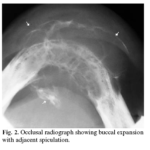
<body><![CDATA[<p><font face="Verdana" size="2">SPB is diagnosed with the absence of clonal plasma cells on a random marrow sample, single osteolytic lesion (no additional lesions on bone survey), M-protein in serum and urine and evidence of systemic myeloma (bone pain, anemia, hypercalcemia, thrombocytopenia, neutropenia and renal failure) (1-5,8,9).</font></p>     <p><font face="Verdana" size="2">In this case report, we discuss a solitary plasmocytoma which is rarely seen in jaws.</font></p>     <p>&nbsp;</p>     <p><B><font face="Verdana">Case report</font></B></p>     <p><font face="Verdana" size="2">A 76-year-old woman consulted our clinic with the primary complaint of slowly developed swelling in her mandible. From her medical history, she had had an operation on the right iliac and right proximal of femoris because of plasmocytoma. Right iliac side had been curetted, proximal of right caput femoris had been excised and a prosthetic restoration had been applied on caput femoris. She was also given radiotherapy course of 25 sessions (dosage was 5000 cGy). During the first radiographic examination, the bone survey showed no evidence of additional lesions, bone marrow puncture and serum electrophoresis were normal.</font></p>     <p><font face="Verdana" size="2">The mandibular swelling had first been noted approximately 6 months after the first operation and initially her total denture was suspected as the cause. Redness and pain also appeared along with the swelling.</font></p>     <p><font face="Verdana" size="2">She was taking glikazid group antidiabteic with a daily dose of 1X1 80 mg (Betanorm<sup>©</sup> ) for type II diabetes mellitus. Except diabetes mellitus, her medical condition was uneventful. Haematological laboratory findings revealed WBC as 3.4×10<sup>3</sup>/uL, RBC 4.10×10<sup>5</sup>/uL, HGB 12.1 g/dL, lymphocytes as 0.5×10<sup>3</sup>/uL, monocytes as 0.2×10<sup>3</sup>/uL and sedimentation levels at 52 mm/h.</font></p>     <p><font face="Verdana" size="2">Clinical examination revealed an indurate and non-tender swelling occluding the region between the lower canines. The lesion was approximately 55 mm in dimension and was covered with intact mucosa and also showed enlargement in the labio-lingual direction. Tongue, lips, buccal and alveolar mucosa were normal in appearance.</font></p>     <p><font face="Verdana" size="2">Panoramic radiography revealed a multilocular radiolucent lesion in the mandibular anterior region, 60×35 mm in dimension which was ill-defined and caused deformation in cortical bone (<a href="#f1">Figure 1</a>).</font></p>     <p align="center"><font face="Verdana" size="2"><a name="f1"><img border="0" src="/img/revistas/medicorpa/v12n1/03i.ht9.gif" width="311" height="229"></a></font></p>     ]]></body>
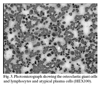
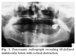
<body><![CDATA[<p>&nbsp;</p>     <p><font face="Verdana" size="2">Expansion of bone in the bucco-lingual direction, cortical bone destruction and periosteal reaction were also realized from the occlusal radiogram (<a href="#f2">Figure 2</a>). In addition, in the second attendance, there were no other bony changes in her skull radiograms.</font></p>     <p align="center"><font face="Verdana" size="2"><a name="f2"><img border="0" src="/img/revistas/medicorpa/v12n1/03i.ht10.gif" width="298" height="297"></a></font></p>     <p><font face="Verdana" size="2">Aspiration cytology was hyper cellular. There were numerous plasma cells having abundant cytoplasm and eccentrically located nuclei. Plasma cells showed slight nucleomegali and irregular chromatin distribution. Some of them were binucleated.</font></p>     <p><font face="Verdana" size="2">Additionally there were some lypmhocytes and osteoclastic giant cells. According to these findings, the histopathological diagnosis was plasmocytoma (<a href="#f3">Figure 3</a>).</font></p>     <p align="center"><font face="Verdana" size="2"><a name="f3"><img border="0" src="/img/revistas/medicorpa/v12n1/03i.ht11.gif" width="333" height="283"></a></font></p>     <p><font face="Verdana" size="2">After the diagnosis of plasmocytoma, the patient was referred to the oncology department for treatment but unfortunately she died before the treatment finished.</font></p>     <p>&nbsp;</p>     <p><B><font face="Verdana">Discussion</font></B></p>     <p><font face="Verdana" size="2">SPB is tumor of plasma cells, which manifests itself as a single osteolytic lesion without plasmocytosis of bone marrow and constitutes approximately 3% of all plasma containing tumors. SPB is more prevalent in males than females, the ratio being 2:1 (1- 8).</font></p>     ]]></body>
<body><![CDATA[<p><font face="Verdana" size="2">The most common sites of SPB are long bones and vertebrae. It rarely involves jaws and when it is seen, only 4.4% of SPB occur in the mandible, most commonly in the bone marrow-rich areas of the body, angulus and ramus mandible (1- 6). As the patient was female and the midline of the mandible as the occurrence site, this was a very rare case.</font></p>     <p><font face="Verdana" size="2">Most common clinical symptoms of SPB are pain in the jaws and teeth, paraesthesia, anesthesia, mobility and migration of the teeth, haemorrhage, swelling in hard and soft tissues and pathological fractures (1-4,6,7). Our case was in harmony with literature since the main complaint was swelling and the lesion’s reddish colour.</font></p>     <p><font face="Verdana" size="2">In our case, the lesion was located in the anterior of the mandible between canines. A study by Pisano et al. (2), containing 13 cases of SPB, showed that nine lesions were located posterior to the premolars and only one case was anterior but distal to the canine. A study carried out by Marick et al. (3) which consisted of 21 cases of SPB showed that in 15 cases, the lesion affected the maxilla more than the mandible and despite the reported female predominance in the literature, males were predominant. The male/female ratio was 2:1. In our case, swelling was the major symptom and pain was present but paraesthesia was not reported.</font></p>     <p><font face="Verdana" size="2">Radiographically, SPB is seen as a well defined, unilocular or multilocular destructive lesion with lack of any sign of bone reaction (1,3). But in our case, there were signs of periosteal reaction which is a rare radiographic sign. This can cause cortical destruction and can spread widely to the surrounding tissues.</font></p>     <p><font face="Verdana" size="2">There is a possibility of transformation of SPB and EMP to MM. This possibility is higher in SPB than EMP. Clinical investigations conclude that 35–85% of SPB cases are transformed to MM in a period of couple of months to a few years. But it is not possible to predict which case may transform, so after treatment, SPB cases must be followed up with routine laboratory monitoring of immunoglobulins and monoclonal proteins in serum and Bence-Jones proteins in urine (1-3). In our case, when the patient first consulted our clinic for the jaw lesion, bone marrow examination results were normal.</font></p>     <p><font face="Verdana" size="2">Plasma cell neoplasms appear as sheets of plasma cells and are indistinguishable histologically. Clinical signs are also the same. Distinguishing one from the other is critical to treatment and survival. According to the description of dysplastic behaviour of plasmocytoma by Sukpaninchnant et al. (10), minimal dysplasia is defined as most of the plasma cells being mature and plasmoblasts (cells with nucleoli) accounting for less than 10% of cells, whereas marked dysplasia is defined as plasmoblasts consisting of greater than 50% of tumour cells. Our case was judged as minimal dysplastic as the histopathological results indicated mature plasma cells.</font></p>     <p><font face="Verdana" size="2">The course of SPB is relatively benign. Nevertheless, prognosis of SPB is poorer than EMP; 58% of cases progress to MM and survival at 10 years is 16% in SPB. The average outliving is over 90 months (6,9). Unfortunately, our patient died during the course of treatment.</font></p>     <p>&nbsp;</p>     <p><B><font face="Verdana">References</font></B></p>     <!-- ref --><p><font face="Verdana" size="2">1. Seoane J, Aguirre-Urizar JM, Esparza-Gomez G, Suarez-Cunqueiro M, Campos-Trapero J, Pomareda M. The spectrum of plasma cell neoplasia in oral pathology. Med Oral 2003;8:269-80.</font>&nbsp;&nbsp;&nbsp;&nbsp;&nbsp;&nbsp;&nbsp;&nbsp;[&#160;<a href="javascript:void(0);" onclick="javascript: window.open('/scielo.php?script=sci_nlinks&ref=2973453&pid=S1698-6946200700010000300001&lng=','','width=640,height=500,resizable=yes,scrollbars=1,menubar=yes,');">Links</a>&#160;]<!-- end-ref --><!-- ref --><p><font face="Verdana" size="2">2. Pisano JJ, Coupland R, Chen SY, Miller AS. Plasmacytoma of the oral cavity and jaws. A clinopathological study of 13 cases. Oral Surg Oral Med Oral Pathol Oral Radiol Endod 1997;83:265-71.</font>&nbsp;&nbsp;&nbsp;&nbsp;&nbsp;&nbsp;&nbsp;&nbsp;[&#160;<a href="javascript:void(0);" onclick="javascript: window.open('/scielo.php?script=sci_nlinks&ref=2973454&pid=S1698-6946200700010000300002&lng=','','width=640,height=500,resizable=yes,scrollbars=1,menubar=yes,');">Links</a>&#160;]<!-- end-ref --><!-- ref --><p><font face="Verdana" size="2">3. Marick EL, Vencio EF, Inwards CY, Unni KK, Nascimento AG. Myeloma of the jaw bones: a clinopathological study of 33 cases. Head Neck 2003;25:373-81.</font>&nbsp;&nbsp;&nbsp;&nbsp;&nbsp;&nbsp;&nbsp;&nbsp;[&#160;<a href="javascript:void(0);" onclick="javascript: window.open('/scielo.php?script=sci_nlinks&ref=2973455&pid=S1698-6946200700010000300003&lng=','','width=640,height=500,resizable=yes,scrollbars=1,menubar=yes,');">Links</a>&#160;]<!-- end-ref --><!-- ref --><p><font face="Verdana" size="2">4. Ozdemir R, Kayiran O, Oruç M, Karaaslan O, Kocer U, Ogün D. Plasmacytoma of the hard palate (brief clinical notes). J Craniofac Surg 2005;16:164-9.</font>&nbsp;&nbsp;&nbsp;&nbsp;&nbsp;&nbsp;&nbsp;&nbsp;[&#160;<a href="javascript:void(0);" onclick="javascript: window.open('/scielo.php?script=sci_nlinks&ref=2973456&pid=S1698-6946200700010000300004&lng=','','width=640,height=500,resizable=yes,scrollbars=1,menubar=yes,');">Links</a>&#160;]<!-- end-ref --><!-- ref --><p><font face="Verdana" size="2">5. Miller FR, Lavertu P, Wanamaker JR, Bonafede J, Wood BG. Plasmacytomas of the head and neck. Otolaryngol Head Neck Surg 1998;119: 614-8.</font>&nbsp;&nbsp;&nbsp;&nbsp;&nbsp;&nbsp;&nbsp;&nbsp;[&#160;<a href="javascript:void(0);" onclick="javascript: window.open('/scielo.php?script=sci_nlinks&ref=2973457&pid=S1698-6946200700010000300005&lng=','','width=640,height=500,resizable=yes,scrollbars=1,menubar=yes,');">Links</a>&#160;]<!-- end-ref --><!-- ref --><p><font face="Verdana" size="2">6. Nofsinger YC, Mirza N, Rowan PT, Lanza D, Weinstein G. Head and neck manifestations of plasma cell neoplasms. The Laryngoscope 1997;107:741-6.</font>&nbsp;&nbsp;&nbsp;&nbsp;&nbsp;&nbsp;&nbsp;&nbsp;[&#160;<a href="javascript:void(0);" onclick="javascript: window.open('/scielo.php?script=sci_nlinks&ref=2973458&pid=S1698-6946200700010000300006&lng=','','width=640,height=500,resizable=yes,scrollbars=1,menubar=yes,');">Links</a>&#160;]<!-- end-ref --><!-- ref --><p><font face="Verdana" size="2">7. Reboiras Lopez MD, Garcia Garcia A, Antunez Lopez J, Blanco Carrion A, Gandara Vila P, Gandara Rey JM. Anesthesia of the right lower hemilip as a first manifestation of multiple myeloma. Presentation of a clinical case. Med Oral 2001;6:168-72.</font>&nbsp;&nbsp;&nbsp;&nbsp;&nbsp;&nbsp;&nbsp;&nbsp;[&#160;<a href="javascript:void(0);" onclick="javascript: window.open('/scielo.php?script=sci_nlinks&ref=2973459&pid=S1698-6946200700010000300007&lng=','','width=640,height=500,resizable=yes,scrollbars=1,menubar=yes,');">Links</a>&#160;]<!-- end-ref --><!-- ref --><p><font face="Verdana" size="2">8. Bolek WT, Marcus RB, Mendenhall NP. Solitary plasmacytoma of bone and soft tissues. Int J Radiation Oncology Biol Phys 1996;36:329-3.</font>&nbsp;&nbsp;&nbsp;&nbsp;&nbsp;&nbsp;&nbsp;&nbsp;[&#160;<a href="javascript:void(0);" onclick="javascript: window.open('/scielo.php?script=sci_nlinks&ref=2973460&pid=S1698-6946200700010000300008&lng=','','width=640,height=500,resizable=yes,scrollbars=1,menubar=yes,');">Links</a>&#160;]<!-- end-ref --><!-- ref --><p><font face="Verdana" size="2">9. Muzio LL, Pannone G, Bucci P. Early clinical diagnosis of solitary plasmocytoma of the jaws: a case report with a six year follow-up. Int J Oral Maxillofac Surg 2001;30:558-60.</font>&nbsp;&nbsp;&nbsp;&nbsp;&nbsp;&nbsp;&nbsp;&nbsp;[&#160;<a href="javascript:void(0);" onclick="javascript: window.open('/scielo.php?script=sci_nlinks&ref=2973461&pid=S1698-6946200700010000300009&lng=','','width=640,height=500,resizable=yes,scrollbars=1,menubar=yes,');">Links</a>&#160;]<!-- end-ref --><!-- ref --><p><font face="Verdana" size="2">10. Sukpaninchnant S, Cousar JB, Leelasiri A, Graber SE, Greer JP, Collins RD. Diagnostic criteria and histological grading in multiple myeloma: histologic and immunohistologic analysis of 176 cases with clinical correlation. Hum Pathol 1994;25:308-18.</font>&nbsp;&nbsp;&nbsp;&nbsp;&nbsp;&nbsp;&nbsp;&nbsp;[&#160;<a href="javascript:void(0);" onclick="javascript: window.open('/scielo.php?script=sci_nlinks&ref=2973462&pid=S1698-6946200700010000300010&lng=','','width=640,height=500,resizable=yes,scrollbars=1,menubar=yes,');">Links</a>&#160;]<!-- end-ref --><p>&nbsp;</p>     ]]></body>
<body><![CDATA[<p>&nbsp;</p>     <p><font face="Verdana" size="2"><a href="#top"><img border="0" src="/img/revistas/medicorpa/v12n1/seta.gif" width="15" height="17"> </a> <a name="bajo"></a><B>Correspondence:</B>    <BR>Dr. Emin Murat Canger    <BR>Ondokuz May&#305;s University Faculty of Dentistry    <BR>Department of Oral Diagnosis and Radiology    <BR>55139 Samsun/Turkey    <BR>E-mail: <a href="mailto:emcanger@omu.edu.tr">emcanger@omu.edu.tr</a></font></p>     <p><font face="Verdana" size="2">Received: 24-02-2006    <BR>Accepted: 7-07-2006</font></p>      ]]></body><back>
<ref-list>
<ref id="B1">
<label>1</label><nlm-citation citation-type="journal">
<person-group person-group-type="author">
<name>
<surname><![CDATA[Seoane]]></surname>
<given-names><![CDATA[J]]></given-names>
</name>
<name>
<surname><![CDATA[Aguirre-Urizar]]></surname>
<given-names><![CDATA[JM]]></given-names>
</name>
<name>
<surname><![CDATA[Esparza-Gomez]]></surname>
<given-names><![CDATA[G]]></given-names>
</name>
<name>
<surname><![CDATA[Suarez-Cunqueiro]]></surname>
<given-names><![CDATA[M]]></given-names>
</name>
<name>
<surname><![CDATA[Campos-Trapero]]></surname>
<given-names><![CDATA[J]]></given-names>
</name>
<name>
<surname><![CDATA[Pomareda]]></surname>
<given-names><![CDATA[M]]></given-names>
</name>
</person-group>
<article-title xml:lang="en"><![CDATA[The spectrum of plasma cell neoplasia in oral pathology]]></article-title>
<source><![CDATA[Med Oral]]></source>
<year>2003</year>
<volume>8</volume>
<page-range>269-80</page-range></nlm-citation>
</ref>
<ref id="B2">
<label>2</label><nlm-citation citation-type="journal">
<person-group person-group-type="author">
<name>
<surname><![CDATA[Pisano]]></surname>
<given-names><![CDATA[JJ]]></given-names>
</name>
<name>
<surname><![CDATA[Coupland]]></surname>
<given-names><![CDATA[R]]></given-names>
</name>
<name>
<surname><![CDATA[Chen]]></surname>
<given-names><![CDATA[SY]]></given-names>
</name>
<name>
<surname><![CDATA[Miller]]></surname>
<given-names><![CDATA[AS]]></given-names>
</name>
</person-group>
<article-title xml:lang="en"><![CDATA[Plasmacytoma of the oral cavity and jaws: A clinopathological study of 13 cases]]></article-title>
<source><![CDATA[Oral Surg Oral Med Oral Pathol Oral Radiol Endod]]></source>
<year>1997</year>
<volume>83</volume>
<page-range>265-71</page-range></nlm-citation>
</ref>
<ref id="B3">
<label>3</label><nlm-citation citation-type="journal">
<person-group person-group-type="author">
<name>
<surname><![CDATA[Marick]]></surname>
<given-names><![CDATA[EL]]></given-names>
</name>
<name>
<surname><![CDATA[Vencio]]></surname>
<given-names><![CDATA[EF]]></given-names>
</name>
<name>
<surname><![CDATA[Inwards]]></surname>
<given-names><![CDATA[CY]]></given-names>
</name>
<name>
<surname><![CDATA[Unni]]></surname>
<given-names><![CDATA[KK]]></given-names>
</name>
<name>
<surname><![CDATA[Nascimento]]></surname>
<given-names><![CDATA[AG]]></given-names>
</name>
</person-group>
<article-title xml:lang="en"><![CDATA[Myeloma of the jaw bones: a clinopathological study of 33 cases]]></article-title>
<source><![CDATA[Head Neck]]></source>
<year>2003</year>
<volume>25</volume>
<page-range>373-81</page-range></nlm-citation>
</ref>
<ref id="B4">
<label>4</label><nlm-citation citation-type="journal">
<person-group person-group-type="author">
<name>
<surname><![CDATA[Ozdemir]]></surname>
<given-names><![CDATA[R]]></given-names>
</name>
<name>
<surname><![CDATA[Kayiran]]></surname>
<given-names><![CDATA[O]]></given-names>
</name>
<name>
<surname><![CDATA[Oruç]]></surname>
<given-names><![CDATA[M]]></given-names>
</name>
<name>
<surname><![CDATA[Karaaslan]]></surname>
<given-names><![CDATA[O]]></given-names>
</name>
<name>
<surname><![CDATA[Kocer]]></surname>
<given-names><![CDATA[U]]></given-names>
</name>
<name>
<surname><![CDATA[Ogün]]></surname>
<given-names><![CDATA[D]]></given-names>
</name>
</person-group>
<article-title xml:lang="en"><![CDATA[Plasmacytoma of the hard palate (brief clinical notes)]]></article-title>
<source><![CDATA[J Craniofac Surg]]></source>
<year>2005</year>
<volume>16</volume>
<page-range>164-9</page-range></nlm-citation>
</ref>
<ref id="B5">
<label>5</label><nlm-citation citation-type="journal">
<person-group person-group-type="author">
<name>
<surname><![CDATA[Miller]]></surname>
<given-names><![CDATA[FR]]></given-names>
</name>
<name>
<surname><![CDATA[Lavertu]]></surname>
<given-names><![CDATA[P]]></given-names>
</name>
<name>
<surname><![CDATA[Wanamaker]]></surname>
<given-names><![CDATA[JR]]></given-names>
</name>
<name>
<surname><![CDATA[Bonafede]]></surname>
<given-names><![CDATA[J]]></given-names>
</name>
<name>
<surname><![CDATA[Wood]]></surname>
<given-names><![CDATA[BG]]></given-names>
</name>
</person-group>
<article-title xml:lang="en"><![CDATA[Plasmacytomas of the head and neck]]></article-title>
<source><![CDATA[Otolaryngol Head Neck Surg]]></source>
<year>1998</year>
<volume>119</volume>
<page-range>614-8</page-range></nlm-citation>
</ref>
<ref id="B6">
<label>6</label><nlm-citation citation-type="journal">
<person-group person-group-type="author">
<name>
<surname><![CDATA[Nofsinger]]></surname>
<given-names><![CDATA[YC]]></given-names>
</name>
<name>
<surname><![CDATA[Mirza]]></surname>
<given-names><![CDATA[N]]></given-names>
</name>
<name>
<surname><![CDATA[Rowan]]></surname>
<given-names><![CDATA[PT]]></given-names>
</name>
<name>
<surname><![CDATA[Lanza]]></surname>
<given-names><![CDATA[D]]></given-names>
</name>
<name>
<surname><![CDATA[Weinstein]]></surname>
<given-names><![CDATA[G]]></given-names>
</name>
</person-group>
<article-title xml:lang="en"><![CDATA[Head and neck manifestations of plasma cell neoplasms]]></article-title>
<source><![CDATA[The Laryngoscope]]></source>
<year>1997</year>
<volume>107</volume>
<page-range>741-6</page-range></nlm-citation>
</ref>
<ref id="B7">
<label>7</label><nlm-citation citation-type="journal">
<person-group person-group-type="author">
<name>
<surname><![CDATA[Reboiras Lopez]]></surname>
<given-names><![CDATA[MD]]></given-names>
</name>
<name>
<surname><![CDATA[Garcia Garcia]]></surname>
<given-names><![CDATA[A]]></given-names>
</name>
<name>
<surname><![CDATA[Antunez Lopez]]></surname>
<given-names><![CDATA[J]]></given-names>
</name>
<name>
<surname><![CDATA[Blanco Carrion]]></surname>
<given-names><![CDATA[A]]></given-names>
</name>
<name>
<surname><![CDATA[Gandara Vila]]></surname>
<given-names><![CDATA[P]]></given-names>
</name>
<name>
<surname><![CDATA[Gandara Rey]]></surname>
<given-names><![CDATA[JM]]></given-names>
</name>
</person-group>
<article-title xml:lang="en"><![CDATA[Anesthesia of the right lower hemilip as a first manifestation of multiple myeloma: Presentation of a clinical case]]></article-title>
<source><![CDATA[Med Oral]]></source>
<year>2001</year>
<volume>6</volume>
<page-range>168-72</page-range></nlm-citation>
</ref>
<ref id="B8">
<label>8</label><nlm-citation citation-type="journal">
<person-group person-group-type="author">
<name>
<surname><![CDATA[Bolek]]></surname>
<given-names><![CDATA[WT]]></given-names>
</name>
<name>
<surname><![CDATA[Marcus]]></surname>
<given-names><![CDATA[RB]]></given-names>
</name>
<name>
<surname><![CDATA[Mendenhall]]></surname>
<given-names><![CDATA[NP]]></given-names>
</name>
</person-group>
<article-title xml:lang="en"><![CDATA[Solitary plasmacytoma of bone and soft tissues]]></article-title>
<source><![CDATA[Int J Radiation Oncology Biol Phys]]></source>
<year>1996</year>
<volume>36</volume>
<page-range>329-3</page-range></nlm-citation>
</ref>
<ref id="B9">
<label>9</label><nlm-citation citation-type="journal">
<person-group person-group-type="author">
<name>
<surname><![CDATA[Muzio]]></surname>
<given-names><![CDATA[LL]]></given-names>
</name>
<name>
<surname><![CDATA[Pannone]]></surname>
<given-names><![CDATA[G]]></given-names>
</name>
<name>
<surname><![CDATA[Bucci]]></surname>
<given-names><![CDATA[P]]></given-names>
</name>
</person-group>
<article-title xml:lang="en"><![CDATA[Early clinical diagnosis of solitary plasmocytoma of the jaws: a case report with a six year follow-up]]></article-title>
<source><![CDATA[Int J Oral Maxillofac Surg]]></source>
<year>2001</year>
<volume>30</volume>
<page-range>558-60</page-range></nlm-citation>
</ref>
<ref id="B10">
<label>10</label><nlm-citation citation-type="journal">
<person-group person-group-type="author">
<name>
<surname><![CDATA[Sukpaninchnant]]></surname>
<given-names><![CDATA[S]]></given-names>
</name>
<name>
<surname><![CDATA[Cousar]]></surname>
<given-names><![CDATA[JB]]></given-names>
</name>
<name>
<surname><![CDATA[Leelasiri]]></surname>
<given-names><![CDATA[A]]></given-names>
</name>
<name>
<surname><![CDATA[Graber]]></surname>
<given-names><![CDATA[SE]]></given-names>
</name>
<name>
<surname><![CDATA[Greer]]></surname>
<given-names><![CDATA[JP]]></given-names>
</name>
<name>
<surname><![CDATA[Collins]]></surname>
<given-names><![CDATA[RD]]></given-names>
</name>
</person-group>
<article-title xml:lang="en"><![CDATA[Diagnostic criteria and histological grading in multiple myeloma: histologic and immunohistologic analysis of 176 cases with clinical correlation]]></article-title>
<source><![CDATA[Hum Pathol]]></source>
<year>1994</year>
<volume>25</volume>
<page-range>308-18</page-range></nlm-citation>
</ref>
</ref-list>
</back>
</article>