<?xml version="1.0" encoding="ISO-8859-1"?><article xmlns:mml="http://www.w3.org/1998/Math/MathML" xmlns:xlink="http://www.w3.org/1999/xlink" xmlns:xsi="http://www.w3.org/2001/XMLSchema-instance">
<front>
<journal-meta>
<journal-id>1698-6946</journal-id>
<journal-title><![CDATA[Medicina Oral, Patología Oral y Cirugía Bucal (Internet)]]></journal-title>
<abbrev-journal-title><![CDATA[Med. oral patol. oral cir.bucal (Internet)]]></abbrev-journal-title>
<issn>1698-6946</issn>
<publisher>
<publisher-name><![CDATA[Sociedad Española de Medicina Oral]]></publisher-name>
</publisher>
</journal-meta>
<article-meta>
<article-id>S1698-69462007000300010</article-id>
<title-group>
<article-title xml:lang="en"><![CDATA[Leiomyoma of the hard palate: A case report]]></article-title>
</title-group>
<contrib-group>
<contrib contrib-type="author">
<name>
<surname><![CDATA[González Sánchez]]></surname>
<given-names><![CDATA[Miguel Angel]]></given-names>
</name>
<xref ref-type="aff" rid="A01"/>
</contrib>
<contrib contrib-type="author">
<name>
<surname><![CDATA[Colorado Bonnin]]></surname>
<given-names><![CDATA[Mar]]></given-names>
</name>
<xref ref-type="aff" rid="A01"/>
</contrib>
<contrib contrib-type="author">
<name>
<surname><![CDATA[Berini Aytés]]></surname>
<given-names><![CDATA[Leonardo]]></given-names>
</name>
<xref ref-type="aff" rid="A01"/>
</contrib>
<contrib contrib-type="author">
<name>
<surname><![CDATA[Gay Escoda]]></surname>
<given-names><![CDATA[Cosme]]></given-names>
</name>
<xref ref-type="aff" rid="A01"/>
<xref ref-type="aff" rid="A02"/>
</contrib>
</contrib-group>
<aff id="A01">
<institution><![CDATA[,University of Barcelona Dental School ]]></institution>
<addr-line><![CDATA[Barcelona ]]></addr-line>
</aff>
<aff id="A02">
<institution><![CDATA[,Teknon Medical Center  ]]></institution>
<addr-line><![CDATA[Barcelona ]]></addr-line>
<country>Spain</country>
</aff>
<pub-date pub-type="pub">
<day>00</day>
<month>05</month>
<year>2007</year>
</pub-date>
<pub-date pub-type="epub">
<day>00</day>
<month>05</month>
<year>2007</year>
</pub-date>
<volume>12</volume>
<numero>3</numero>
<fpage>221</fpage>
<lpage>224</lpage>
<copyright-statement/>
<copyright-year/>
<self-uri xlink:href="http://scielo.isciii.es/scielo.php?script=sci_arttext&amp;pid=S1698-69462007000300010&amp;lng=en&amp;nrm=iso"></self-uri><self-uri xlink:href="http://scielo.isciii.es/scielo.php?script=sci_abstract&amp;pid=S1698-69462007000300010&amp;lng=en&amp;nrm=iso"></self-uri><self-uri xlink:href="http://scielo.isciii.es/scielo.php?script=sci_pdf&amp;pid=S1698-69462007000300010&amp;lng=en&amp;nrm=iso"></self-uri><abstract abstract-type="short" xml:lang="en"><p><![CDATA[Leiomyoma is a benign smooth muscle tumor that is very rarely located in the oral cavity. It is accepted that the smooth muscle giving rise to this tumor corresponds to the tunica media of the arteries. The tumor can develop at any age, with no clear gender predilection, and typically manifests as a slow-growing, asymptomatic lesion. The diagnosis is based on the histopathological study, with the use of specific staining techniques, including immunohistochemical markers. Treatment consists of complete resection of the lesion, with due safety margins. We present the case of a 57-year-old male with a tumor located for the previous four months in the region of the hard palate, adjacent to the left upper premolars. After surgical resection with safety margins, hematoxylin-eosin staining and immunohistochemical study confirmed the diagnosis of leiomyoma.]]></p></abstract>
<abstract abstract-type="short" xml:lang="es"><p><![CDATA[El leiomioma es un tumor benigno del músculo liso muy poco frecuente a nivel de la cavidad bucal. Se acepta que la fuente de músculo liso originaria de este tumor es la túnica media de los vasos arteriales. Se trata de un tumor que puede aparecer a cualquier edad, sin presentar una clara predilección por el sexo y normalmente lo hace en forma de tumoración asintomática de crecimiento lento. Su diagnóstico está basado en el estudio anatomopatológico, utilizándose tinciones específicas como podrían ser las técnicas de inmunohistoquímica. Su tratamiento consiste en la extirpación completa de la lesión con márgenes de seguridad. En este artículo se expone el caso de un paciente hombre de 57 años que presentaba una lesión en el paladar duro, adyacente a los premolares superiores del lado izquierdo, de 4 meses de evolución. Tras la exéresis quirúrgica con márgenes de seguridad de dicha lesión, se realizó su estudio anatomopatológico. La tinción con hematoxilina y eosina junto con el estudio inmunohistoquímico de la muestra confirmaron el diagnóstico de leiomioma.]]></p></abstract>
<kwd-group>
<kwd lng="en"><![CDATA[Leiomyoma]]></kwd>
<kwd lng="en"><![CDATA[smooth muscle tumor]]></kwd>
<kwd lng="en"><![CDATA[palatal tumor]]></kwd>
<kwd lng="es"><![CDATA[Leiomioma]]></kwd>
<kwd lng="es"><![CDATA[tumor de músculo liso]]></kwd>
<kwd lng="es"><![CDATA[tumor de paladar]]></kwd>
</kwd-group>
</article-meta>
</front><body><![CDATA[ <p>&nbsp;</p>     <p>&nbsp;</p>     <p><B><font face="Verdana" size="2"><a name="top"></a></font> <font face="Verdana" size="4">Leiomyoma of the hard palate: A case report</font></B></p>     <p>&nbsp;</p>     <p>&nbsp;</p>     <p><font face="Verdana" size="2"><B>Miguel Angel González Sánchez<sup>1</sup>, Mar Colorado Bonnin<sup>2</sup>, Leonardo Berini Aytés<sup>3</sup>, Cosme Gay Escoda<sup>4</sup></B></font></p>     <p><font face="Verdana" size="2">(1) DDS and resident of the Master of Oral Surgery and Implantology University of Barcelona Dental School    <BR>(2) DDS and Master of Oral Surgery and Implantology University of Barcelona Dental School    <BR>(3) DDS, MD. Assistant Professor of Oral Surgery. Professor of the Master of Oral Surgery and Implantology. University of Barcelona Dental School    <BR>(4) DDS, MD, PhD. Chairman of Oral and Maxillofacial Surgery. Director of the Master of Oral Surgery and Implantology.    ]]></body>
<body><![CDATA[<br> University of Barcelona Dental School. Oral and maxillofacial surgeon of the Teknon Medical Center, Barcelona (Spain)</font></p>     <p><font face="Verdana" size="2"><a href="#back">Correspondence</a></font></p>     <p>&nbsp;</p>     <p>&nbsp;</p>  <hr size="1">      <p><font face="Verdana" size="2"><B>ABSTRACT</B></font></p>     <p><font face="Verdana" size="2">Leiomyoma is a benign smooth muscle tumor that is very rarely located in the oral cavity. It is accepted that the smooth muscle giving rise to this tumor corresponds to the tunica media of the arteries. The tumor can develop at any age, with no clear gender predilection, and typically manifests as a slow-growing, asymptomatic lesion. The diagnosis is based on the histopathological study, with the use of specific staining techniques, including immunohistochemical markers. Treatment consists of complete resection of the lesion, with due safety margins.    <BR>We present the case of a 57-year-old male with a tumor located for the previous four months in the region of the hard palate, adjacent to the left upper premolars. After surgical resection with safety margins, hematoxylin-eosin staining and immunohistochemical study confirmed the diagnosis of leiomyoma.</font></p>     <p><font face="Verdana" size="2"><B>Key words:</B> Leiomyoma, smooth muscle tumor, palatal tumor.</font></p> <hr size="1">     <p><font face="Verdana" size="2"><B>RESUMEN</B></font></p>     <p><font face="Verdana" size="2">El leiomioma es un tumor benigno del músculo liso muy poco frecuente a nivel de la cavidad bucal. Se acepta que la fuente de músculo liso originaria de este tumor es la túnica media de los vasos arteriales. Se trata de un tumor que puede aparecer a cualquier edad, sin presentar una clara predilección por el sexo y normalmente lo hace en forma de tumoración asintomática de crecimiento lento. Su diagnóstico está basado en el estudio anatomopatológico, utilizándose tinciones específicas como podrían ser las técnicas de inmunohistoquímica. Su tratamiento consiste en la extirpación completa de la lesión con márgenes de seguridad.    ]]></body>
<body><![CDATA[<br> En este artículo se expone el caso de un paciente hombre de 57 años que presentaba una lesión en el paladar duro, adyacente a los premolares superiores del lado izquierdo, de 4 meses de evolución. Tras la exéresis quirúrgica con márgenes de seguridad de dicha lesión, se realizó su estudio anatomopatológico. La tinción con hematoxilina y eosina junto con el estudio inmunohistoquímico de la muestra confirmaron el diagnóstico de leiomioma.</font></p>     <p><font face="Verdana" size="2"><B>Palabras clave:</B> Leiomioma, tumor de músculo liso, tumor de paladar.</font></p> <hr size="1">     <p>&nbsp;</p>     <p><font face="Verdana"><B>Introduction</B></font></p>     <p><font face="Verdana" size="2">Leiomyoma is a benign smooth muscle tumor that may appear in any location, though it is more common in the uterus, gastrointestinal tract and skin. It is rarely found in the oral cavity (0.065%), due to the scarcity of smooth muscle in this territory (1).</font></p>     <p><font face="Verdana" size="2">Leiomyoma is usually seen in adults and shows no gender predilection. The most frequent oral locations are the lips, tongue, hard and soft palate, and the cheeks.</font></p>     <p><font face="Verdana" size="2">The tumor generally manifests as a slow-growing painless lesion, often of a purplish color. The diagnosis is exclusively based on the histological findings. Clinically, a differential diagnosis must be established with lesions of the oral mucosa or connective tissue, such as fibromas, lipomas, salivary gland neoplasms, vascular tumors such as lymphangioma or hemangioma, etc. The differential diagnosis moreover must also include the malignant form of leiomyoma, i.e., leiomyosarcoma.</font></p>     <p><font face="Verdana" size="2">Treatment consists of complete resection, with due safety margins and periodic controls to ensure early identification of possible tumor relapse.</font></p>     <p><font face="Verdana" size="2">The present article describes a new case of oral leiomyoma, located in the palatal region, and evaluates the clinical and histological characteristics of the lesion, with a view to including the latter in the routine differential diagnosis of oral mucosal lesions.</font></p>     <p>&nbsp;</p>     ]]></body>
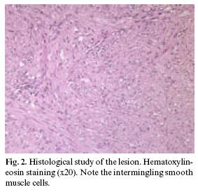
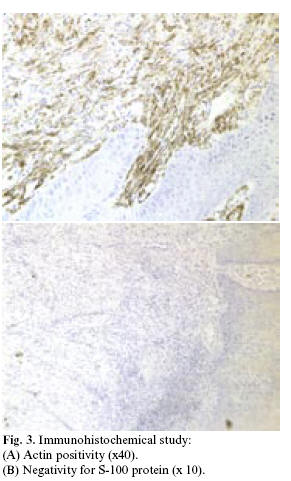
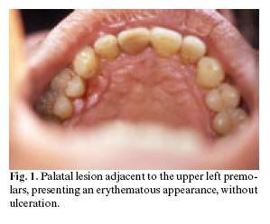
<body><![CDATA[<p><font face="Verdana"><B>Clinical case</B></font></p>     <p><font face="Verdana" size="2">A 57-year-old male presented with a history of arterial  hypertension controlled with medication (telmisartan 80 mg/day via the oral  route). There were no surgical antecedents, and no known drug allergies. The  patient had stopped smoking (30 cigarettes and a cigar daily) 16 years before,  and referred moderate alcohol consumption.</font></p>     <p><font face="Verdana" size="2">The patient was referred to our Service by his personal  dentist for evaluation of an erythematous lesion on the hard palate, adjacent to  the palatal surface of the left upper premolars (<a href="#f1">Figure 1</a>). The lesion had been  present for the previous four months and persisted despite treatment by the  dentist with antiseptic rinses and antibiotics (spiramycin and metronidazole).</font></p>     <p align="center"><font face="Verdana" size="2"><a name="f1"><img border="0" src="/img/revistas/medicorpa/v12n3/10_medora72.jpg" width="299" height="249"></a></font></p>     <p><font face="Verdana" size="2">We compiled a case history, and the physical examination  revealed a lesion measuring about 5 mm in diameter, located adjacent to the  palatal surface of the upper premolars in the second quadrant. The lesion was of  a red color, similar to an ulceration, painless and non-hemorrhagic in response  to palpation. Orthopantomography revealed no alterations in the region of the  lesion. Based on the clinical and radiological findings, moderate chronic  periodontitis was diagnosed - the lesion thus being suspected to have a  periodontal origin.</font></p>     <p><font face="Verdana" size="2">Periodontal treatment with an excisional biopsy of the lesion  under local anesthetic was decided. A cold scalpel was used to remove the entire  lesion, leaving safety margins, with the inclusion of periosteum. The surgical  wound was allowed to heal by second intention. There were no intra- or  postoperative complications.</font></p>     <p><font face="Verdana" size="2">The histopathological study diagnosed oral leiomyoma.  Hematoxylin-eosin staining (<a href="#f2">Figure 2</a>) revealed the presence of intermingling  smooth muscle bands separated by cellular fibrous connective tissue.  Immunohistochemical techniques were also applied, involving monoclonal  antibodies against actin, vimentin, cytokeratin and the S-100 protein (<a href="#f3">Figure  3</a>). Positivity was observed for actin and vimentin, with negative results for  the rest of markers - thus confirming the diagnosis of leiomyoma. Computed  tomography (CT) ruled out bone involvement, thus confirming the exclusively  mucosal distribution of the leiomyoma.</font></p>     <p align="center"><font face="Verdana" size="2"><a name="f2"><img border="0" src="/img/revistas/medicorpa/v12n3/10_medora73.jpg" width="283" height="274"></a></font></p>     <p align="center"><font face="Verdana" size="2">    <br> <a name="f3"><img border="0" src="/img/revistas/medicorpa/v12n3/10_medora74.jpg" width="291" height="481"></a></font></p>     ]]></body>
<body><![CDATA[<p><font face="Verdana" size="2">Periodic controls were made every 6 months the first year,  and yearly thereafter. No evidence of tumor relapse was observed after 28 months  of follow-up.</font></p>     <p>&nbsp;</p>     <p><font face="Verdana"><B>Discussion</B></font></p>     <p><font face="Verdana" size="2">Leiomyoma is infrequent in the oral cavity, due to the  scarcity of smooth muscle in this territory (1). It possible origin has been  commented by a number of authors (2,3). Thus, Stout (3) suggested the smooth  muscle of the tunica media of the arteries to be the probable origin of oral  leiomyomas. Other authors in turn consider leiomyomas to derive from the remains  of embryonic tissue such as the lingual duct or circumvallate papilla of the  tongue (2).</font></p>     <p><font face="Verdana" size="2">The first case of oral leiomyoma was described in 1884, and  since then a number of additional cases have been documented (1). In the review  published by Farman (4) in 1975, involving 7748 smooth muscle tumors located  throughout the body, only 5 cases (0.065%) corresponded to the oral cavity. The  most common location was the female genitourinary tract, representing 95% of the  total cases. Hachisuga et al. (5) recorded 15 cases (2.7%) of oral  angioleiomyomas in a series of 562 angioleiomyomas registered in a General  Pathology Department. Brooks et al. (6) in turn published a retrospective study  of 12 angioleiomyomas, one solid leiomyoma and one leiomyosarcoma, out of a  total of 76,412 biopsies / oral lesions registered in a Department of Oral  Medicine in the period between 1963 and 2001. The incidence of angioleiomyoma  was 0.016%, and represented 92.3% of all benign smooth muscle tumors located in  the oral cavity.</font></p>     <p><font face="Verdana" size="2">Leiomyoma is usually seen in adults - the greatest incidence  corresponding to the 40-59 years age interval. Regarding gender distribution,  some authors consider both males and females to be affected in equal proportion  (7,8), though considerable controversy exists on this point, since other  investigators have reported a 2:1 female predilection (6), while others consider  males to outnumber females 2:1 (1). Our case corresponded to a 57-year-old male,  and coincides with the typical presentations reported in the literature.</font></p>     <p><font face="Verdana" size="2">The most frequent oral locations are the lips, tongue, hard  and soft palate, and the cheeks (9). In the series described by Svane et al.  (9), 21% of the leiomyomas were located in the palate. Of these, 91% were  angioleiomyomas, while the remaining 9% corresponded to solid leiomyomas. Brooks  et al. (6) recorded two angioleiomyomas at palatal level (16.7%) - one in the  hard palate and the other in the soft palate.</font></p>     <p><font face="Verdana" size="2">The World Health Organization (10) classifies leiomyomas into  three histological types: leiomyoma (solid), angiomyoma (vascular leiomyoma) and  epithelial leiomyoma (leiomyoblastoma). Solid leiomyoma is a well delimited  tumor not associated to the vascular smooth muscle, and which consists of a  network of fusiform cells with a variable presence of collagen. Angioleiomyomas  derive from the smooth muscle of the blood vessels. The smooth muscle fibers of  the vessels exhibit a circular distribution around the lumen, and are associated  to collagen fibers. Lastly, leiomyoblastoma is composed of round or polygonal  cells with clear areas surrounding the nucleus, and an acidophilic cytoplasm.  Smooth muscle fibers are rarely found. The most frequent presentation is  angioleiomyoma, which accounts for 74% of all oral leiomyomas. In comparison,  solid leiomyoma represents 25%, and only one case of leiomyoblastoma has been  documented to date (9). According to other authors, angioleiomyomas represent  64-66.2% of all types of oral leiomyomas (6). In a review of 142 oral leiomyomas,  67.0% were vascular leiomyomas, 31.7% corresponded to solid leiomyomas, and 1.3%  were leiomyoblastomas (11). This is probably because the main source of smooth  muscle tissue in the oral cavity is tunica media of the arteries (7). Duhig and  Ayer (12) suggested that vascular leiomyoma represents only a stage within a  continuous process of smooth muscle maturation. The maturation sequence would be  as follows: hemangioma, angioma, vascular leiomyoma, leiomyoma and solid  leiomyoma. According to Damm and Neville (13), solid leiomyoma is histologically  very different from angioleiomyoma, and the two entities therefore should be  regarded as separate tumors.</font></p>     <p><font face="Verdana" size="2">Leiomyoma tends to manifest as a smooth-surfaced submucosal  nodule. The overlying epithelium rarely ulcerates, though in some cases there is  histological evidence of ulceration. The color of the lesions depends on their  vascularization and depth. However, although their origin is related to the  blood vessels, only 55.9% are red, blue or purple in color. The rest show the  appearance of normal mucosa, or have a grayish tone (6). At palpation, the  tumors appear firm and are generally well delimited, with free displacement  within the lax tissues of the lip and oral mucosa (14). The lesions tend to grow  slowly, with a size ranging from a few millimeters to 3 cm (7). In the cases  described by Brooks et al., all the lesions were between 2-10 mm in size (6).  The tumor in our patient was 7 mm in diameter, and presented a pink-purple  color, as commented above.</font></p>     <p><font face="Verdana" size="2">Most oral leiomyomas present as asymptomatic lesions, though  different authors have described clinical symptoms - most often pain in response  to palpation, chewing and swallowing difficulties, and tooth mobility (1). Since  most leiomyomas are asymptomatic, months or years may elapse before the patient  seeks medical help (6).</font></p>     ]]></body>
<body><![CDATA[<p><font face="Verdana" size="2">The diagnosis of leiomyoma is relatively difficult to  establish, due to the similarity with other fusiform cell tumors. The  differential diagnosis must include other mesenchymal tumors (fibroma,  neurofibroma, lipoma, etc.), salivary gland neoplasms (mucocele, pleomorphic  adenoma, etc.), vascular tumors (lymphangioma, hemangioma, pyogenic granuloma,  etc.), and soft tissue cysts such as dermoid cysts (6,7). When located in the  region of the hard palate, adjacent to teeth (as in our case), the tumor can be  confused with a periodontal lesion.</font></p>     <p><font face="Verdana" size="2">The definitive diagnosis of leiomyoma is therefore based on  the histological study of the lesion. Leiomyomas are composed of fusiform smooth  muscle cells with elongated nuclei, similar to fibroblasts. The cells are  distributed in parallel bundles, and the lesions are encapsulated or well  delimited within the surrounding tissue. No fibrous stroma is noted - only small  capillaries among the tumor cells (14). In order to differentiate leiomyoma from  the rest of fusiform cell tumors, specific stains are used to identify collagen  and muscle cells, such as the Van Gieson, Masson trichromic and Mallory  phosphotungstic acid-hematoxylin (PTAH) stains. Van Gieson staining is  recommended for muscle. The Masson trichromic stain differentiates the  cytoplasmic elements of the smooth muscle cells, which stain red, from collagen  and fibroblasts, which stain blue or green. However, both the Van Gieson and  Masson trichromic stains can give rise to false positive results for muscle and  collagen fibers; it is therefore advisable to confirm the presence of myofibrils  by using the Mallory PTAH stain (13). Immunohistochemical techniques can also be  used, as in our patient. In this context, specific monoclonal antibodies for  actin (a smooth muscle marker) are useful for confirming the diagnosis of  leiomyoma.</font></p>     <p><font face="Verdana" size="2">Leiomyoma must be carefully differentiated from  leiomyosarcoma, particularly low-grade leiomyosarcoma. To this effect, a  determinant factor is the presence of mitotic figures. In the presence of over  10 mitoses per high-magnification field (x40), the lesion is considered to have  a malignant behavior, while fewer than two mitotic figures per 10 high-magnification  fields is indicative of a good prognosis (1). The presence of ulceration is also  considered to be indicative of malignancy. Immunohistochemical techniques and  molecular markers such as PCNA, bcl-2, CDK4, p53 and MDM2 are correlated to  malignant lesions; the diagnostic procedure for differentiating muscle tumors is  therefore based on these methods (15).</font></p>     <p><font face="Verdana" size="2">The treatment of choice is local resection, including an  adequate safety margin of normal-appearing tissue. Despite the vascular origin  of these lesions, important bleeding after exeresis is rare. Likewise, these  benign smooth muscle tumors rarely relapse. Nevertheless, Brooks et al. (6)  documented relapse two weeks and 9 months after resecting two hard palate  leiomyomas.</font></p>     <p><font face="Verdana" size="2">It can be concluded that leiomyoma is a benign tumor of scant  incidence in the oral cavity, and with a good prognosis, though it must be  included in the differential diagnosis of oral mucosal lesions. The treatment of  choice is surgical resection with adequate safety margins in all cases, due to  the high incidence of malignancy of this tumor within the oral cavity, when  compared with the rest of anatomical locations. The possibility of relapse  moreover requires periodic patient controls after resection.</font></p>     <p>&nbsp;</p>     <p><font face="Verdana"><B>References</B></font></p>     <!-- ref --><p><font face="Verdana" size="2">1. Lloria-Benet M, Bagán JV, Lloria de Miguel E, Borja-Morant AB, Alonso S. Leiomioma oral: A propósito de un caso clínico. Med Oral 2003;8:215-9.</font>&nbsp;&nbsp;&nbsp;&nbsp;&nbsp;&nbsp;&nbsp;&nbsp;[&#160;<a href="javascript:void(0);" onclick="javascript: window.open('/scielo.php?script=sci_nlinks&ref=2978226&pid=S1698-6946200700030001000001&lng=','','width=640,height=500,resizable=yes,scrollbars=1,menubar=yes,');">Links</a>&#160;]<!-- end-ref --><!-- ref --><p> <font face="Verdana" size="2">2. Praal FR, Ioannides CA, Jan van Beek G, Van de Molengraft F. Oral leiomyomas. J Maxillofac Surg 1982;10:229-35.</font>&nbsp;&nbsp;&nbsp;&nbsp;&nbsp;&nbsp;&nbsp;&nbsp;[&#160;<a href="javascript:void(0);" onclick="javascript: window.open('/scielo.php?script=sci_nlinks&ref=2978227&pid=S1698-6946200700030001000002&lng=','','width=640,height=500,resizable=yes,scrollbars=1,menubar=yes,');">Links</a>&#160;]<!-- end-ref --><!-- ref --><p> <font face="Verdana" size="2">3. Stout AP. Solitary cutaneous and subcutaneous leiomyoma. Am J Cancer 1937;29:435.</font>&nbsp;&nbsp;&nbsp;&nbsp;&nbsp;&nbsp;&nbsp;&nbsp;[&#160;<a href="javascript:void(0);" onclick="javascript: window.open('/scielo.php?script=sci_nlinks&ref=2978228&pid=S1698-6946200700030001000003&lng=','','width=640,height=500,resizable=yes,scrollbars=1,menubar=yes,');">Links</a>&#160;]<!-- end-ref --><!-- ref --><p> <font face="Verdana" size="2">4. Farman AG. Benign smooth muscle tumours. S Afr Med J 1975;49:1333-40.</font>&nbsp;&nbsp;&nbsp;&nbsp;&nbsp;&nbsp;&nbsp;&nbsp;[&#160;<a href="javascript:void(0);" onclick="javascript: window.open('/scielo.php?script=sci_nlinks&ref=2978229&pid=S1698-6946200700030001000004&lng=','','width=640,height=500,resizable=yes,scrollbars=1,menubar=yes,');">Links</a>&#160;]<!-- end-ref --><!-- ref --><p> <font face="Verdana" size="2">5. Hachisuga T, Hashimoto H, Enjoji M. Angioleiomyoma: A clinicopathologic reappraisal of 562 cases. Cancer 1984;54:126-30.</font>&nbsp;&nbsp;&nbsp;&nbsp;&nbsp;&nbsp;&nbsp;&nbsp;[&#160;<a href="javascript:void(0);" onclick="javascript: window.open('/scielo.php?script=sci_nlinks&ref=2978230&pid=S1698-6946200700030001000005&lng=','','width=640,height=500,resizable=yes,scrollbars=1,menubar=yes,');">Links</a>&#160;]<!-- end-ref --><!-- ref --><p> <font face="Verdana" size="2">6. Brooks JK, Nikitakis NG, Goodman NJ, Levy BA. Clinicopathology characterization of oral angioleiomyomas. Oral Surg Oral Med Oral Pathol Oral Radiol Endod 2002;94:221-7.</font>&nbsp;&nbsp;&nbsp;&nbsp;&nbsp;&nbsp;&nbsp;&nbsp;[&#160;<a href="javascript:void(0);" onclick="javascript: window.open('/scielo.php?script=sci_nlinks&ref=2978231&pid=S1698-6946200700030001000006&lng=','','width=640,height=500,resizable=yes,scrollbars=1,menubar=yes,');">Links</a>&#160;]<!-- end-ref --><!-- ref --><p> <font face="Verdana" size="2">7. Leung KW, Wong DY, Li WY. Oral Leiomyoma: Case Report. J Oral Maxillofac Surg 1990;48:735-8.</font>&nbsp;&nbsp;&nbsp;&nbsp;&nbsp;&nbsp;&nbsp;&nbsp;[&#160;<a href="javascript:void(0);" onclick="javascript: window.open('/scielo.php?script=sci_nlinks&ref=2978232&pid=S1698-6946200700030001000007&lng=','','width=640,height=500,resizable=yes,scrollbars=1,menubar=yes,');">Links</a>&#160;]<!-- end-ref --><!-- ref --><p> <font face="Verdana" size="2">8. Natiella JR, Neiders ME, Greene GW. Oral leiomyoma: Report of six cases and a review of the literature. J Oral Pathol 1982;11:353-65.</font>&nbsp;&nbsp;&nbsp;&nbsp;&nbsp;&nbsp;&nbsp;&nbsp;[&#160;<a href="javascript:void(0);" onclick="javascript: window.open('/scielo.php?script=sci_nlinks&ref=2978233&pid=S1698-6946200700030001000008&lng=','','width=640,height=500,resizable=yes,scrollbars=1,menubar=yes,');">Links</a>&#160;]<!-- end-ref --><!-- ref --><p> <font face="Verdana" size="2">9. Svane TJ, Smith BR, Cosentino BJ, Cundiff EJ, Ceravolo JJ. Oral leiomyomas: Review of the literature and report of a case of palatal angioleiomyoma. J Periodontol 1986;7:433-5.</font>&nbsp;&nbsp;&nbsp;&nbsp;&nbsp;&nbsp;&nbsp;&nbsp;[&#160;<a href="javascript:void(0);" onclick="javascript: window.open('/scielo.php?script=sci_nlinks&ref=2978234&pid=S1698-6946200700030001000009&lng=','','width=640,height=500,resizable=yes,scrollbars=1,menubar=yes,');">Links</a>&#160;]<!-- end-ref --><!-- ref --><p> <font face="Verdana" size="2">10. Enzinger F, Lattes R, Torloni H. Histological typing of soft tissues and tumors. Geneva, World Health Organization 1969:30-1.</font>&nbsp;&nbsp;&nbsp;&nbsp;&nbsp;&nbsp;&nbsp;&nbsp;[&#160;<a href="javascript:void(0);" onclick="javascript: window.open('/scielo.php?script=sci_nlinks&ref=2978235&pid=S1698-6946200700030001000010&lng=','','width=640,height=500,resizable=yes,scrollbars=1,menubar=yes,');">Links</a>&#160;]<!-- end-ref --><!-- ref --><p> <font face="Verdana" size="2">11. Baden E, Doyle JL, Lederman DA. Leiomyoma of the oral cavity: A light microscopic and immunohistochemical study with review of the literature from 1884-1992. Oral Oncol Eur J Cancer 1994;30:1-7.</font>&nbsp;&nbsp;&nbsp;&nbsp;&nbsp;&nbsp;&nbsp;&nbsp;[&#160;<a href="javascript:void(0);" onclick="javascript: window.open('/scielo.php?script=sci_nlinks&ref=2978236&pid=S1698-6946200700030001000011&lng=','','width=640,height=500,resizable=yes,scrollbars=1,menubar=yes,');">Links</a>&#160;]<!-- end-ref --><!-- ref --><p> <font face="Verdana" size="2">12. Duhig JT, Ayer JP. Vascular leiomyoma: A study of sixty-one cases. Arch Pathol 1959;68:424-30.</font>&nbsp;&nbsp;&nbsp;&nbsp;&nbsp;&nbsp;&nbsp;&nbsp;[&#160;<a href="javascript:void(0);" onclick="javascript: window.open('/scielo.php?script=sci_nlinks&ref=2978237&pid=S1698-6946200700030001000012&lng=','','width=640,height=500,resizable=yes,scrollbars=1,menubar=yes,');">Links</a>&#160;]<!-- end-ref --><!-- ref --><p> <font face="Verdana" size="2">13. Damm DD, Neville BW. Oral leiomyomas. Oral Surg 1979;47:343-7.</font>&nbsp;&nbsp;&nbsp;&nbsp;&nbsp;&nbsp;&nbsp;&nbsp;[&#160;<a href="javascript:void(0);" onclick="javascript: window.open('/scielo.php?script=sci_nlinks&ref=2978238&pid=S1698-6946200700030001000013&lng=','','width=640,height=500,resizable=yes,scrollbars=1,menubar=yes,');">Links</a>&#160;]<!-- end-ref --><!-- ref --><p> <font face="Verdana" size="2">14. Sapp J, Eversole L, Wysocki G. Patología oral y maxilofacial contemporánea. Madrid: Elsevier; 2005. p. 314-5.</font>&nbsp;&nbsp;&nbsp;&nbsp;&nbsp;&nbsp;&nbsp;&nbsp;[&#160;<a href="javascript:void(0);" onclick="javascript: window.open('/scielo.php?script=sci_nlinks&ref=2978239&pid=S1698-6946200700030001000014&lng=','','width=640,height=500,resizable=yes,scrollbars=1,menubar=yes,');">Links</a>&#160;]<!-- end-ref --><!-- ref --><p> <font face="Verdana" size="2">15. Nikitakis NG, Lopes M, Bailey J, Blanchaert R, Ord R, Sauk J. Oral leiomyosarcoma: Review of the literature and report of two cases with assessment of the prognosis and diagnostic significance of immunohistochemical and moleculars markers. Oral Oncol 2002;38:201-8.</font>&nbsp;&nbsp;&nbsp;&nbsp;&nbsp;&nbsp;&nbsp;&nbsp;[&#160;<a href="javascript:void(0);" onclick="javascript: window.open('/scielo.php?script=sci_nlinks&ref=2978240&pid=S1698-6946200700030001000015&lng=','','width=640,height=500,resizable=yes,scrollbars=1,menubar=yes,');">Links</a>&#160;]<!-- end-ref --><p> &nbsp;</p>     <p> &nbsp;</p>     <p> <a href="#top"><img border="0" src="/img/revistas/medicorpa/v12n3/seta.gif" width="15" height="17"></a><font face="Verdana" size="2"><B><a name="back"></a>Correspondence:</B>    <BR>Prof. Cosme Gay Escoda    <BR>Centro Médico Teknon    <BR>C/Vilana,12.    <BR>08022. Barcelona    <br> E-mail: <a href="mailto:cgay@ub.edu">cgay@ub.edu</a></font></p>     ]]></body>
<body><![CDATA[<p> <font face="Verdana" size="2">Received: 5-08-2006    <BR>Accepted: 08-03-2007</font></p>       ]]></body><back>
<ref-list>
<ref id="B1">
<label>1</label><nlm-citation citation-type="journal">
<person-group person-group-type="author">
<name>
<surname><![CDATA[Lloria-Benet]]></surname>
<given-names><![CDATA[M]]></given-names>
</name>
<name>
<surname><![CDATA[Bagán]]></surname>
<given-names><![CDATA[JV]]></given-names>
</name>
<name>
<surname><![CDATA[Lloria de Miguel]]></surname>
<given-names><![CDATA[E]]></given-names>
</name>
<name>
<surname><![CDATA[Borja-Morant]]></surname>
<given-names><![CDATA[AB]]></given-names>
</name>
<name>
<surname><![CDATA[Alonso]]></surname>
<given-names><![CDATA[S]]></given-names>
</name>
</person-group>
<article-title xml:lang="es"><![CDATA[Leiomioma oral: A propósito de un caso clínico]]></article-title>
<source><![CDATA[Med Oral]]></source>
<year>2003</year>
<volume>8</volume>
<page-range>215-9</page-range></nlm-citation>
</ref>
<ref id="B2">
<label>2</label><nlm-citation citation-type="journal">
<person-group person-group-type="author">
<name>
<surname><![CDATA[Praal]]></surname>
<given-names><![CDATA[FR]]></given-names>
</name>
<name>
<surname><![CDATA[Ioannides]]></surname>
<given-names><![CDATA[CA]]></given-names>
</name>
<name>
<surname><![CDATA[Jan van Beek]]></surname>
<given-names><![CDATA[G]]></given-names>
</name>
<name>
<surname><![CDATA[Van de Molengraft]]></surname>
<given-names><![CDATA[F]]></given-names>
</name>
</person-group>
<article-title xml:lang="en"><![CDATA[Oral leiomyomas]]></article-title>
<source><![CDATA[J Maxillofac Surg]]></source>
<year>1982</year>
<volume>10</volume>
<page-range>229-35</page-range></nlm-citation>
</ref>
<ref id="B3">
<label>3</label><nlm-citation citation-type="journal">
<person-group person-group-type="author">
<name>
<surname><![CDATA[Stout]]></surname>
<given-names><![CDATA[AP]]></given-names>
</name>
</person-group>
<article-title xml:lang="en"><![CDATA[Solitary cutaneous and subcutaneous leiomyoma]]></article-title>
<source><![CDATA[Am J Cancer]]></source>
<year>1937</year>
<volume>29</volume>
<page-range>435</page-range></nlm-citation>
</ref>
<ref id="B4">
<label>4</label><nlm-citation citation-type="journal">
<person-group person-group-type="author">
<name>
<surname><![CDATA[Farman]]></surname>
<given-names><![CDATA[AG]]></given-names>
</name>
</person-group>
<article-title xml:lang="en"><![CDATA[Benign smooth muscle tumours]]></article-title>
<source><![CDATA[S Afr Med J]]></source>
<year>1975</year>
<volume>49</volume>
<page-range>1333-40</page-range></nlm-citation>
</ref>
<ref id="B5">
<label>5</label><nlm-citation citation-type="journal">
<person-group person-group-type="author">
<name>
<surname><![CDATA[Hachisuga]]></surname>
<given-names><![CDATA[T]]></given-names>
</name>
<name>
<surname><![CDATA[Hashimoto]]></surname>
<given-names><![CDATA[H]]></given-names>
</name>
<name>
<surname><![CDATA[Enjoji]]></surname>
<given-names><![CDATA[M]]></given-names>
</name>
</person-group>
<article-title xml:lang="en"><![CDATA[Angioleiomyoma: A clinicopathologic reappraisal of 562 cases]]></article-title>
<source><![CDATA[Cancer]]></source>
<year>1984</year>
<volume>54</volume>
<page-range>126-30</page-range></nlm-citation>
</ref>
<ref id="B6">
<label>6</label><nlm-citation citation-type="journal">
<person-group person-group-type="author">
<name>
<surname><![CDATA[Brooks]]></surname>
<given-names><![CDATA[JK]]></given-names>
</name>
<name>
<surname><![CDATA[Nikitakis]]></surname>
<given-names><![CDATA[NG]]></given-names>
</name>
<name>
<surname><![CDATA[Goodman]]></surname>
<given-names><![CDATA[NJ]]></given-names>
</name>
<name>
<surname><![CDATA[Levy]]></surname>
<given-names><![CDATA[BA]]></given-names>
</name>
</person-group>
<article-title xml:lang="en"><![CDATA[Clinicopathology characterization of oral angioleiomyomas]]></article-title>
<source><![CDATA[Oral Surg Oral Med Oral Pathol Oral Radiol Endod]]></source>
<year>2002</year>
<volume>94</volume>
<page-range>221-7</page-range></nlm-citation>
</ref>
<ref id="B7">
<label>7</label><nlm-citation citation-type="journal">
<person-group person-group-type="author">
<name>
<surname><![CDATA[Leung]]></surname>
<given-names><![CDATA[KW]]></given-names>
</name>
<name>
<surname><![CDATA[Wong]]></surname>
<given-names><![CDATA[DY]]></given-names>
</name>
<name>
<surname><![CDATA[Li]]></surname>
<given-names><![CDATA[WY]]></given-names>
</name>
</person-group>
<article-title xml:lang="en"><![CDATA[Oral Leiomyoma: Case Report]]></article-title>
<source><![CDATA[J Oral Maxillofac Surg]]></source>
<year>1990</year>
<volume>48</volume>
<page-range>735-8</page-range></nlm-citation>
</ref>
<ref id="B8">
<label>8</label><nlm-citation citation-type="journal">
<person-group person-group-type="author">
<name>
<surname><![CDATA[Natiella]]></surname>
<given-names><![CDATA[JR]]></given-names>
</name>
<name>
<surname><![CDATA[Neiders]]></surname>
<given-names><![CDATA[ME]]></given-names>
</name>
<name>
<surname><![CDATA[Greene]]></surname>
<given-names><![CDATA[GW]]></given-names>
</name>
</person-group>
<article-title xml:lang="en"><![CDATA[Oral leiomyoma: Report of six cases and a review of the literature]]></article-title>
<source><![CDATA[J Oral Pathol]]></source>
<year>1982</year>
<volume>11</volume>
<page-range>353-65</page-range></nlm-citation>
</ref>
<ref id="B9">
<label>9</label><nlm-citation citation-type="journal">
<person-group person-group-type="author">
<name>
<surname><![CDATA[Svane]]></surname>
<given-names><![CDATA[TJ]]></given-names>
</name>
<name>
<surname><![CDATA[Smith]]></surname>
<given-names><![CDATA[BR]]></given-names>
</name>
<name>
<surname><![CDATA[Cosentino]]></surname>
<given-names><![CDATA[BJ]]></given-names>
</name>
<name>
<surname><![CDATA[Cundiff]]></surname>
<given-names><![CDATA[EJ]]></given-names>
</name>
<name>
<surname><![CDATA[Ceravolo]]></surname>
<given-names><![CDATA[JJ]]></given-names>
</name>
</person-group>
<article-title xml:lang="en"><![CDATA[Oral leiomyomas: Review of the literature and report of a case of palatal angioleiomyoma]]></article-title>
<source><![CDATA[J Periodontol]]></source>
<year>1986</year>
<volume>7</volume>
<page-range>433-5</page-range></nlm-citation>
</ref>
<ref id="B10">
<label>10</label><nlm-citation citation-type="book">
<person-group person-group-type="author">
<name>
<surname><![CDATA[Enzinger]]></surname>
<given-names><![CDATA[F]]></given-names>
</name>
<name>
<surname><![CDATA[Lattes]]></surname>
<given-names><![CDATA[R]]></given-names>
</name>
<name>
<surname><![CDATA[Torloni]]></surname>
<given-names><![CDATA[H]]></given-names>
</name>
</person-group>
<source><![CDATA[Histological typing of soft tissues and tumors]]></source>
<year>1969</year>
<page-range>30-1</page-range><publisher-loc><![CDATA[Geneva ]]></publisher-loc>
<publisher-name><![CDATA[World Health Organization]]></publisher-name>
</nlm-citation>
</ref>
<ref id="B11">
<label>11</label><nlm-citation citation-type="journal">
<person-group person-group-type="author">
<name>
<surname><![CDATA[Baden]]></surname>
<given-names><![CDATA[E]]></given-names>
</name>
<name>
<surname><![CDATA[Doyle]]></surname>
<given-names><![CDATA[JL]]></given-names>
</name>
<name>
<surname><![CDATA[Lederman]]></surname>
<given-names><![CDATA[DA]]></given-names>
</name>
</person-group>
<article-title xml:lang="en"><![CDATA[Leiomyoma of the oral cavity: A light microscopic and immunohistochemical study with review of the literature from 1884-1992]]></article-title>
<source><![CDATA[Oral Oncol Eur J Cancer]]></source>
<year>1994</year>
<volume>30</volume>
<page-range>1-7</page-range></nlm-citation>
</ref>
<ref id="B12">
<label>12</label><nlm-citation citation-type="journal">
<person-group person-group-type="author">
<name>
<surname><![CDATA[Duhig]]></surname>
<given-names><![CDATA[JT]]></given-names>
</name>
<name>
<surname><![CDATA[Ayer]]></surname>
<given-names><![CDATA[JP]]></given-names>
</name>
</person-group>
<article-title xml:lang="en"><![CDATA[Vascular leiomyoma: A study of sixty-one cases]]></article-title>
<source><![CDATA[Arch Pathol]]></source>
<year>1959</year>
<volume>68</volume>
<page-range>424-30</page-range></nlm-citation>
</ref>
<ref id="B13">
<label>13</label><nlm-citation citation-type="journal">
<person-group person-group-type="author">
<name>
<surname><![CDATA[Damm]]></surname>
<given-names><![CDATA[DD]]></given-names>
</name>
<name>
<surname><![CDATA[Neville]]></surname>
<given-names><![CDATA[BW]]></given-names>
</name>
</person-group>
<article-title xml:lang="en"><![CDATA[Oral leiomyomas]]></article-title>
<source><![CDATA[Oral Surg]]></source>
<year>1979</year>
<volume>47</volume>
<page-range>343-7</page-range></nlm-citation>
</ref>
<ref id="B14">
<label>14</label><nlm-citation citation-type="book">
<person-group person-group-type="author">
<name>
<surname><![CDATA[Sapp]]></surname>
<given-names><![CDATA[J]]></given-names>
</name>
<name>
<surname><![CDATA[Eversole]]></surname>
<given-names><![CDATA[L]]></given-names>
</name>
<name>
<surname><![CDATA[Wysocki]]></surname>
<given-names><![CDATA[G]]></given-names>
</name>
</person-group>
<source><![CDATA[Patología oral y maxilofacial contemporánea]]></source>
<year>2005</year>
<page-range>314-5</page-range><publisher-loc><![CDATA[Madrid ]]></publisher-loc>
<publisher-name><![CDATA[Elsevier]]></publisher-name>
</nlm-citation>
</ref>
<ref id="B15">
<label>15</label><nlm-citation citation-type="journal">
<person-group person-group-type="author">
<name>
<surname><![CDATA[Nikitakis]]></surname>
<given-names><![CDATA[NG]]></given-names>
</name>
<name>
<surname><![CDATA[Lopes]]></surname>
<given-names><![CDATA[M]]></given-names>
</name>
<name>
<surname><![CDATA[Bailey]]></surname>
<given-names><![CDATA[J]]></given-names>
</name>
<name>
<surname><![CDATA[Blanchaert]]></surname>
<given-names><![CDATA[R]]></given-names>
</name>
<name>
<surname><![CDATA[Ord]]></surname>
<given-names><![CDATA[R]]></given-names>
</name>
<name>
<surname><![CDATA[Sauk]]></surname>
<given-names><![CDATA[J]]></given-names>
</name>
</person-group>
<article-title xml:lang="en"><![CDATA[Oral leiomyosarcoma: Review of the literature and report of two cases with assessment of the prognosis and diagnostic significance of immunohistochemical and moleculars markers]]></article-title>
<source><![CDATA[Oral Oncol]]></source>
<year>2002</year>
<volume>38</volume>
<page-range>201-8</page-range></nlm-citation>
</ref>
</ref-list>
</back>
</article>