<?xml version="1.0" encoding="ISO-8859-1"?><article xmlns:mml="http://www.w3.org/1998/Math/MathML" xmlns:xlink="http://www.w3.org/1999/xlink" xmlns:xsi="http://www.w3.org/2001/XMLSchema-instance">
<front>
<journal-meta>
<journal-id>1698-6946</journal-id>
<journal-title><![CDATA[Medicina Oral, Patología Oral y Cirugía Bucal (Internet)]]></journal-title>
<abbrev-journal-title><![CDATA[Med. oral patol. oral cir.bucal (Internet)]]></abbrev-journal-title>
<issn>1698-6946</issn>
<publisher>
<publisher-name><![CDATA[Sociedad Española de Medicina Oral]]></publisher-name>
</publisher>
</journal-meta>
<article-meta>
<article-id>S1698-69462007000500012</article-id>
<title-group>
<article-title xml:lang="es"><![CDATA[Sporadic oral angiomyolipoma: Case report]]></article-title>
</title-group>
<contrib-group>
<contrib contrib-type="author">
<name>
<surname><![CDATA[Álvarez Alvarez]]></surname>
<given-names><![CDATA[Carlos]]></given-names>
</name>
<xref ref-type="aff" rid="A01"/>
</contrib>
<contrib contrib-type="author">
<name>
<surname><![CDATA[Fernández Sanromán]]></surname>
<given-names><![CDATA[Jacinto]]></given-names>
</name>
<xref ref-type="aff" rid="A02"/>
</contrib>
<contrib contrib-type="author">
<name>
<surname><![CDATA[Fernández Castilla]]></surname>
<given-names><![CDATA[Manuel]]></given-names>
</name>
<xref ref-type="aff" rid="A03"/>
</contrib>
<contrib contrib-type="author">
<name>
<surname><![CDATA[Antón Badiola]]></surname>
<given-names><![CDATA[Iosu]]></given-names>
</name>
<xref ref-type="aff" rid="A01"/>
</contrib>
</contrib-group>
<aff id="A01">
<institution><![CDATA[,Hospital Montecelo Department of Pathology ]]></institution>
<addr-line><![CDATA[ ]]></addr-line>
</aff>
<aff id="A02">
<institution><![CDATA[,Hospital Montecelo Department of Maxillofacial Surgery ]]></institution>
<addr-line><![CDATA[ ]]></addr-line>
</aff>
<aff id="A03">
<institution><![CDATA[,Hospital Montecelo Department of Radiology ]]></institution>
<addr-line><![CDATA[ ]]></addr-line>
</aff>
<pub-date pub-type="pub">
<day>00</day>
<month>09</month>
<year>2007</year>
</pub-date>
<pub-date pub-type="epub">
<day>00</day>
<month>09</month>
<year>2007</year>
</pub-date>
<volume>12</volume>
<numero>5</numero>
<fpage>391</fpage>
<lpage>393</lpage>
<copyright-statement/>
<copyright-year/>
<self-uri xlink:href="http://scielo.isciii.es/scielo.php?script=sci_arttext&amp;pid=S1698-69462007000500012&amp;lng=en&amp;nrm=iso"></self-uri><self-uri xlink:href="http://scielo.isciii.es/scielo.php?script=sci_abstract&amp;pid=S1698-69462007000500012&amp;lng=en&amp;nrm=iso"></self-uri><self-uri xlink:href="http://scielo.isciii.es/scielo.php?script=sci_pdf&amp;pid=S1698-69462007000500012&amp;lng=en&amp;nrm=iso"></self-uri><abstract abstract-type="short" xml:lang="en"><p><![CDATA[Angiomyolipoma (AML) is a rare, benign tumour composed of a variable proportion of lipocytes, smooth muscle and thick-walled blood vessels. AML is part of a family of tumours arising from perivascular epithelioid cells (PEComas), and many cases are associated with tuberous sclerosis, with the kidney being the most frequent site involved. We report a case of sporadic AML in the hard palate of a 52-year-old male, an extremely unusual location for this tumour. Differentiation from other benign and malignant oral mesenchymal lesions depends on recognition of the three histologic components, and immunohistochemical techniques may be helpful. AML occurring in the head and neck do not express HMB-45, an antibody that identifies immature melanosomes, conversely to the usual immunopositivity shown in AMLs from kidney and liver, suggesting that there are differences among them. A wide surgical excision is considered curative, as this tumour usually behaves in a benign fashion.]]></p></abstract>
<abstract abstract-type="short" xml:lang="es"><p><![CDATA[El angiomiolipoma (AML) es un tumor benigno infrecuente compuesto por una proporción variable de lipocitos, músculo liso y vasos de paredes gruesas. Forma parte de la familia de tumores originados en las células epitelioides perivasculares (PEComas), y muchos casos se asocian a esclerosis tuberosa, siendo el riñón la localización más frecuente. Presentamos un caso de AML esporádico en el paladar duro de un varón de 52 años, una localización extremadamente rara para este tumor. El diagnóstico diferencial con otras lesiones mesenquimales tanto benignas como malignas de la zona se basa en la identificación histológica de los 3 componentes, siendo de ayuda las tinciones inmunohistoquímicas. Los AMLs localizados en cabeza y cuello no expresan HMB-45, un anticuerpo que identifica melanosomas inmaduros, mientras que los renales y hepáticos sí lo hacen, lo que sugiere que existen diferencias entre ambos AMLs. El tratamiento de elección es la exéresis quirúrgica completa, ya que estos tumores suelen tener un comportamiento benigno.]]></p></abstract>
<kwd-group>
<kwd lng="en"><![CDATA[Angiomyolipoma]]></kwd>
<kwd lng="en"><![CDATA[palate]]></kwd>
<kwd lng="en"><![CDATA[HMB-45]]></kwd>
<kwd lng="en"><![CDATA[angioleiomyoma]]></kwd>
<kwd lng="es"><![CDATA[Angiomiolipoma]]></kwd>
<kwd lng="es"><![CDATA[paladar]]></kwd>
<kwd lng="es"><![CDATA[HMB-45]]></kwd>
<kwd lng="es"><![CDATA[angioleiomioma]]></kwd>
</kwd-group>
</article-meta>
</front><body><![CDATA[ <p>&nbsp;</p>     <p>&nbsp;</p>     <p><font face="Verdana" size="4"><B><a name="top"></a>Sporadic oral angiomyolipoma. Case report</B></font></p>     <p>&nbsp;</p>     <p>&nbsp;</p>     <p><font face="Verdana" size="2"><B>Carlos Álvarez Alvarez<sup>1</sup>, Jacinto Fernández Sanromán<sup>2</sup>, Manuel Fernández Castilla<sup>3</sup>, Iosu Antón Badiola<sup>4</sup></B></font></p>     <p><font face="Verdana" size="2">(1) M.D. Department of Pathology    <BR>(2) M.D. Department of Maxillofacial Surgery    <BR>(3) M.D. Department of Radiology    <BR>(4) M.D. Head of Department of Pathology</font></p>     ]]></body>
<body><![CDATA[<p><font face="Verdana" size="2"><a href="#bajo">Correspondence</a></font></p>     <p>&nbsp;</p>     <p>&nbsp;</p><hr size="1">     <p><font face="Verdana" size="2"><b>ABSTRACT</b></font></p>     <p><font face="Verdana" size="2">Angiomyolipoma (AML) is a rare, benign tumour composed of a variable proportion of lipocytes, smooth muscle and thick-walled blood vessels. AML is part of a family of tumours arising from perivascular epithelioid cells (PEComas), and many cases are associated with tuberous sclerosis, with the kidney being the most frequent site involved. We report a case of sporadic AML in the hard palate of a 52-year-old male, an extremely unusual location for this tumour. Differentiation from other benign and malignant oral mesenchymal lesions depends on recognition of the three histologic components, and immunohistochemical techniques may be helpful. AML occurring in the head and neck do not express HMB-45, an antibody that identifies immature melanosomes, conversely to the usual immunopositivity shown in AMLs from kidney and liver, suggesting that there are differences among them. A wide surgical excision is considered curative, as this tumour usually behaves in a benign fashion.</font></p>     <p><font face="Verdana" size="2"><B>Key words:</B> Angiomyolipoma, palate, HMB-45, angioleiomyoma.</font></p> <hr size="1">     <p><font face="Verdana" size="2"><B>RESUMEN</B></font></p>     <p><font face="Verdana" size="2">El angiomiolipoma (AML) es un tumor benigno infrecuente compuesto por una proporción variable de lipocitos, músculo liso y vasos de paredes gruesas. Forma parte de la familia de tumores originados en las células epitelioides perivasculares (PEComas), y muchos casos se asocian a esclerosis tuberosa, siendo el riñón la localización más frecuente. Presentamos un caso de AML esporádico en el paladar duro de un varón de 52 años, una localización extremadamente rara para este tumor. El diagnóstico diferencial con otras lesiones mesenquimales tanto benignas como malignas de la zona se basa en la identificación histológica de los 3 componentes, siendo de ayuda las tinciones inmunohistoquímicas. Los AMLs localizados en cabeza y cuello no expresan HMB-45, un anticuerpo que identifica melanosomas inmaduros, mientras que los renales y hepáticos sí lo hacen, lo que sugiere que existen diferencias entre ambos AMLs. El tratamiento de elección es la exéresis quirúrgica completa, ya que estos tumores suelen tener un comportamiento benigno.</font></p>     <p><font face="Verdana" size="2"><B>Palabras clave:</B> Angiomiolipoma, paladar, HMB-45, angioleiomioma.</font></p> <hr size="1">     <p>&nbsp;</p>     ]]></body>
<body><![CDATA[<p><font face="Verdana"><B>Introduction</B></font></p>     <p><font face="Verdana" size="2">Angiomyolipoma (AML) is a rare benign mesenchymal tumour that occurs most commonly in the kidney. Generally considered benign, it contains variable amounts of smooth muscle, adipose tissue and blood vessels. Some AML occur in the setting of tuberous sclerosis (TS), and others are sporadic cases (1,2). We present the clinical, radiographic, and pathologic findings of an AML of the hard palate in a 52-year-old man without TS, an extremely unusual location for this tumour. The correct diagnosis rests on identifying the triad of fat, vessels, and smooth muscle.</font></p>     <p>&nbsp;</p>     <p><font face="Verdana"><B>Case report</B></font></p>     <p><font face="Verdana" size="2">
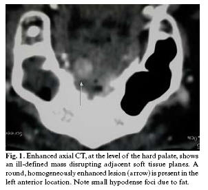
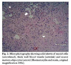
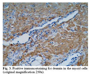
A 52-year-old male with no clinical evidence of TS noticed a small painless lump in the hard palate. Grossly, a 6-mm polypoid and reddish tumour was observed. Contrast-enhanced computed tomography (CT) showed an ill-defined, round, homogeneously enhanced lesion in the hard palate disrupting adjacent soft tissue planes, without lymphadenopathy (<a href="#f1">Fig. 1</a>). A hemangiomatous lesion was suggested, and the tumour was widely excised. On microscopic examination the mucosa appeared bulged by a well-circumscribed but unencapsulated mass composed of an admixture of smooth muscle cells and thick-walled vessels. Irregularly dispersed among the smooth muscle cells were scarce adipocytes singly and in small clusters (<a href="#f2">Fig. 2</a>). Smooth muscle cells stained positively for desmin (<a href="#f3">Fig. 3</a>), muscle specific actin, and h-caldesmon, and adipocytes were positive for S-100 protein. No immunostaining was seen for cytokeratins or HMB45. Histopathological diagnosis was AML.</font></p>     <p align="center"><font face="Verdana" size="2"><a name="f1"><img border="0" src="/img/revistas/medicorpa/v12n5/12.htm34.jpg" width="302" height="276"></a></font></p>     <p align="center"><font face="Verdana" size="2"><a name="f2"><img border="0" src="/img/revistas/medicorpa/v12n5/12.htm35.jpg" width="294" height="269"></a></font></p>     <p align="center"><font face="Verdana" size="2"><a name="f3"><img border="0" src="/img/revistas/medicorpa/v12n5/12.htm36.jpg" width="298" height="244"></a></font></p>     <p>&nbsp;</p>     <p><font face="Verdana"><B>Discussion</B></font></p>     ]]></body>
<body><![CDATA[<p><font face="Verdana" size="2">AML is considered part of a family of tumours arising from perivascular epithelioid cells (PEComas) that show co-expression for melanocytic and muscle markers (1). AML is the most common benign tumour of the kidney, and has been encountered less often in other locations, the liver being the most common. The most frequently reported sites in the head and neck region were the lips, the palate, and buccal mucosa (2-4).</font></p>     <p><font face="Verdana" size="2">AML can occur in association with TS complex, or as an isolated disorder. In about half of the cases renal AMLs are associated with TS, and these tumours are often multiple, bilateral, and associated with cysts. In the oral cavity, only 11 cases, including the present one, have been reported, and only one of them was multiple and associated with TS (4). They differ from renal and hepatic AMLs in that they lack epithelioid cells (2,3,5) and are negative for the monoclonal antibody against immature melanosomes HMB-45 (2,4), conversely to the usual immunopositivity shown in AMLs from other sites (1).</font></p>     <p><font face="Verdana" size="2">Clinically, oral AML could be confused with any other exophytic tumour. AML seldom has been included in the differential diagnosis of a mass of the oral region before surgical excision, but preoperative radiological diagnosis of AML could be possible if intratumoral fat can be demonstrated. The presence of myoid cells, adipose tissue and blood vessels are the characteristic histopathological clues.</font></p>     <p><font face="Verdana" size="2">As the proportions of the components vary considerably from patient to patient, a careful search for adipocytes and abnormal blood vessels may help in the diagnosis because there may be scarce adipose tissue. Strongly positive smooth muscle actin and desmin immunostaining confirm the presence of smooth muscle. From a histological point of view, AML should be distinguished from angiolipoma and angioleiomyoma, which is an encapsulated tumour characterised by vascular spaces surrounded by thick smooth muscle walls. Although small groups of mature fat cells have been rarely reported in some angioleiomyomas, these tumours tend to occur in women, whereas AMLs usually affect older men and are unencapsulated tumours.</font></p>     <p><font face="Verdana" size="2">A wide surgical excision is considered curative, as this tumour usually behaves in a benign fashion (2-5).</font></p>     <p>&nbsp;</p>     <p><font face="Verdana"><b>References</b></font></p>     <!-- ref --><p><font face="Verdana" size="2">1. Eble JN. Angiomyolipoma of kidney. Semin Diagn Pathol 1998;15:21-40</font>&nbsp;&nbsp;&nbsp;&nbsp;&nbsp;&nbsp;&nbsp;&nbsp;[&#160;<a href="javascript:void(0);" onclick="javascript: window.open('/scielo.php?script=sci_nlinks&ref=2976600&pid=S1698-6946200700050001200001&lng=','','width=640,height=500,resizable=yes,scrollbars=1,menubar=yes,');">Links</a>&#160;]<!-- end-ref --><!-- ref --><p><font face="Verdana" size="2">2. Watanabe K, Suzuki T. Mucocutaneous angiomyolipoma. A report of 2 cases arising in the nasal cavity. Arch Pathol Lab Med 1999;123:789-92.</font>&nbsp;&nbsp;&nbsp;&nbsp;&nbsp;&nbsp;&nbsp;&nbsp;[&#160;<a href="javascript:void(0);" onclick="javascript: window.open('/scielo.php?script=sci_nlinks&ref=2976601&pid=S1698-6946200700050001200002&lng=','','width=640,height=500,resizable=yes,scrollbars=1,menubar=yes,');">Links</a>&#160;]<!-- end-ref --><!-- ref --><p><font face="Verdana" size="2">3. Piattelli A, Fioroni M, Rubini C, Fiera E. Angiomyolipoma of the palate. Report of a case. Oral Oncol 2001;37:323-5.</font>&nbsp;&nbsp;&nbsp;&nbsp;&nbsp;&nbsp;&nbsp;&nbsp;[&#160;<a href="javascript:void(0);" onclick="javascript: window.open('/scielo.php?script=sci_nlinks&ref=2976602&pid=S1698-6946200700050001200003&lng=','','width=640,height=500,resizable=yes,scrollbars=1,menubar=yes,');">Links</a>&#160;]<!-- end-ref --><!-- ref --><p><font face="Verdana" size="2">4. López-López J, Rodríguez de Rivera E, Marqués MS, Finestres F, Chimenos E, Roselló X. Tuberous sclerosis and its oral manifestations. A clinical case. Med Oral 2004;9:216-23</font>&nbsp;&nbsp;&nbsp;&nbsp;&nbsp;&nbsp;&nbsp;&nbsp;[&#160;<a href="javascript:void(0);" onclick="javascript: window.open('/scielo.php?script=sci_nlinks&ref=2976603&pid=S1698-6946200700050001200004&lng=','','width=640,height=500,resizable=yes,scrollbars=1,menubar=yes,');">Links</a>&#160;]<!-- end-ref --><!-- ref --><p><font face="Verdana" size="2">5. Tardio JC, Martin-Fragueiro LM. Angiomyolipoma of the nasal cavity Histopathology 2002;41:174-5.</font>&nbsp;&nbsp;&nbsp;&nbsp;&nbsp;&nbsp;&nbsp;&nbsp;[&#160;<a href="javascript:void(0);" onclick="javascript: window.open('/scielo.php?script=sci_nlinks&ref=2976604&pid=S1698-6946200700050001200005&lng=','','width=640,height=500,resizable=yes,scrollbars=1,menubar=yes,');">Links</a>&#160;]<!-- end-ref --><p>&nbsp;</p>     <p>&nbsp;</p>     <p><font face="Verdana" size="2"><b><a href="#top"><img border="0" src="/img/revistas/medicorpa/v12n5/seta.gif" width="15" height="17"></a> <a name="bajo"></a>Correspondence:</b>    <BR>Dr. Carlos Alvarez Alvarez    <BR>Servicio de Anatomía Patológica. Hospital Montecelo    <BR>Mourente s/n    <BR>36071 Pontevedra.España    <BR>E-mail: <a href="mailto:capialvarez@yahoo.com">capialvarez@yahoo.com</a></font></p>     ]]></body>
<body><![CDATA[<p><font face="Verdana" size="2">Received: 8-02-2006    <BR>Accepted: 21-05-2006</font></p>      ]]></body><back>
<ref-list>
<ref id="B1">
<label>1</label><nlm-citation citation-type="journal">
<person-group person-group-type="author">
<name>
<surname><![CDATA[Eble]]></surname>
<given-names><![CDATA[JN]]></given-names>
</name>
</person-group>
<article-title xml:lang="en"><![CDATA[Angiomyolipoma of kidney]]></article-title>
<source><![CDATA[Semin Diagn Pathol]]></source>
<year>1998</year>
<volume>15</volume>
<page-range>21-40</page-range></nlm-citation>
</ref>
<ref id="B2">
<label>2</label><nlm-citation citation-type="journal">
<person-group person-group-type="author">
<name>
<surname><![CDATA[Watanabe]]></surname>
<given-names><![CDATA[K]]></given-names>
</name>
<name>
<surname><![CDATA[Suzuki]]></surname>
<given-names><![CDATA[T]]></given-names>
</name>
</person-group>
<article-title xml:lang="en"><![CDATA[Mucocutaneous angiomyolipoma: A report of 2 cases arising in the nasal cavity]]></article-title>
<source><![CDATA[Arch Pathol Lab Med]]></source>
<year>1999</year>
<volume>123</volume>
<page-range>789-92</page-range></nlm-citation>
</ref>
<ref id="B3">
<label>3</label><nlm-citation citation-type="journal">
<person-group person-group-type="author">
<name>
<surname><![CDATA[Piattelli]]></surname>
<given-names><![CDATA[A]]></given-names>
</name>
<name>
<surname><![CDATA[Fioroni]]></surname>
<given-names><![CDATA[M]]></given-names>
</name>
<name>
<surname><![CDATA[Rubini]]></surname>
<given-names><![CDATA[C]]></given-names>
</name>
<name>
<surname><![CDATA[Fiera]]></surname>
<given-names><![CDATA[E]]></given-names>
</name>
</person-group>
<article-title xml:lang="en"><![CDATA[Angiomyolipoma of the palate: Report of a case]]></article-title>
<source><![CDATA[Oral Oncol]]></source>
<year>2001</year>
<volume>37</volume>
<page-range>323-5</page-range></nlm-citation>
</ref>
<ref id="B4">
<label>4</label><nlm-citation citation-type="journal">
<person-group person-group-type="author">
<name>
<surname><![CDATA[López-López]]></surname>
<given-names><![CDATA[J]]></given-names>
</name>
<name>
<surname><![CDATA[Rodríguez de Rivera]]></surname>
<given-names><![CDATA[E]]></given-names>
</name>
<name>
<surname><![CDATA[Marqués]]></surname>
<given-names><![CDATA[MS]]></given-names>
</name>
<name>
<surname><![CDATA[Finestres]]></surname>
<given-names><![CDATA[F]]></given-names>
</name>
<name>
<surname><![CDATA[Chimenos]]></surname>
<given-names><![CDATA[E]]></given-names>
</name>
<name>
<surname><![CDATA[Roselló]]></surname>
<given-names><![CDATA[X]]></given-names>
</name>
</person-group>
<article-title xml:lang="en"><![CDATA[Tuberous sclerosis and its oral manifestations: A clinical case]]></article-title>
<source><![CDATA[Med Oral]]></source>
<year>2004</year>
<volume>9</volume>
<page-range>216-23</page-range></nlm-citation>
</ref>
<ref id="B5">
<label>5</label><nlm-citation citation-type="journal">
<person-group person-group-type="author">
<name>
<surname><![CDATA[Tardio]]></surname>
<given-names><![CDATA[JC]]></given-names>
</name>
<name>
<surname><![CDATA[Martin-Fragueiro]]></surname>
<given-names><![CDATA[LM]]></given-names>
</name>
</person-group>
<article-title xml:lang="en"><![CDATA[Angiomyolipoma of the nasal cavity]]></article-title>
<source><![CDATA[Histopathology]]></source>
<year>2002</year>
<volume>41</volume>
<page-range>174-5</page-range></nlm-citation>
</ref>
</ref-list>
</back>
</article>
