<?xml version="1.0" encoding="ISO-8859-1"?><article xmlns:mml="http://www.w3.org/1998/Math/MathML" xmlns:xlink="http://www.w3.org/1999/xlink" xmlns:xsi="http://www.w3.org/2001/XMLSchema-instance">
<front>
<journal-meta>
<journal-id>1130-0108</journal-id>
<journal-title><![CDATA[Revista Española de Enfermedades Digestivas]]></journal-title>
<abbrev-journal-title><![CDATA[Rev. esp. enferm. dig.]]></abbrev-journal-title>
<issn>1130-0108</issn>
<publisher>
<publisher-name><![CDATA[Sociedad Española de Patología Digestiva]]></publisher-name>
</publisher>
</journal-meta>
<article-meta>
<article-id>S1130-01082011000300003</article-id>
<title-group>
<article-title xml:lang="en"><![CDATA[Definitive diagnosis of neuroendocrine tumors using fine-needle aspiration-puncture guided by endoscopic ultrasonography]]></article-title>
<article-title xml:lang="es"><![CDATA[Diagnóstico definitivo de los tumores neuroendocrinos (TNE) mediante PAAF ecodirigida por ultrasonografía endoscópica (USE)]]></article-title>
</title-group>
<contrib-group>
<contrib contrib-type="author">
<name>
<surname><![CDATA[Gornals]]></surname>
<given-names><![CDATA[Joan]]></given-names>
</name>
<xref ref-type="aff" rid="A01"/>
<xref ref-type="aff" rid="A02"/>
<xref ref-type="aff" rid="A03"/>
</contrib>
<contrib contrib-type="author">
<name>
<surname><![CDATA[Varas]]></surname>
<given-names><![CDATA[Modesto]]></given-names>
</name>
<xref ref-type="aff" rid="A03"/>
</contrib>
<contrib contrib-type="author">
<name>
<surname><![CDATA[Catalá]]></surname>
<given-names><![CDATA[Isabel]]></given-names>
</name>
<xref ref-type="aff" rid="A01"/>
</contrib>
<contrib contrib-type="author">
<name>
<surname><![CDATA[Maisterra]]></surname>
<given-names><![CDATA[Sandra]]></given-names>
</name>
<xref ref-type="aff" rid="A01"/>
</contrib>
<contrib contrib-type="author">
<name>
<surname><![CDATA[Pons]]></surname>
<given-names><![CDATA[Carlos]]></given-names>
</name>
<xref ref-type="aff" rid="A01"/>
</contrib>
<contrib contrib-type="author">
<name>
<surname><![CDATA[Bargalló]]></surname>
<given-names><![CDATA[Domingo]]></given-names>
</name>
<xref ref-type="aff" rid="A02"/>
</contrib>
<contrib contrib-type="author">
<name>
<surname><![CDATA[Serrano]]></surname>
<given-names><![CDATA[Teresa]]></given-names>
</name>
<xref ref-type="aff" rid="A01"/>
</contrib>
<contrib contrib-type="author">
<name>
<surname><![CDATA[Fabregat]]></surname>
<given-names><![CDATA[Joan]]></given-names>
</name>
<xref ref-type="aff" rid="A01"/>
</contrib>
</contrib-group>
<aff id="A01">
<institution><![CDATA[,Hospital Universitario de Bellvitge Service of Digestive Diseases, Pathology, and Digestive and General Surgery Department of Enchoendoscopy]]></institution>
<addr-line><![CDATA[Hospitalet de Llobregat ]]></addr-line>
<country>Spain</country>
</aff>
<aff id="A02">
<institution><![CDATA[,Centro Médico Delfos Department of Echoendoscopy ]]></institution>
<addr-line><![CDATA[Barcelona ]]></addr-line>
<country>Spain</country>
</aff>
<aff id="A03">
<institution><![CDATA[,Centro Médico Teknon  ]]></institution>
<addr-line><![CDATA[Barcelona ]]></addr-line>
<country>Spain</country>
</aff>
<pub-date pub-type="pub">
<day>00</day>
<month>03</month>
<year>2011</year>
</pub-date>
<pub-date pub-type="epub">
<day>00</day>
<month>03</month>
<year>2011</year>
</pub-date>
<volume>103</volume>
<numero>3</numero>
<fpage>123</fpage>
<lpage>128</lpage>
<copyright-statement/>
<copyright-year/>
<self-uri xlink:href="http://scielo.isciii.es/scielo.php?script=sci_arttext&amp;pid=S1130-01082011000300003&amp;lng=en&amp;nrm=iso"></self-uri><self-uri xlink:href="http://scielo.isciii.es/scielo.php?script=sci_abstract&amp;pid=S1130-01082011000300003&amp;lng=en&amp;nrm=iso"></self-uri><self-uri xlink:href="http://scielo.isciii.es/scielo.php?script=sci_pdf&amp;pid=S1130-01082011000300003&amp;lng=en&amp;nrm=iso"></self-uri><abstract abstract-type="short" xml:lang="en"><p><![CDATA[Background: the detection and diagnosis of neuroendocrine tumors (NETs) is challenging. Endoscopic ultrasonography (EUS) has a significant role in the detection of NETs suspected from clinical manifestations or imaging techniques, as well as in their precise localization and cytological confirmation using EUS-Fine-needle aspiration-puncture (FNA). Objective: to assess the usefulness and precision of EUS-FNAP in the differential diagnosis and confirmation of NETs, in a retrospective review of our experience. Patients and methods: in a total of 55 patients with suspected NETs who underwent radial or sectorial EUS, 42 tumors were detected in 40 cases. EUS-FNA using a 22G needle was performed for 16 cases with suspected functional (hormonal disorders: 6 cases) and non-functional NETs (10 cases). Ki 67 or immunocytochemistry (ICC) testing was performed for all. There was confirmation in 9 cases (5 female and 4 male) with a mean age of 51 years (range: 41-81 years). All tumors were located in the pancreas except for one in the mediastinum and one in the rectum, with a mean size of 19 mm (range: 10-40 mm). Results: there were no complications attributable to FNA. Sensitivity was 100% and both precision and PPV were 89%, as a false positive result suggested a diagnosis with NET during cytology that surgery finally revealed to be a pancreatic pseudopapillary solid tumor. Conclusions: EUS-FNA with a 22G needle for NETs has high sensitivity and PPV at cytological confirmation with few complications.]]></p></abstract>
<abstract abstract-type="short" xml:lang="es"><p><![CDATA[Introducción: la localización y diagnóstico de los tumores neuroendocrinos (TNE) es difícil. La ultrasonografía endoscópica (USE) tiene un papel significativo en la detección de TNE sospechados por la clínica u otras técnicas de imagen, así como en la localización exacta y confirmación citológica mediante USE-PAAF. Objetivo: valorar la utilidad y precisión de la PAAF-USE en el diagnóstico diferencial y de confirmación de los TNE, en una revisión retrospectiva de la experiencia de nuestro grupo. Pacientes y métodos: de un total de 55 enfermos con sospecha de TNE a los que se le practicó USE radial o sectorial, se detectaron 42 tumores en 40 casos. En 16 casos con sospecha de TNE funcionantes (trastornos hormonales: 6 casos) y no funcionantes (10 casos), se les practicó USE-PAAF con 22 G. En todos se efectuó Ki 67 o inmunocitoquímica (ICQ). Hubo confirmación quirúrgica en 9 casos (5 M y 4 V), con una media de edad de 51 años (rango: 41-81 años). Los tumores se localizaban todos en el páncreas, excepto uno en el mediastino y uno en el recto, con un tamaño medio de 19 mm (rango: 10-40 mm). Resultados: no hubo complicaciones atribuibles a la PAAF. La sensibilidad fue del 100% (8/8), y la precisión y el VPP fue del 89% (8/9), ya que hubo un falso positivo que en el estudio citológico sugirió el diagnóstico de TNE pero que su resección quirúrgica confirmó el diagnóstico de tumor sólido seudopapilar del páncreas. Conclusiones: la USE-PAAF con 22 G de los TNE posee una alta sensibilidad y VPP en la confirmación citológica de estos pacientes, con muy escasas complicaciones.]]></p></abstract>
<kwd-group>
<kwd lng="en"><![CDATA[Fine-needle aspiration-puncture (FNA) guided by endoscopic ultrasonography (EUS) or echoendoscopy, neuroendocrine tumors (NETs)]]></kwd>
<kwd lng="en"><![CDATA[Pancreatic endocrine tumors (PETs)]]></kwd>
<kwd lng="en"><![CDATA[Immunocytochemistry]]></kwd>
<kwd lng="en"><![CDATA[Immunohistochemistry]]></kwd>
<kwd lng="en"><![CDATA[Chromogranin]]></kwd>
<kwd lng="en"><![CDATA[Synaptophysin]]></kwd>
<kwd lng="en"><![CDATA[Cytokeratin 19]]></kwd>
<kwd lng="en"><![CDATA[Vimentin]]></kwd>
<kwd lng="en"><![CDATA[Ki 67]]></kwd>
<kwd lng="en"><![CDATA[CD56]]></kwd>
<kwd lng="es"><![CDATA[Punción aspiración con aguja fina (PAAF) por ultrasonografía endoscópica (USE) o por ecoendoscopia. Tumores neuroendocrinos (TNE)]]></kwd>
<kwd lng="es"><![CDATA[Tumores endocrinos pancreáticos (TEP)]]></kwd>
<kwd lng="es"><![CDATA[Inmunocitoquímica]]></kwd>
<kwd lng="es"><![CDATA[Inmunohistoquímica]]></kwd>
<kwd lng="es"><![CDATA[Cromogranina]]></kwd>
<kwd lng="es"><![CDATA[Sinaptofisina]]></kwd>
<kwd lng="es"><![CDATA[Citoqueratina 19]]></kwd>
<kwd lng="es"><![CDATA[Vimentina]]></kwd>
<kwd lng="es"><![CDATA[Ki 67]]></kwd>
<kwd lng="es"><![CDATA[CD56]]></kwd>
</kwd-group>
</article-meta>
</front><body><![CDATA[ <p><a name="top"></a><font face="Verdana" size="2"><b>ORIGINAL PAPERS</b></font></p>     <p>&nbsp;</p>     <p><font face="Verdana" size="4"><b>Definitive diagnosis of neuroendocrine tumors using fine-needle aspiration-puncture guided by endoscopic ultrasonography</b></font></p>     <p><font face="Verdana" size="4"><b>Diagn&oacute;stico definitivo de los tumores neuroendocrinos (TNE) mediante PAAF ecodirigida por ultrasonograf&iacute;a endosc&oacute;pica (USE)</b></font></p>     <p>&nbsp;</p>     <p>&nbsp;</p>     <p><font face="Verdana" size="2"><b>Joan Gornals<sup>1,2,3</sup>, Modesto Varas<sup>3</sup>, Isabel Catal&aacute;<sup>1</sup>, Sandra Maisterra<sup>1</sup>, Carlos Pons<sup>1</sup>, Domingo Bargall&oacute;<sup>2</sup>, Teresa Serrano<sup>1</sup> and Joan Fabregat<sup>1</sup></b></font></p>     <p><font face="Verdana" size="2"><sup>1</sup>Department of Enchoendoscopy. Service of Digestive Diseases, Pathology, and Digestive and General Surgery. Hospital Universitario de Bellvitge. Hospitalet de Llobregat, Barcelona. Spain.    <br><sup>2</sup>Department of Echoendoscopy. Centro M&eacute;dico Delfos. Barcelona, Spain.    <br><sup>3</sup>Centro M&eacute;dico Teknon. Barcelona, Spain</font></p>     ]]></body>
<body><![CDATA[<p><font face="Verdana" size="2"><a href="#bajo">Correspondence</a></font></p>     <p>&nbsp;</p>     <p>&nbsp;</p> <hr size="1">     <p><font face="Verdana" size="2"><b>ABSTRACT</b></font></p>     <p><font face="Verdana" size="2"><b>Background:</b> the detection and diagnosis of neuroendocrine tumors (NETs) is challenging. Endoscopic ultrasonography (EUS) has a significant role in the detection of NETs suspected from clinical manifestations or imaging techniques, as well as in their precise localization and cytological confirmation using EUS-Fine-needle aspiration-puncture (FNA).    <br><b>Objective:</b> to assess the usefulness and precision of EUS-FNAP in the differential diagnosis and confirmation of NETs, in a retrospective review of our experience.    <br><b>Patients and methods:</b> in a total of 55 patients with suspected NETs who underwent radial or sectorial EUS, 42 tumors were detected in 40 cases. EUS-FNA using a 22G needle was performed for 16 cases with suspected functional (hormonal disorders: 6 cases) and non-functional NETs (10 cases). Ki 67 or immunocytochemistry (ICC) testing was performed for all.    <br>There was confirmation in 9 cases (5 female and 4 male) with a mean age of 51 years (range: 41-81 years).    <br>All tumors were located in the pancreas except for one in the mediastinum and one in the rectum, with a mean size of 19 mm (range: 10-40 mm).    <br><b>Results:</b> there were no complications attributable to FNA. Sensitivity was 100% and both precision and PPV were 89%, as a false positive result suggested a diagnosis with NET during cytology that surgery finally revealed to be a pancreatic pseudopapillary solid tumor.    ]]></body>
<body><![CDATA[<br><b>Conclusions:</b> EUS-FNA with a 22G needle for NETs has high sensitivity and PPV at cytological confirmation with few complications.</font></p>     <p><font face="Verdana" size="2"><b>Key words:</b> Fine-needle aspiration-puncture (FNA) guided by endoscopic ultrasonography (EUS) or echoendoscopy, neuroendocrine tumors (NETs). Pancreatic endocrine tumors (PETs). Immunocytochemistry. Immunohistochemistry. Chromogranin. Synaptophysin. Cytokeratin 19. Vimentin. Ki 67. CD56.</font></p> <hr size="1">     <p><font face="Verdana" size="2"><b>RESUMEN</b></font></p>     <p><font face="Verdana" size="2"><b>Introducci&oacute;n:</b> la localizaci&oacute;n y diagn&oacute;stico de los tumores neuroendocrinos (TNE) es dif&iacute;cil. La ultrasonograf&iacute;a endosc&oacute;pica (USE) tiene un papel significativo en la detecci&oacute;n de TNE sospechados por la cl&iacute;nica u otras t&eacute;cnicas de imagen, as&iacute; como en la localizaci&oacute;n exacta y confirmaci&oacute;n citol&oacute;gica mediante USE-PAAF.    <br><b>Objetivo:</b> valorar la utilidad y precisi&oacute;n de la PAAF-USE en el diagn&oacute;stico diferencial y de confirmaci&oacute;n de los TNE, en una revisi&oacute;n retrospectiva de la experiencia de nuestro grupo.    <br><b>Pacientes y m&eacute;todos:</b> de un total de 55 enfermos con sospecha de TNE a los que se le practic&oacute; USE radial o sectorial, se detectaron 42 tumores en 40 casos. En 16 casos con sospecha de TNE funcionantes (trastornos hormonales: 6 casos) y no funcionantes (10 casos), se les practic&oacute; USE-PAAF con 22 G. En todos se efectu&oacute; Ki 67 o inmunocitoqu&iacute;mica (ICQ).    <br>Hubo confirmaci&oacute;n quir&uacute;rgica en 9 casos (5 M y 4 V), con una media de edad de 51 a&ntilde;os (rango: 41-81 a&ntilde;os).    <br>Los tumores se localizaban todos en el p&aacute;ncreas, excepto uno en el mediastino y uno en el recto, con un tama&ntilde;o medio de 19 mm (rango: 10-40 mm).    <br><b>Resultados:</b> no hubo complicaciones atribuibles a la PAAF. La sensibilidad fue del 100% (8/8), y la precisi&oacute;n y el VPP fue del 89% (8/9), ya que hubo un falso positivo que en el estudio citol&oacute;gico sugiri&oacute; el diagn&oacute;stico de TNE pero que su resecci&oacute;n quir&uacute;rgica confirm&oacute; el diagn&oacute;stico de tumor s&oacute;lido seudopapilar del p&aacute;ncreas.    <br><b>Conclusiones:</b> la USE-PAAF con 22 G de los TNE posee una alta sensibilidad y VPP en la confirmaci&oacute;n citol&oacute;gica de estos pacientes, con muy escasas complicaciones.</font></p>     ]]></body>
<body><![CDATA[<p><font face="Verdana" size="2"><b>Palabras clave:</b> Punci&oacute;n aspiraci&oacute;n con aguja fina (PAAF) por ultrasonograf&iacute;a endosc&oacute;pica (USE) o por ecoendoscopia. Tumores neuroendocrinos (TNE). Tumores endocrinos pancre&aacute;ticos (TEP). Inmunocitoqu&iacute;mica. Inmunohistoqu&iacute;mica. Cromogranina. Sinaptofisina. Citoqueratina 19. Vimentina. Ki 67. CD56.</font></p> <hr size="1">     <p>&nbsp;</p>     <p><font face="Verdana" size="2"><b>Introduction</b></font></p>     <p><font face="Verdana" size="2">The preoperative diagnosis and precise localization of neuroendocrine tumors (NETs), particularly pancreatic NETs (PNETs), is challenging, and vital for a definitive cure of patients (1). For non-functioning cases, confirmation by histology is most necessary because of potential differential diagnoses. PNETs share histological properties with carcinoids: both are considered to derive from the diffuse endocrine cell system; they unusually exhibit mitotic features (assessable using the Ki-67 -index); they usually show electrodense granules that contain hormones and various peptides, chromogranins (A, B, C), neuron-specific enolase (NSE), and synaptophysin (2,3).</font></p>     <p><font face="Verdana" size="2">PNETs are clinically classified as functional (Zollinger-Ellison syndrome, etc.) and non-functional. The clinical diagnosis of functional PNETs is relatively straightforward.</font></p>     <p><font face="Verdana" size="2">Most are benign (no metastases) and small, and may be associated with multiple endocrine neoplasia (MEN). Non-functional tumors are most common among PNETs, and have a high incidence of metastatic disease.</font></p>     <p><font face="Verdana" size="2">Their precise localization in the pancreas is difficult. Echoendoscopy or endoscopic ultrasonography (EUS) is a rather recently introduced diagnostic technique, and may diagnose tumors smaller than 1 cm (up to 3 mm) in the pancreas head and body with a sensitivity above 85% (93% in the larger series), whereas those in the tail are harder to assess (1).</font></p>     <p><font face="Verdana" size="2">EUS allows fine-needle aspiration-puncture (FNA) under ultrasound (US) guidance (4), and the collection of material for cytology and histology with a yield nearing 90%. In addition, immunocytochemistry (ICC) and immunohistochemistry (IHC) tests may be performed on obtained samples for chromogranin (C-A), synaptophysin, cytokeratin 19, and various hormones or peptides, with diagnoses that may reach 100% for cystic PNETs (5).</font></p>     <p><font face="Verdana" size="2">A recent classification proposed by WHO (2) assigned three categories to NETs: well-differentiated tumor, well-differentiated carcinoma, and poorly differentiated carcinoma based on histology, size (limit: 2 cm), and proliferation index (Ki-67 = 2%).</font></p>     <p><font face="Verdana" size="2">A TNM (tumor, node, and metastasis) classification has also been suggested for PNETs based on the WHO classification (3).</font></p>     ]]></body>
<body><![CDATA[<p>&nbsp;</p>     <p><font face="Verdana" size="2"><b>Objective</b></font></p>     <p><font face="Verdana" size="2">To assess the usefulness and precision of EUS-FNA in the differential and confirmatory diagnosis of NETs using a retrospective review of our team's experience.</font></p>     <p>&nbsp;</p>     <p><font face="Verdana" size="2"><b>Patients and method</b></font></p>     <p><font face="Verdana" size="2">For a total of 55 patients with suspected PNETs who underwent radial or sectorial EUS, 42 tumors were identified in 40 patients. Inclusion criteria for EUS-FNA: patients with presumed NET diagnosis with EUS, uncertain or non-functional.</font></p>     <p><font face="Verdana" size="2">For 16 cases (8 women and 8 men with a mean age of 56, range: 41-92 years with suspected functional (6 cases) and non-functional (10 cases) tumors, none of them cystic, EUS-FNA was performed using a 22 G needle (Echotip Ultra, Cook Medical) with conventional technique. All cases underwent Ki67 testing or immunocytochemistry for chromogranin, synaptophysin, and various hormones or peptides.</font></p>     <p><font face="Verdana" size="2">There was surgical confirmation (the gold standard) in 9 patients; in the remaining cases imaging techniques and 12-month follow-up (the gold standard) were used to reach a definitive diagnosis.</font></p>     <p><font face="Verdana" size="2">From all 16 patients 9 (5 women, 4 men) were selected with a mean age of 51 years (range: 41-81 years).</font></p>     <p><font face="Verdana" size="2">All tumors were in the pancreas, and one was in the mediastinum and one in the rectum, with a mean size of 19 mm (range: 10 to 40 mm) (<a target="_blank" href="/img/revistas/diges/v103n3/original2_t1.jpg">Table I</a>).</font></p>     ]]></body>
<body><![CDATA[<p><font face="Verdana" size="2">Regarding pancreatic tumors, three were in the head, two in the tail, and two in the body. Only two patients had metastases.</font></p>     <p><font face="Verdana" size="2">All examinations (EUS-FNA) were performed after collecting an informed consent, with prior coagulation testing, and using sedation (propofol) by an anesthetist.</font></p>     <p><font face="Verdana" size="2">A cytologist was in all cases present in the examination room where EUS-FNA procedures were carried out.</font></p>     <p><font face="Verdana" size="2">Diagnostic precision (P), sensitivity (S), specificity (Sp), positive predictive value (PPV) and negative predictive value (NPV) were all analyzed using standard formulas.</font></p>     <p>&nbsp;</p>     <p><font face="Verdana" size="2"><b>Results</b></font></p>     <p><font face="Verdana" size="2">There were no EUS-FNA-related complications (hemorrhage and perforation).</font></p>     <p><font face="Verdana" size="2">In the total series (16 cases) S was 100% with a Sp of 67%, P and PPV of 93 and 92%, respectively.</font></p>     <p><font face="Verdana" size="2">In patients with surgical confirmation (9 cases) sensitivity (S) was 100%, and precision (P) and PPV were 89%, as cytology yielded a false positive result that was eventually diagnosed as a solid pancreatic pseudopapillary tumor following surgical excision and tail pancreatectomy plus IHC.</font></p>     <p>&nbsp;</p>     ]]></body>
<body><![CDATA[<p><font face="Verdana" size="2"><b>Discussion</b></font></p>     <p><font face="Verdana" size="2">EUS-FNA has been performed for PNETs for slightly over 10 years now. In earlier works both sensitivity and precision were low, with a specificity of 100% (6); however, they gradually increased, and sensitivity reached about 90% (94% in the most extensive series in the literature) (6-22) (<a target="_blank" href="/img/revistas/diges/v103n3/original2_t2.jpg">Table II</a>).</font></p>     <p><font face="Verdana" size="2">Our findings are consistent with those in the literature (S: 100%).</font></p>     <p><font face="Verdana" size="2">Typical EUS findings include homogeneous pancreatic nodules or lesions that are hypoechogenic, solid, hypervascular, and encapsulated with well-delimited borders (1,22,29), even non-functional ones (most of them) (22). NFPETs show the greatest sizes and are more advanced (<a target="_blank" href="/img/revistas/diges/v103n3/original2_f1.jpg">Fig. 1</a>).</font></p>     <p><font face="Verdana" size="2">
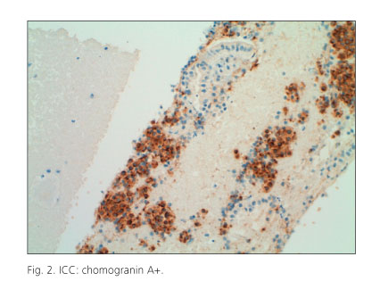
The use of ICC techniques (chromogranin, synaptophysin, etc.) (cytokeratin 19) (23) considerably improves sensitivity on cytology material (<a href="#f2">Fig. 2</a>).</font></p>     <p align="center"><font face="Verdana" size="2"><a name="f2"><img src="/img/revistas/diges/v103n3/original2_f2.jpg" width="426" height="334"></a></font></p>     <p><font face="Verdana" size="2">The Ki 67 index (24-26) and microsatellite instability have also been assessed in samples (27,28) to establish the benign or malignant nature of tumors, and hence their prognosis.</font></p>     <p><font face="Verdana" size="2">Algorithms are similar for PNETs and pancreatic cancers (PCs) (4,29) (<a target="_blank" href="/img/revistas/diges/v103n3/original2_f3.jpg">Fig. 3</a>).</font></p>     <p><font face="Verdana" size="2">When a tumor is resectable according to computed tomography plus EUS, and both clinical and morphological features are consistent, laparoscopic or open surgery may be readily performed. For uncertain or non-functioning tumors EUS-FNA may be used to confirm diagnostic suspicion.</font></p>     <p><font face="Verdana" size="2">Sometimes a histological differential diagnosis is -difficult between pancreatic endocrine tumors, solid pseudopapillary tumor, acinar cell carcinomas, mucinous tumors, and lymphoma/plasmocytoma. In recent years various cases of solid pseudopapillary tumor have been described where ICC reached the right diagnosis on EUS-FNA-collected samples (30-33): most were vimentin+ and cytokeratin+, whereas chomogranin and NSE were negative (they may be focally positive though) (34).</font></p>     ]]></body>
<body><![CDATA[<p><font face="Verdana" size="2">In this multicenter study in 28 patients with pseudopapillary tumors (34) a preoperative diagnosis was reached for 21 cases (75%); vimentin, alfa1-antitrypsin, CD10, and beta-catenin were positive in all cases, whereas chromogranin was positive in just 1/20 (5%) and synaptophysin in 10/17 (59%); however, the best marker to tell endocrine -tumors from solid pseudopapillary tumors was E-cadherin/B-catenin/CD10 according to a recent study (35).</font></p>     <p><font face="Verdana" size="2">In our case with a solid pancreatic pseudopapillary tumor IHC was key for a definitive diagnosis. False positive results have been described in other series (12,18).</font></p>     <p><font face="Verdana" size="2">A recent Japanese study (36) reviewed 455 pancreatic FNA procedures: 28 were rare pancreatic tumors (no ductal adenocarcinomas). EUS-FNA with cytology, cell-block, and immunocytochemistry correctly diagnosed tumor type in 19 patients 19 (68%).</font></p>     <p><font face="Verdana" size="2">In differentiating benign from malignant tumors it had a sensitivity of 69%, a specificity of 100%, a PPV of 100%, a NPV of 79%, and a precision of 86%. None of the three malignant pancreatic endocrine tumors was diagnosed as such. EUS-FNA changed the presumed diag-nosis in 11 cases (39%).</font></p>     <p><font face="Verdana" size="2">Four cases have been recently reported (37) where small (8-16 mm), non-functioning pancreatic endocrine tumors were found together with intraductal papillary mucinous neoplasms. PNETs remained undetected by common imaging techniques (CT and MRI); 3/4 were diag-nosed using EUS, and only 1/3 using EUS-FNA.</font></p>     <p><font face="Verdana" size="2">To conclude, ICC on cytology samples collected by EUS-FNA is key for a definitive diagnosis of PNETs. Our study (S: 100%) (PPV: 89%) confirmed the findings in the literature (mean sensitivity of 94%, mean specificity of 95%) (<a target="_blank" href="/img/revistas/diges/v103n3/original2_t2.jpg">Table II</a>).</font></p>     <p><font face="Verdana" size="2">Notwithstanding, the diagnostic panel is increasingly greater, and novel markers emerge including SERPINB8 (38), which is as sensitive as C-A and synaptophysin, or CDX-2, PDX-1, NESP-55 and TTF-1, which may help in the differential diagnosis between gastrointestinal and pulmonary carcinoids, and pancreatic endocrine tumors (39), with CK 19 being an independent prognostic factor for PNETs, particularly non-insulinomas according to a recent review (40). However, chromogranin and synaptophysin remain the key markers since many years ago (43) to this day (44).</font></p>     <p><font face="Verdana" size="2">Thus, believe that ICC is key for a definitive diagnosis of NETs (45), a statement not fully shared by other teams (44).</font></p>     <p>&nbsp;</p>     <p><font face="Verdana" size="2"><b>References</b></font></p>     ]]></body>
<body><![CDATA[<!-- ref --><p><font face="Verdana" size="2">1. Varas MJ, Miquel JM, Maluenda MD, et al. Preoperative detection of gastrointestinal neuroendocrine tumors using endoscopic ultrasonography. Rev Esp Enferm Dig 2006;98:828-36.    &nbsp;&nbsp;&nbsp;&nbsp;&nbsp;&nbsp;&nbsp;&nbsp;[&#160;<a href="javascript:void(0);" onclick="javascript: window.open('/scielo.php?script=sci_nlinks&ref=5310355&pid=S1130-0108201100030000300001&lng=','','width=640,height=500,resizable=yes,scrollbars=1,menubar=yes,');">Links</a>&#160;]<!-- end-ref --></font></p>    <!-- ref --><p><font face="Verdana" size="2">2. Kloppel G. Tumour biology and histopathology of neuroendocrine tumours. Best Pract Res Clin Endocrinol Metab 2007;21:15-31.    &nbsp;&nbsp;&nbsp;&nbsp;&nbsp;&nbsp;&nbsp;&nbsp;[&#160;<a href="javascript:void(0);" onclick="javascript: window.open('/scielo.php?script=sci_nlinks&ref=5310357&pid=S1130-0108201100030000300002&lng=','','width=640,height=500,resizable=yes,scrollbars=1,menubar=yes,');">Links</a>&#160;]<!-- end-ref --></font></p>    <!-- ref --><p><font face="Verdana" size="2">3. Metz DC, Jensen RT. Gastrointestinal neuroendocrine tumors: pancreatic endocrine tumors. Gastroenterology 2008;135:1469-92.    &nbsp;&nbsp;&nbsp;&nbsp;&nbsp;&nbsp;&nbsp;&nbsp;[&#160;<a href="javascript:void(0);" onclick="javascript: window.open('/scielo.php?script=sci_nlinks&ref=5310359&pid=S1130-0108201100030000300003&lng=','','width=640,height=500,resizable=yes,scrollbars=1,menubar=yes,');">Links</a>&#160;]<!-- end-ref --></font></p>    <!-- ref --><p><font face="Verdana" size="2">4. McLean AM, Fairclough PD. Endoscopic ultrasound in the localization of pancreatic islet cell tumours. Best Pract Res Clin Endocrinol Metab 2005; 19:177-93.    &nbsp;&nbsp;&nbsp;&nbsp;&nbsp;&nbsp;&nbsp;&nbsp;[&#160;<a href="javascript:void(0);" onclick="javascript: window.open('/scielo.php?script=sci_nlinks&ref=5310361&pid=S1130-0108201100030000300004&lng=','','width=640,height=500,resizable=yes,scrollbars=1,menubar=yes,');">Links</a>&#160;]<!-- end-ref --></font></p>    <!-- ref --><p><font face="Verdana" size="2">5. Varas MJ. Neuroendocrine tumors -fascination and infrequency. Rev Esp Enferm Dig 2009;101:195-208.    &nbsp;&nbsp;&nbsp;&nbsp;&nbsp;&nbsp;&nbsp;&nbsp;[&#160;<a href="javascript:void(0);" onclick="javascript: window.open('/scielo.php?script=sci_nlinks&ref=5310363&pid=S1130-0108201100030000300005&lng=','','width=640,height=500,resizable=yes,scrollbars=1,menubar=yes,');">Links</a>&#160;]<!-- end-ref --></font></p>    ]]></body>
<body><![CDATA[<!-- ref --><p><font face="Verdana" size="2">6. Ciaccia D, Harada N, Wiersema MJ, et al. Preoperative localization and diagnosis of pancreatic and pancreatic islet cell tumors using endoscopic ultrasound guided fine needle aspiration: a multicenter experience. Gastrointest Endosc 1997;45:AB170.    &nbsp;&nbsp;&nbsp;&nbsp;&nbsp;&nbsp;&nbsp;&nbsp;[&#160;<a href="javascript:void(0);" onclick="javascript: window.open('/scielo.php?script=sci_nlinks&ref=5310365&pid=S1130-0108201100030000300006&lng=','','width=640,height=500,resizable=yes,scrollbars=1,menubar=yes,');">Links</a>&#160;]<!-- end-ref --></font></p>    <!-- ref --><p><font face="Verdana" size="2">7. Voss M, Hammel P, Molas G, et al. Value of endoscopic ultrasound guided fine needle aspiration biopsy in the diagnosis of solid pancreatic masses. Gut 2000;46:244-9.    &nbsp;&nbsp;&nbsp;&nbsp;&nbsp;&nbsp;&nbsp;&nbsp;[&#160;<a href="javascript:void(0);" onclick="javascript: window.open('/scielo.php?script=sci_nlinks&ref=5310367&pid=S1130-0108201100030000300007&lng=','','width=640,height=500,resizable=yes,scrollbars=1,menubar=yes,');">Links</a>&#160;]<!-- end-ref --></font></p>    <!-- ref --><p><font face="Verdana" size="2">8. Gress PG, Barawi M, Kim D, et al. Preoperative localization of a neuroendocrine tumors of the pancreas with EUS-guided fine needle tattooing. Gastrointest Endosc 2002;55:594-7.    &nbsp;&nbsp;&nbsp;&nbsp;&nbsp;&nbsp;&nbsp;&nbsp;[&#160;<a href="javascript:void(0);" onclick="javascript: window.open('/scielo.php?script=sci_nlinks&ref=5310369&pid=S1130-0108201100030000300008&lng=','','width=640,height=500,resizable=yes,scrollbars=1,menubar=yes,');">Links</a>&#160;]<!-- end-ref --></font></p>    <!-- ref --><p><font face="Verdana" size="2">9. Jhala D, Eloubeidi M, Chhieng DC, et al. Fine needle aspiration biopsy of the islet cell tumor of pancreas: a comparison between computerized axial tomography and endoscopic ultrasound-guided fine needle aspiration biopsy. Ann Diagn Pathol 2002;6:106-12.    &nbsp;&nbsp;&nbsp;&nbsp;&nbsp;&nbsp;&nbsp;&nbsp;[&#160;<a href="javascript:void(0);" onclick="javascript: window.open('/scielo.php?script=sci_nlinks&ref=5310371&pid=S1130-0108201100030000300009&lng=','','width=640,height=500,resizable=yes,scrollbars=1,menubar=yes,');">Links</a>&#160;]<!-- end-ref --></font></p>    <!-- ref --><p><font face="Verdana" size="2">10. Gin&egrave;s A, Vázquez-Sequeiros E, Soria MT, et al. Usefulness of EUS-guided fine needle aspiration (EUS-FNA) in the diagnosis of functioning neuroendocrine tumors. Gastrointest Endosc 2002;56: 291-6.    &nbsp;&nbsp;&nbsp;&nbsp;&nbsp;&nbsp;&nbsp;&nbsp;[&#160;<a href="javascript:void(0);" onclick="javascript: window.open('/scielo.php?script=sci_nlinks&ref=5310373&pid=S1130-0108201100030000300010&lng=','','width=640,height=500,resizable=yes,scrollbars=1,menubar=yes,');">Links</a>&#160;]<!-- end-ref --></font></p>    ]]></body>
<body><![CDATA[<!-- ref --><p><font face="Verdana" size="2">11. Santo E, Kariv R, Monges G, et al. The role of linear array endoscopic ultrasound with fine-needle aspiration in the diagnosis and preoperative evaluation of pancreatic neuroendocrine tumors -experience with 76 cases. Gastrointest Endosc 2002;56:S118.    &nbsp;&nbsp;&nbsp;&nbsp;&nbsp;&nbsp;&nbsp;&nbsp;[&#160;<a href="javascript:void(0);" onclick="javascript: window.open('/scielo.php?script=sci_nlinks&ref=5310375&pid=S1130-0108201100030000300011&lng=','','width=640,height=500,resizable=yes,scrollbars=1,menubar=yes,');">Links</a>&#160;]<!-- end-ref --></font></p>    <!-- ref --><p><font face="Verdana" size="2">12. Ardengh JC, de Paulo GA, Ferrari AP. EUS-guided FNA in the diagnosis of pancreatic neuroendocrine tumors before surgery. Gastrointest Endosc 2004;60:378-84.    &nbsp;&nbsp;&nbsp;&nbsp;&nbsp;&nbsp;&nbsp;&nbsp;[&#160;<a href="javascript:void(0);" onclick="javascript: window.open('/scielo.php?script=sci_nlinks&ref=5310377&pid=S1130-0108201100030000300012&lng=','','width=640,height=500,resizable=yes,scrollbars=1,menubar=yes,');">Links</a>&#160;]<!-- end-ref --></font></p>    <!-- ref --><p><font face="Verdana" size="2">13. Gu M, Ghafari S, Lin F, et al. Cytological diagnosis of endocrine tumors of the pancreas by endoscopic ultrasound-guided fine-needle aspiration biopsy. Diagn Cytopathol 2005;32:204-10.    &nbsp;&nbsp;&nbsp;&nbsp;&nbsp;&nbsp;&nbsp;&nbsp;[&#160;<a href="javascript:void(0);" onclick="javascript: window.open('/scielo.php?script=sci_nlinks&ref=5310379&pid=S1130-0108201100030000300013&lng=','','width=640,height=500,resizable=yes,scrollbars=1,menubar=yes,');">Links</a>&#160;]<!-- end-ref --></font></p>    <!-- ref --><p><font face="Verdana" size="2">14. Chang F, Vu C, Chandra A, et al. Endoscopic ultrasound-guided fine needle aspiration cytology of pancreatic neuroendocrine tumours: cytomorphological and immunocytochemical evaluation. Diagn Cytopathol 2006;17:10-7.    &nbsp;&nbsp;&nbsp;&nbsp;&nbsp;&nbsp;&nbsp;&nbsp;[&#160;<a href="javascript:void(0);" onclick="javascript: window.open('/scielo.php?script=sci_nlinks&ref=5310381&pid=S1130-0108201100030000300014&lng=','','width=640,height=500,resizable=yes,scrollbars=1,menubar=yes,');">Links</a>&#160;]<!-- end-ref --></font></p>    <!-- ref --><p><font face="Verdana" size="2">15. Baker MS, Knuth JL, DeWitt J, et al. Pancreatic cystic neuroendocrine tumors: preoperative diagnosis with endoscopic ultrasound and fine-needle immunocytology. J Gastrointestinal Surg 2008;12: 450-6.    &nbsp;&nbsp;&nbsp;&nbsp;&nbsp;&nbsp;&nbsp;&nbsp;[&#160;<a href="javascript:void(0);" onclick="javascript: window.open('/scielo.php?script=sci_nlinks&ref=5310383&pid=S1130-0108201100030000300015&lng=','','width=640,height=500,resizable=yes,scrollbars=1,menubar=yes,');">Links</a>&#160;]<!-- end-ref --></font></p>    ]]></body>
<body><![CDATA[<!-- ref --><p><font face="Verdana" size="2">16. Pais SA, Mcgreevy K, Leblanc JK, et al. Utility of EUS-FNA in the diagnosis of pancreatic neuroendocrine tumors: correlation with histopathology in 76 patients. Gastrointest Endosc 2007; 65: AB 304.    &nbsp;&nbsp;&nbsp;&nbsp;&nbsp;&nbsp;&nbsp;&nbsp;[&#160;<a href="javascript:void(0);" onclick="javascript: window.open('/scielo.php?script=sci_nlinks&ref=5310385&pid=S1130-0108201100030000300016&lng=','','width=640,height=500,resizable=yes,scrollbars=1,menubar=yes,');">Links</a>&#160;]<!-- end-ref --></font></p>    <!-- ref --><p><font face="Verdana" size="2">17. Jani N, Khalid A, Kaushik N, et al. EUS-guided FNA diagnosis of pancreatic endocrine tumors: new trends identified. Gastrointest Endosc 2008;67:44-50.    &nbsp;&nbsp;&nbsp;&nbsp;&nbsp;&nbsp;&nbsp;&nbsp;[&#160;<a href="javascript:void(0);" onclick="javascript: window.open('/scielo.php?script=sci_nlinks&ref=5310387&pid=S1130-0108201100030000300017&lng=','','width=640,height=500,resizable=yes,scrollbars=1,menubar=yes,');">Links</a>&#160;]<!-- end-ref --></font></p>    <!-- ref --><p><font face="Verdana" size="2">18. Chatzipantelis P, Salla C, Kostantinou P, et al. Endoscopic ultrasound-guided fine-needle aspiration cytology of pancreatic neuroendocrine tumors: a study of 48 cases. Cancer 2008;114:255-62.    &nbsp;&nbsp;&nbsp;&nbsp;&nbsp;&nbsp;&nbsp;&nbsp;[&#160;<a href="javascript:void(0);" onclick="javascript: window.open('/scielo.php?script=sci_nlinks&ref=5310389&pid=S1130-0108201100030000300018&lng=','','width=640,height=500,resizable=yes,scrollbars=1,menubar=yes,');">Links</a>&#160;]<!-- end-ref --></font></p>    <!-- ref --><p><font face="Verdana" size="2">19. Kongham P, Al-Haddad M, Attasaranya S, et al. EUS and clinical characteristics of cystic pancreatic neuroendocrine tumors. Endoscopy 2008;40:602-5.    &nbsp;&nbsp;&nbsp;&nbsp;&nbsp;&nbsp;&nbsp;&nbsp;[&#160;<a href="javascript:void(0);" onclick="javascript: window.open('/scielo.php?script=sci_nlinks&ref=5310391&pid=S1130-0108201100030000300019&lng=','','width=640,height=500,resizable=yes,scrollbars=1,menubar=yes,');">Links</a>&#160;]<!-- end-ref --></font></p>    <!-- ref --><p><font face="Verdana" size="2">20. Alsohaibani F, Bigam D, Kneteman N, et al. The impact of preoperative endoscopic ultrasound on the surgical management of pancreatic neuroendocrine tumours. Can J Gastroenterol 2008;22:817-20.    &nbsp;&nbsp;&nbsp;&nbsp;&nbsp;&nbsp;&nbsp;&nbsp;[&#160;<a href="javascript:void(0);" onclick="javascript: window.open('/scielo.php?script=sci_nlinks&ref=5310393&pid=S1130-0108201100030000300020&lng=','','width=640,height=500,resizable=yes,scrollbars=1,menubar=yes,');">Links</a>&#160;]<!-- end-ref --></font></p>    ]]></body>
<body><![CDATA[<!-- ref --><p><font face="Verdana" size="2">21. Charfi S, Marcy M, Bories E, et al. Cystic pancreatic endocrine tumors: an endoscopic ultrasound-guided fine-needle aspiration biopsy study with histologic correlation. Cancer Cytopathol 2009;117:203-10.    &nbsp;&nbsp;&nbsp;&nbsp;&nbsp;&nbsp;&nbsp;&nbsp;[&#160;<a href="javascript:void(0);" onclick="javascript: window.open('/scielo.php?script=sci_nlinks&ref=5310395&pid=S1130-0108201100030000300021&lng=','','width=640,height=500,resizable=yes,scrollbars=1,menubar=yes,');">Links</a>&#160;]<!-- end-ref --></font></p>    <!-- ref --><p><font face="Verdana" size="2">22. Figueiredo F, Giovannini M, Monges G, et al. EUS-FNA predicts 5-year survival in pancreatic endocrine tumors. Gastrointest Endosc 2009;70:907-14.    &nbsp;&nbsp;&nbsp;&nbsp;&nbsp;&nbsp;&nbsp;&nbsp;[&#160;<a href="javascript:void(0);" onclick="javascript: window.open('/scielo.php?script=sci_nlinks&ref=5310397&pid=S1130-0108201100030000300022&lng=','','width=640,height=500,resizable=yes,scrollbars=1,menubar=yes,');">Links</a>&#160;]<!-- end-ref --></font></p>    <!-- ref --><p><font face="Verdana" size="2">23. La Rosa S, Rigoli E, Uccella S, et al. Prognostic and biological significance of cytokeratin 19 in pancreatic endocrine tumours. Histopathology 2007;50:597-606.    &nbsp;&nbsp;&nbsp;&nbsp;&nbsp;&nbsp;&nbsp;&nbsp;[&#160;<a href="javascript:void(0);" onclick="javascript: window.open('/scielo.php?script=sci_nlinks&ref=5310399&pid=S1130-0108201100030000300023&lng=','','width=640,height=500,resizable=yes,scrollbars=1,menubar=yes,');">Links</a>&#160;]<!-- end-ref --></font></p>    <!-- ref --><p><font face="Verdana" size="2">24. Piani C, Franchi GM, Cappelleti Ch, et al. Cytological Ki 67 in pancreatic endocrine tumours: an opportunity for pre-operative grading. Endocrine-related. Cancer 2008;15:175-81.    &nbsp;&nbsp;&nbsp;&nbsp;&nbsp;&nbsp;&nbsp;&nbsp;[&#160;<a href="javascript:void(0);" onclick="javascript: window.open('/scielo.php?script=sci_nlinks&ref=5310401&pid=S1130-0108201100030000300024&lng=','','width=640,height=500,resizable=yes,scrollbars=1,menubar=yes,');">Links</a>&#160;]<!-- end-ref --></font></p>    <!-- ref --><p><font face="Verdana" size="2">25. Alesiev BA, Darwin PE, Goloubeva O, et al. Proliferative rate in endoscopic ultrasound fine-needle aspiration on pancreatic endocrine tumors: correlation with clinical behavior. Cancer Cytopathol 2009; 117:40-5.    &nbsp;&nbsp;&nbsp;&nbsp;&nbsp;&nbsp;&nbsp;&nbsp;[&#160;<a href="javascript:void(0);" onclick="javascript: window.open('/scielo.php?script=sci_nlinks&ref=5310403&pid=S1130-0108201100030000300025&lng=','','width=640,height=500,resizable=yes,scrollbars=1,menubar=yes,');">Links</a>&#160;]<!-- end-ref --></font></p>    ]]></body>
<body><![CDATA[<!-- ref --><p><font face="Verdana" size="2">26. Chatzipantelis P, Konstantinou P, Kaklamanos M, et al. The role of cytomorphology and proliferative activity in predicting biologic behavior of pancreatic neuroendocrine tumors: a study by endoscopic ultrasound-guided fine-needle aspiration cytology. Cancer Cytopathol 2009;117:211-6.    &nbsp;&nbsp;&nbsp;&nbsp;&nbsp;&nbsp;&nbsp;&nbsp;[&#160;<a href="javascript:void(0);" onclick="javascript: window.open('/scielo.php?script=sci_nlinks&ref=5310405&pid=S1130-0108201100030000300026&lng=','','width=640,height=500,resizable=yes,scrollbars=1,menubar=yes,');">Links</a>&#160;]<!-- end-ref --></font></p>    <!-- ref --><p><font face="Verdana" size="2">27. Nodit L, McGrath KM, Zahid M, et al. Endoscopic ultrasound-guided fine needle aspirate microsatellite loss analysis and pancreatic endocrine tumor outcome. Clin Gastroenterol and Hepatol 2006;4: 1474-8.    &nbsp;&nbsp;&nbsp;&nbsp;&nbsp;&nbsp;&nbsp;&nbsp;[&#160;<a href="javascript:void(0);" onclick="javascript: window.open('/scielo.php?script=sci_nlinks&ref=5310407&pid=S1130-0108201100030000300027&lng=','','width=640,height=500,resizable=yes,scrollbars=1,menubar=yes,');">Links</a>&#160;]<!-- end-ref --></font></p>    <!-- ref --><p><font face="Verdana" size="2">28. Fasanella KE, McGrath KM, Sanders M, et al. Pancreatic endocrine tumor EUS-guided FNA DNA microsatellite loss and mortality. Gastrointest Endosc 2009;69:1074-80.    &nbsp;&nbsp;&nbsp;&nbsp;&nbsp;&nbsp;&nbsp;&nbsp;[&#160;<a href="javascript:void(0);" onclick="javascript: window.open('/scielo.php?script=sci_nlinks&ref=5310409&pid=S1130-0108201100030000300028&lng=','','width=640,height=500,resizable=yes,scrollbars=1,menubar=yes,');">Links</a>&#160;]<!-- end-ref --></font></p>    <!-- ref --><p><font face="Verdana" size="2">29. Varas MJ. Ultrasonograf&iacute;a endosc&oacute;pica. Aplicaciones diagn&oacute;sticas y terap&eacute;uticas. Madrid: Ed. Medica Panamericana; 2008.    &nbsp;&nbsp;&nbsp;&nbsp;&nbsp;&nbsp;&nbsp;&nbsp;[&#160;<a href="javascript:void(0);" onclick="javascript: window.open('/scielo.php?script=sci_nlinks&ref=5310411&pid=S1130-0108201100030000300029&lng=','','width=640,height=500,resizable=yes,scrollbars=1,menubar=yes,');">Links</a>&#160;]<!-- end-ref --></font></p>    <!-- ref --><p><font face="Verdana" size="2">30. Mergener K, Detweller SE, Traverso LW. Solid pseudopapillary tumor of the pancreas: diagnosis by EUS-guided fine-needle aspiration. Endoscopy 2003;35:1083-4.    &nbsp;&nbsp;&nbsp;&nbsp;&nbsp;&nbsp;&nbsp;&nbsp;[&#160;<a href="javascript:void(0);" onclick="javascript: window.open('/scielo.php?script=sci_nlinks&ref=5310413&pid=S1130-0108201100030000300030&lng=','','width=640,height=500,resizable=yes,scrollbars=1,menubar=yes,');">Links</a>&#160;]<!-- end-ref --></font></p>    ]]></body>
<body><![CDATA[<!-- ref --><p><font face="Verdana" size="2">31. Master SS, Savides TJ. Diagnosis of solid-pseudopapillary neoplasm of the pancreas by EUS-guided FNA. Gastrointest Endosc 2003; 57: 965-8.    &nbsp;&nbsp;&nbsp;&nbsp;&nbsp;&nbsp;&nbsp;&nbsp;[&#160;<a href="javascript:void(0);" onclick="javascript: window.open('/scielo.php?script=sci_nlinks&ref=5310415&pid=S1130-0108201100030000300031&lng=','','width=640,height=500,resizable=yes,scrollbars=1,menubar=yes,');">Links</a>&#160;]<!-- end-ref --></font></p>    <!-- ref --><p><font face="Verdana" size="2">32. Bardales RH, Centeno B, Mallery JS, et al. Endoscopic ultrasound-guided fine-needle aspiration cytology diagnosis of solid-pseuopapillary tumor of the pancreas. A rare neoplasm of elusive origin but characteristic cytomorphologic features. Am J Clin Pathol 2004;121: 654-62.    &nbsp;&nbsp;&nbsp;&nbsp;&nbsp;&nbsp;&nbsp;&nbsp;[&#160;<a href="javascript:void(0);" onclick="javascript: window.open('/scielo.php?script=sci_nlinks&ref=5310417&pid=S1130-0108201100030000300032&lng=','','width=640,height=500,resizable=yes,scrollbars=1,menubar=yes,');">Links</a>&#160;]<!-- end-ref --></font></p>    <!-- ref --><p><font face="Verdana" size="2">33. Salla C, Chatzipantelis P, Konstantinou P, et al. Endoscopic ultrasound-guided fine-needle aspiration cytology diagnosis of solid pseudopapillary tumor of the pancreas: a case report and literature review. World J Gastroenterol 2007;13:5158-63.    &nbsp;&nbsp;&nbsp;&nbsp;&nbsp;&nbsp;&nbsp;&nbsp;[&#160;<a href="javascript:void(0);" onclick="javascript: window.open('/scielo.php?script=sci_nlinks&ref=5310419&pid=S1130-0108201100030000300033&lng=','','width=640,height=500,resizable=yes,scrollbars=1,menubar=yes,');">Links</a>&#160;]<!-- end-ref --></font></p>    <!-- ref --><p><font face="Verdana" size="2">34. Jani N, Dewitt J, Eloubeidi M, et al. Endoscopic ultrasound-guided fine-needle aspiration for diagnosis of solid pseudopapillary tumors of the pancreas: a multicenter experience. Endoscopy 2008;40:200-3.    &nbsp;&nbsp;&nbsp;&nbsp;&nbsp;&nbsp;&nbsp;&nbsp;[&#160;<a href="javascript:void(0);" onclick="javascript: window.open('/scielo.php?script=sci_nlinks&ref=5310421&pid=S1130-0108201100030000300034&lng=','','width=640,height=500,resizable=yes,scrollbars=1,menubar=yes,');">Links</a>&#160;]<!-- end-ref --></font></p>    <!-- ref --><p><font face="Verdana" size="2">35. Burford H, Baloch Z, Liu X, et al. E-cadherin/(beta)-Catenin and CD10: A limited immunohistochemical panel to distinguish pancreatic endocrine neoplasm from solid pseudopapillary neoplasm of the pancreas on endoscopic ultrasound-guided fine-needle aspirates of the pancreas. Am J Clin Pathol 2009;132:831-9.    &nbsp;&nbsp;&nbsp;&nbsp;&nbsp;&nbsp;&nbsp;&nbsp;[&#160;<a href="javascript:void(0);" onclick="javascript: window.open('/scielo.php?script=sci_nlinks&ref=5310423&pid=S1130-0108201100030000300035&lng=','','width=640,height=500,resizable=yes,scrollbars=1,menubar=yes,');">Links</a>&#160;]<!-- end-ref --></font></p>    ]]></body>
<body><![CDATA[<!-- ref --><p><font face="Verdana" size="2">36. Imaoka H, Yamao K, Bhatia V, et al. Rare pancreatic neoplasms: the utility of endoscopic ultrasound-guided fine-needle aspiration -a large single center study. J Gastroenterol 2009;44:146-53.    &nbsp;&nbsp;&nbsp;&nbsp;&nbsp;&nbsp;&nbsp;&nbsp;[&#160;<a href="javascript:void(0);" onclick="javascript: window.open('/scielo.php?script=sci_nlinks&ref=5310425&pid=S1130-0108201100030000300036&lng=','','width=640,height=500,resizable=yes,scrollbars=1,menubar=yes,');">Links</a>&#160;]<!-- end-ref --></font></p>    <!-- ref --><p><font face="Verdana" size="2">37. Gill KRS, Scimeca D, Stauffer J, et al. Pancreatic neuroendocrine tumors among patients with intraductal papillary mucinous neoplasms: real association or just a coincidence? J Pancreas (on line) 2009; 10(5): 515-7.    &nbsp;&nbsp;&nbsp;&nbsp;&nbsp;&nbsp;&nbsp;&nbsp;[&#160;<a href="javascript:void(0);" onclick="javascript: window.open('/scielo.php?script=sci_nlinks&ref=5310427&pid=S1130-0108201100030000300037&lng=','','width=640,height=500,resizable=yes,scrollbars=1,menubar=yes,');">Links</a>&#160;]<!-- end-ref --></font></p>    <!-- ref --><p><font face="Verdana" size="2">38. De Koning PJ, Bovenschen N, Broekhuizen R, et al. Serine protease inhibitor 8 is a novel immunohistochemical marker for neuroendocrine tumors of the pancreas. Pancreas 2009;38:461-7.    &nbsp;&nbsp;&nbsp;&nbsp;&nbsp;&nbsp;&nbsp;&nbsp;[&#160;<a href="javascript:void(0);" onclick="javascript: window.open('/scielo.php?script=sci_nlinks&ref=5310429&pid=S1130-0108201100030000300038&lng=','','width=640,height=500,resizable=yes,scrollbars=1,menubar=yes,');">Links</a>&#160;]<!-- end-ref --></font></p>    <!-- ref --><p><font face="Verdana" size="2">39. Srivastava A, Hornick JL. Immunohistochemical staining for CDX-2, PDX-1, NESP-55, and TTF-1 can help distinguish gastrointestinal carcinoid tumors from pancreatic endocrine and pulmonary carcinoid tumors. Am J Surg Pathol 2009;33:626-32.    &nbsp;&nbsp;&nbsp;&nbsp;&nbsp;&nbsp;&nbsp;&nbsp;[&#160;<a href="javascript:void(0);" onclick="javascript: window.open('/scielo.php?script=sci_nlinks&ref=5310431&pid=S1130-0108201100030000300039&lng=','','width=640,height=500,resizable=yes,scrollbars=1,menubar=yes,');">Links</a>&#160;]<!-- end-ref --></font></p>    <!-- ref --><p><font face="Verdana" size="2">40. Jain R, Fischer S, Serra S, et al. The use of Cytokeratin (CK 19) immunohistochemistry in lesions of the pancreas, gastrointestinal tract, and liver. Appl Immunohistochem Mol Morphol 2010;18:9-15.    &nbsp;&nbsp;&nbsp;&nbsp;&nbsp;&nbsp;&nbsp;&nbsp;[&#160;<a href="javascript:void(0);" onclick="javascript: window.open('/scielo.php?script=sci_nlinks&ref=5310433&pid=S1130-0108201100030000300040&lng=','','width=640,height=500,resizable=yes,scrollbars=1,menubar=yes,');">Links</a>&#160;]<!-- end-ref --></font></p>    ]]></body>
<body><![CDATA[<!-- ref --><p><font face="Verdana" size="2">41. Varas MJ, Mu&ntilde;oz F, Espin&oacute;s JC, et al. Tumores carcinoides digestivos. Rev Esp Enferm Dig 2010;102:533-7.    &nbsp;&nbsp;&nbsp;&nbsp;&nbsp;&nbsp;&nbsp;&nbsp;[&#160;<a href="javascript:void(0);" onclick="javascript: window.open('/scielo.php?script=sci_nlinks&ref=5310435&pid=S1130-0108201100030000300041&lng=','','width=640,height=500,resizable=yes,scrollbars=1,menubar=yes,');">Links</a>&#160;]<!-- end-ref --></font></p>    <!-- ref --><p><font face="Verdana" size="2">42. Varas MJ, Gornals JB, Pons C, et al. Utilidad de la ultrasonograf&iacute;a endosc&oacute;pica (USE) para seleccionar tumores carcinoides como candidatos a una resecci&oacute;n endosc&oacute;pica. Rev Esp Enferm Dig 2010; 102:577-82.    &nbsp;&nbsp;&nbsp;&nbsp;&nbsp;&nbsp;&nbsp;&nbsp;[&#160;<a href="javascript:void(0);" onclick="javascript: window.open('/scielo.php?script=sci_nlinks&ref=5310437&pid=S1130-0108201100030000300042&lng=','','width=640,height=500,resizable=yes,scrollbars=1,menubar=yes,');">Links</a>&#160;]<!-- end-ref --></font></p>    <!-- ref --><p><font face="Verdana" size="2">43. Lam KY, Lo CY. Pancreatic endocrine tumor: a 22-year clinico-pathological experience with morphological, immunohistochemical observation and review of the literature. Eur J Surg Oncol 1997;23: 36-42.    &nbsp;&nbsp;&nbsp;&nbsp;&nbsp;&nbsp;&nbsp;&nbsp;[&#160;<a href="javascript:void(0);" onclick="javascript: window.open('/scielo.php?script=sci_nlinks&ref=5310439&pid=S1130-0108201100030000300043&lng=','','width=640,height=500,resizable=yes,scrollbars=1,menubar=yes,');">Links</a>&#160;]<!-- end-ref --></font></p>    <!-- ref --><p><font face="Verdana" size="2">44. Pais SA, Al-Haddad M, Mohamadnejad M, et al. EUS for pancreatic neuroendocrine tumors: a single-center, 11-year experience. Gastrointest Endosc 2010;71:1185-93.    &nbsp;&nbsp;&nbsp;&nbsp;&nbsp;&nbsp;&nbsp;&nbsp;[&#160;<a href="javascript:void(0);" onclick="javascript: window.open('/scielo.php?script=sci_nlinks&ref=5310441&pid=S1130-0108201100030000300044&lng=','','width=640,height=500,resizable=yes,scrollbars=1,menubar=yes,');">Links</a>&#160;]<!-- end-ref --></font></p>    <!-- ref --><p><font face="Verdana" size="2">45. Hosoda W, Takagi T, Mizuno N, et al. Diagnostic approach to pancreatic tumors with the specimens of endoscopic ultrasound-guided fine needle aspiration. Pathol Int 2010; 60: 358-64.    &nbsp;&nbsp;&nbsp;&nbsp;&nbsp;&nbsp;&nbsp;&nbsp;[&#160;<a href="javascript:void(0);" onclick="javascript: window.open('/scielo.php?script=sci_nlinks&ref=5310443&pid=S1130-0108201100030000300045&lng=','','width=640,height=500,resizable=yes,scrollbars=1,menubar=yes,');">Links</a>&#160;]<!-- end-ref --></font></p>     ]]></body>
<body><![CDATA[<p>&nbsp;</p>     <p>&nbsp;</p>     <p><font face="Verdana" size="2"><a href="#top"><img border="0" src="/img/revistas/diges/v103n3/seta.gif" width="15" height="17"></a><a name="bajo"></a><b>Correspondence:</b>    <br>M. Varas Lorenzo.    <br>C. M. Teknon.    <br>C/ Marquesa de Vilallonga 12.    <br>08017 Barcelona, Spain.    <br>e-mail:  <a href="mailto:varas@dr.teknon.es">varas@dr.teknon.es</a></font></p>     <p><font face="Verdana" size="2">Received: 19-07-10.    <br>Accepted: 30-09-10.</font></p>     ]]></body>
<body><![CDATA[ ]]></body><back>
<ref-list>
<ref id="B1">
<label>1</label><nlm-citation citation-type="journal">
<person-group person-group-type="author">
<name>
<surname><![CDATA[Varas]]></surname>
<given-names><![CDATA[MJ]]></given-names>
</name>
<name>
<surname><![CDATA[Miquel]]></surname>
<given-names><![CDATA[JM]]></given-names>
</name>
<name>
<surname><![CDATA[Maluenda]]></surname>
<given-names><![CDATA[MD]]></given-names>
</name>
</person-group>
<article-title xml:lang="en"><![CDATA[Preoperative detection of gastrointestinal neuroendocrine tumors using endoscopic ultrasonography]]></article-title>
<source><![CDATA[Rev Esp Enferm Dig]]></source>
<year>2006</year>
<volume>98</volume>
<page-range>828-36</page-range></nlm-citation>
</ref>
<ref id="B2">
<label>2</label><nlm-citation citation-type="journal">
<person-group person-group-type="author">
<name>
<surname><![CDATA[Kloppel]]></surname>
<given-names><![CDATA[G]]></given-names>
</name>
</person-group>
<article-title xml:lang="en"><![CDATA[Tumour biology and histopathology of neuroendocrine tumours]]></article-title>
<source><![CDATA[Best Pract Res Clin Endocrinol Metab]]></source>
<year>2007</year>
<volume>21</volume>
<page-range>15-31</page-range></nlm-citation>
</ref>
<ref id="B3">
<label>3</label><nlm-citation citation-type="journal">
<person-group person-group-type="author">
<name>
<surname><![CDATA[Metz]]></surname>
<given-names><![CDATA[DC]]></given-names>
</name>
<name>
<surname><![CDATA[Jensen]]></surname>
<given-names><![CDATA[RT]]></given-names>
</name>
</person-group>
<article-title xml:lang="en"><![CDATA[Gastrointestinal neuroendocrine tumors: pancreatic endocrine tumors]]></article-title>
<source><![CDATA[Gastroenterology]]></source>
<year>2008</year>
<volume>135</volume>
<page-range>1469-92</page-range></nlm-citation>
</ref>
<ref id="B4">
<label>4</label><nlm-citation citation-type="journal">
<person-group person-group-type="author">
<name>
<surname><![CDATA[McLean]]></surname>
<given-names><![CDATA[AM]]></given-names>
</name>
<name>
<surname><![CDATA[Fairclough]]></surname>
<given-names><![CDATA[PD]]></given-names>
</name>
</person-group>
<article-title xml:lang="en"><![CDATA[Endoscopic ultrasound in the localization of pancreatic islet cell tumours]]></article-title>
<source><![CDATA[Best Pract Res Clin Endocrinol Metab]]></source>
<year>2005</year>
<volume>19</volume>
<page-range>177-93</page-range></nlm-citation>
</ref>
<ref id="B5">
<label>5</label><nlm-citation citation-type="journal">
<person-group person-group-type="author">
<name>
<surname><![CDATA[Varas]]></surname>
<given-names><![CDATA[MJ]]></given-names>
</name>
</person-group>
<article-title xml:lang="en"><![CDATA[Neuroendocrine tumors: fascination and infrequency]]></article-title>
<source><![CDATA[Rev Esp Enferm Dig]]></source>
<year>2009</year>
<volume>101</volume>
<page-range>195-208</page-range></nlm-citation>
</ref>
<ref id="B6">
<label>6</label><nlm-citation citation-type="journal">
<person-group person-group-type="author">
<name>
<surname><![CDATA[Ciaccia]]></surname>
<given-names><![CDATA[D]]></given-names>
</name>
<name>
<surname><![CDATA[Harada]]></surname>
<given-names><![CDATA[N]]></given-names>
</name>
<name>
<surname><![CDATA[Wiersema]]></surname>
<given-names><![CDATA[MJ]]></given-names>
</name>
</person-group>
<article-title xml:lang="en"><![CDATA[Preoperative localization and diagnosis of pancreatic and pancreatic islet cell tumors using endoscopic ultrasound guided fine needle aspiration: a multicenter experience]]></article-title>
<source><![CDATA[Gastrointest Endosc]]></source>
<year>1997</year>
<volume>45</volume>
<page-range>AB170</page-range></nlm-citation>
</ref>
<ref id="B7">
<label>7</label><nlm-citation citation-type="journal">
<person-group person-group-type="author">
<name>
<surname><![CDATA[Voss]]></surname>
<given-names><![CDATA[M]]></given-names>
</name>
<name>
<surname><![CDATA[Hammel]]></surname>
<given-names><![CDATA[P]]></given-names>
</name>
<name>
<surname><![CDATA[Molas]]></surname>
<given-names><![CDATA[G]]></given-names>
</name>
</person-group>
<article-title xml:lang="en"><![CDATA[Value of endoscopic ultrasound guided fine needle aspiration biopsy in the diagnosis of solid pancreatic masses]]></article-title>
<source><![CDATA[Gut]]></source>
<year>2000</year>
<volume>46</volume>
<page-range>244-9</page-range></nlm-citation>
</ref>
<ref id="B8">
<label>8</label><nlm-citation citation-type="journal">
<person-group person-group-type="author">
<name>
<surname><![CDATA[Gress]]></surname>
<given-names><![CDATA[PG]]></given-names>
</name>
<name>
<surname><![CDATA[Barawi]]></surname>
<given-names><![CDATA[M]]></given-names>
</name>
<name>
<surname><![CDATA[Kim]]></surname>
<given-names><![CDATA[D]]></given-names>
</name>
</person-group>
<article-title xml:lang="en"><![CDATA[Preoperative localization of a neuroendocrine tumors of the pancreas with EUS-guided fine needle tattooing]]></article-title>
<source><![CDATA[Gastrointest Endosc]]></source>
<year>2002</year>
<volume>55</volume>
<page-range>594-7</page-range></nlm-citation>
</ref>
<ref id="B9">
<label>9</label><nlm-citation citation-type="journal">
<person-group person-group-type="author">
<name>
<surname><![CDATA[Jhala]]></surname>
<given-names><![CDATA[D]]></given-names>
</name>
<name>
<surname><![CDATA[Eloubeidi]]></surname>
<given-names><![CDATA[M]]></given-names>
</name>
<name>
<surname><![CDATA[Chhieng]]></surname>
<given-names><![CDATA[DC]]></given-names>
</name>
</person-group>
<article-title xml:lang="en"><![CDATA[Fine needle aspiration biopsy of the islet cell tumor of pancreas: a comparison between computerized axial tomography and endoscopic ultrasound-guided fine needle aspiration biopsy]]></article-title>
<source><![CDATA[Ann Diagn Pathol]]></source>
<year>2002</year>
<volume>6</volume>
<page-range>106-12</page-range></nlm-citation>
</ref>
<ref id="B10">
<label>10</label><nlm-citation citation-type="journal">
<person-group person-group-type="author">
<name>
<surname><![CDATA[Ginès]]></surname>
<given-names><![CDATA[A]]></given-names>
</name>
<name>
<surname><![CDATA[Vázquez-Sequeiros]]></surname>
<given-names><![CDATA[E]]></given-names>
</name>
<name>
<surname><![CDATA[Soria]]></surname>
<given-names><![CDATA[MT]]></given-names>
</name>
</person-group>
<article-title xml:lang="en"><![CDATA[Usefulness of EUS-guided fine needle aspiration (EUS-FNA) in the diagnosis of functioning neuroendocrine tumors]]></article-title>
<source><![CDATA[Gastrointest Endosc]]></source>
<year>2002</year>
<volume>56</volume>
<page-range>291-6</page-range></nlm-citation>
</ref>
<ref id="B11">
<label>11</label><nlm-citation citation-type="journal">
<person-group person-group-type="author">
<name>
<surname><![CDATA[Santo]]></surname>
<given-names><![CDATA[E]]></given-names>
</name>
<name>
<surname><![CDATA[Kariv]]></surname>
<given-names><![CDATA[R]]></given-names>
</name>
<name>
<surname><![CDATA[Monges]]></surname>
<given-names><![CDATA[G]]></given-names>
</name>
</person-group>
<article-title xml:lang="en"><![CDATA[The role of linear array endoscopic ultrasound with fine-needle aspiration in the diagnosis and preoperative evaluation of pancreatic neuroendocrine tumors: experience with 76 cases]]></article-title>
<source><![CDATA[Gastrointest Endosc]]></source>
<year>2002</year>
<volume>56</volume>
<page-range>S118</page-range></nlm-citation>
</ref>
<ref id="B12">
<label>12</label><nlm-citation citation-type="journal">
<person-group person-group-type="author">
<name>
<surname><![CDATA[Ardengh]]></surname>
<given-names><![CDATA[JC]]></given-names>
</name>
<name>
<surname><![CDATA[de Paulo]]></surname>
<given-names><![CDATA[GA]]></given-names>
</name>
<name>
<surname><![CDATA[Ferrari]]></surname>
<given-names><![CDATA[AP]]></given-names>
</name>
</person-group>
<article-title xml:lang="en"><![CDATA[EUS-guided FNA in the diagnosis of pancreatic neuroendocrine tumors before surgery]]></article-title>
<source><![CDATA[Gastrointest Endosc]]></source>
<year>2004</year>
<volume>60</volume>
<page-range>378-84</page-range></nlm-citation>
</ref>
<ref id="B13">
<label>13</label><nlm-citation citation-type="journal">
<person-group person-group-type="author">
<name>
<surname><![CDATA[Gu]]></surname>
<given-names><![CDATA[M]]></given-names>
</name>
<name>
<surname><![CDATA[Ghafari]]></surname>
<given-names><![CDATA[S]]></given-names>
</name>
<name>
<surname><![CDATA[Lin]]></surname>
<given-names><![CDATA[F]]></given-names>
</name>
</person-group>
<article-title xml:lang="en"><![CDATA[Cytological diagnosis of endocrine tumors of the pancreas by endoscopic ultrasound-guided fine-needle aspiration biopsy]]></article-title>
<source><![CDATA[Diagn Cytopathol]]></source>
<year>2005</year>
<volume>32</volume>
<page-range>204-10</page-range></nlm-citation>
</ref>
<ref id="B14">
<label>14</label><nlm-citation citation-type="journal">
<person-group person-group-type="author">
<name>
<surname><![CDATA[Chang]]></surname>
<given-names><![CDATA[F]]></given-names>
</name>
<name>
<surname><![CDATA[Vu]]></surname>
<given-names><![CDATA[C]]></given-names>
</name>
<name>
<surname><![CDATA[Chandra]]></surname>
<given-names><![CDATA[A]]></given-names>
</name>
</person-group>
<article-title xml:lang="en"><![CDATA[Endoscopic ultrasound-guided fine needle aspiration cytology of pancreatic neuroendocrine tumours: cytomorphological and immunocytochemical evaluation]]></article-title>
<source><![CDATA[Diagn Cytopathol]]></source>
<year>2006</year>
<volume>17</volume>
<page-range>10-7</page-range></nlm-citation>
</ref>
<ref id="B15">
<label>15</label><nlm-citation citation-type="journal">
<person-group person-group-type="author">
<name>
<surname><![CDATA[Baker]]></surname>
<given-names><![CDATA[MS]]></given-names>
</name>
<name>
<surname><![CDATA[Knuth]]></surname>
<given-names><![CDATA[JL]]></given-names>
</name>
<name>
<surname><![CDATA[DeWitt]]></surname>
<given-names><![CDATA[J]]></given-names>
</name>
</person-group>
<article-title xml:lang="en"><![CDATA[Pancreatic cystic neuroendocrine tumors: preoperative diagnosis with endoscopic ultrasound and fine-needle immunocytology]]></article-title>
<source><![CDATA[J Gastrointestinal Surg]]></source>
<year>2008</year>
<volume>12</volume>
<page-range>450-6</page-range></nlm-citation>
</ref>
<ref id="B16">
<label>16</label><nlm-citation citation-type="journal">
<person-group person-group-type="author">
<name>
<surname><![CDATA[Pais]]></surname>
<given-names><![CDATA[SA]]></given-names>
</name>
<name>
<surname><![CDATA[Mcgreevy]]></surname>
<given-names><![CDATA[K]]></given-names>
</name>
<name>
<surname><![CDATA[Leblanc]]></surname>
<given-names><![CDATA[JK]]></given-names>
</name>
</person-group>
<article-title xml:lang="en"><![CDATA[Utility of EUS-FNA in the diagnosis of pancreatic neuroendocrine tumors: correlation with histopathology in 76 patients]]></article-title>
<source><![CDATA[Gastrointest Endosc]]></source>
<year>2007</year>
<volume>65</volume>
<page-range>AB 304</page-range></nlm-citation>
</ref>
<ref id="B17">
<label>17</label><nlm-citation citation-type="journal">
<person-group person-group-type="author">
<name>
<surname><![CDATA[Jani]]></surname>
<given-names><![CDATA[N]]></given-names>
</name>
<name>
<surname><![CDATA[Khalid]]></surname>
<given-names><![CDATA[A]]></given-names>
</name>
<name>
<surname><![CDATA[Kaushik]]></surname>
<given-names><![CDATA[N]]></given-names>
</name>
</person-group>
<article-title xml:lang="en"><![CDATA[EUS-guided FNA diagnosis of pancreatic endocrine tumors: new trends identified]]></article-title>
<source><![CDATA[Gastrointest Endosc]]></source>
<year>2008</year>
<volume>67</volume>
<page-range>44-50</page-range></nlm-citation>
</ref>
<ref id="B18">
<label>18</label><nlm-citation citation-type="journal">
<person-group person-group-type="author">
<name>
<surname><![CDATA[Chatzipantelis]]></surname>
<given-names><![CDATA[P]]></given-names>
</name>
<name>
<surname><![CDATA[Salla]]></surname>
<given-names><![CDATA[C]]></given-names>
</name>
<name>
<surname><![CDATA[Kostantinou]]></surname>
<given-names><![CDATA[P]]></given-names>
</name>
</person-group>
<article-title xml:lang="en"><![CDATA[Endoscopic ultrasound-guided fine-needle aspiration cytology of pancreatic neuroendocrine tumors: a study of 48 cases]]></article-title>
<source><![CDATA[Cancer]]></source>
<year>2008</year>
<volume>114</volume>
<page-range>255-62</page-range></nlm-citation>
</ref>
<ref id="B19">
<label>19</label><nlm-citation citation-type="journal">
<person-group person-group-type="author">
<name>
<surname><![CDATA[Kongham]]></surname>
<given-names><![CDATA[P]]></given-names>
</name>
<name>
<surname><![CDATA[Al-Haddad]]></surname>
<given-names><![CDATA[M]]></given-names>
</name>
<name>
<surname><![CDATA[Attasaranya]]></surname>
<given-names><![CDATA[S]]></given-names>
</name>
</person-group>
<article-title xml:lang="en"><![CDATA[EUS and clinical characteristics of cystic pancreatic neuroendocrine tumors]]></article-title>
<source><![CDATA[Endoscopy]]></source>
<year>2008</year>
<volume>40</volume>
<page-range>602-5</page-range></nlm-citation>
</ref>
<ref id="B20">
<label>20</label><nlm-citation citation-type="journal">
<person-group person-group-type="author">
<name>
<surname><![CDATA[Alsohaibani]]></surname>
<given-names><![CDATA[F]]></given-names>
</name>
<name>
<surname><![CDATA[Bigam]]></surname>
<given-names><![CDATA[D]]></given-names>
</name>
<name>
<surname><![CDATA[Kneteman]]></surname>
<given-names><![CDATA[N]]></given-names>
</name>
</person-group>
<article-title xml:lang="en"><![CDATA[The impact of preoperative endoscopic ultrasound on the surgical management of pancreatic neuroendocrine tumours]]></article-title>
<source><![CDATA[Can J Gastroenterol]]></source>
<year>2008</year>
<volume>22</volume>
<page-range>817-20</page-range></nlm-citation>
</ref>
<ref id="B21">
<label>21</label><nlm-citation citation-type="journal">
<person-group person-group-type="author">
<name>
<surname><![CDATA[Charfi]]></surname>
<given-names><![CDATA[S]]></given-names>
</name>
<name>
<surname><![CDATA[Marcy]]></surname>
<given-names><![CDATA[M]]></given-names>
</name>
<name>
<surname><![CDATA[Bories]]></surname>
<given-names><![CDATA[E]]></given-names>
</name>
</person-group>
<article-title xml:lang="en"><![CDATA[Cystic pancreatic endocrine tumors: an endoscopic ultrasound-guided fine-needle aspiration biopsy study with histologic correlation]]></article-title>
<source><![CDATA[Cancer Cytopathol]]></source>
<year>2009</year>
<volume>117</volume>
<page-range>203-10</page-range></nlm-citation>
</ref>
<ref id="B22">
<label>22</label><nlm-citation citation-type="journal">
<person-group person-group-type="author">
<name>
<surname><![CDATA[Figueiredo]]></surname>
<given-names><![CDATA[F]]></given-names>
</name>
<name>
<surname><![CDATA[Giovannini]]></surname>
<given-names><![CDATA[M]]></given-names>
</name>
<name>
<surname><![CDATA[Monges]]></surname>
<given-names><![CDATA[G]]></given-names>
</name>
</person-group>
<article-title xml:lang="en"><![CDATA[EUS-FNA predicts 5-year survival in pancreatic endocrine tumors]]></article-title>
<source><![CDATA[Gastrointest Endosc]]></source>
<year>2009</year>
<volume>70</volume>
<page-range>907-14</page-range></nlm-citation>
</ref>
<ref id="B23">
<label>23</label><nlm-citation citation-type="journal">
<person-group person-group-type="author">
<name>
<surname><![CDATA[La Rosa]]></surname>
<given-names><![CDATA[S]]></given-names>
</name>
<name>
<surname><![CDATA[Rigoli]]></surname>
<given-names><![CDATA[E]]></given-names>
</name>
<name>
<surname><![CDATA[Uccella]]></surname>
<given-names><![CDATA[S]]></given-names>
</name>
</person-group>
<article-title xml:lang="en"><![CDATA[Prognostic and biological significance of cytokeratin 19 in pancreatic endocrine tumours]]></article-title>
<source><![CDATA[Histopathology]]></source>
<year>2007</year>
<volume>50</volume>
<page-range>597-606</page-range></nlm-citation>
</ref>
<ref id="B24">
<label>24</label><nlm-citation citation-type="journal">
<person-group person-group-type="author">
<name>
<surname><![CDATA[Piani]]></surname>
<given-names><![CDATA[C]]></given-names>
</name>
<name>
<surname><![CDATA[Franchi]]></surname>
<given-names><![CDATA[GM]]></given-names>
</name>
<name>
<surname><![CDATA[Cappelleti]]></surname>
<given-names><![CDATA[Ch]]></given-names>
</name>
</person-group>
<article-title xml:lang="en"><![CDATA[Cytological Ki 67 in pancreatic endocrine tumours: an opportunity for pre-operative grading]]></article-title>
<source><![CDATA[Endocrine-related. Cancer]]></source>
<year>2008</year>
<volume>15</volume>
<page-range>175-81</page-range></nlm-citation>
</ref>
<ref id="B25">
<label>25</label><nlm-citation citation-type="journal">
<person-group person-group-type="author">
<name>
<surname><![CDATA[Alesiev]]></surname>
<given-names><![CDATA[BA]]></given-names>
</name>
<name>
<surname><![CDATA[Darwin]]></surname>
<given-names><![CDATA[PE]]></given-names>
</name>
<name>
<surname><![CDATA[Goloubeva]]></surname>
<given-names><![CDATA[O]]></given-names>
</name>
</person-group>
<article-title xml:lang="en"><![CDATA[Proliferative rate in endoscopic ultrasound fine-needle aspiration on pancreatic endocrine tumors: correlation with clinical behavior]]></article-title>
<source><![CDATA[Cancer Cytopathol]]></source>
<year>2009</year>
<volume>117</volume>
<page-range>40-5</page-range></nlm-citation>
</ref>
<ref id="B26">
<label>26</label><nlm-citation citation-type="journal">
<person-group person-group-type="author">
<name>
<surname><![CDATA[Chatzipantelis]]></surname>
<given-names><![CDATA[P]]></given-names>
</name>
<name>
<surname><![CDATA[Konstantinou]]></surname>
<given-names><![CDATA[P]]></given-names>
</name>
<name>
<surname><![CDATA[Kaklamanos]]></surname>
<given-names><![CDATA[M]]></given-names>
</name>
</person-group>
<article-title xml:lang="en"><![CDATA[The role of cytomorphology and proliferative activity in predicting biologic behavior of pancreatic neuroendocrine tumors: a study by endoscopic ultrasound-guided fine-needle aspiration cytology]]></article-title>
<source><![CDATA[Cancer Cytopathol]]></source>
<year>2009</year>
<volume>117</volume>
<page-range>211-6</page-range></nlm-citation>
</ref>
<ref id="B27">
<label>27</label><nlm-citation citation-type="journal">
<person-group person-group-type="author">
<name>
<surname><![CDATA[Nodit]]></surname>
<given-names><![CDATA[L]]></given-names>
</name>
<name>
<surname><![CDATA[McGrath]]></surname>
<given-names><![CDATA[KM]]></given-names>
</name>
<name>
<surname><![CDATA[Zahid]]></surname>
<given-names><![CDATA[M]]></given-names>
</name>
</person-group>
<article-title xml:lang="en"><![CDATA[Endoscopic ultrasound-guided fine needle aspirate microsatellite loss analysis and pancreatic endocrine tumor outcome]]></article-title>
<source><![CDATA[Clin Gastroenterol and Hepatol]]></source>
<year>2006</year>
<volume>4</volume>
<page-range>1474-8</page-range></nlm-citation>
</ref>
<ref id="B28">
<label>28</label><nlm-citation citation-type="journal">
<person-group person-group-type="author">
<name>
<surname><![CDATA[Fasanella]]></surname>
<given-names><![CDATA[KE]]></given-names>
</name>
<name>
<surname><![CDATA[McGrath]]></surname>
<given-names><![CDATA[KM]]></given-names>
</name>
<name>
<surname><![CDATA[Sanders]]></surname>
<given-names><![CDATA[M]]></given-names>
</name>
</person-group>
<article-title xml:lang="en"><![CDATA[Pancreatic endocrine tumor EUS-guided FNA DNA microsatellite loss and mortality]]></article-title>
<source><![CDATA[Gastrointest Endosc]]></source>
<year>2009</year>
<volume>69</volume>
<page-range>1074-80</page-range></nlm-citation>
</ref>
<ref id="B29">
<label>29</label><nlm-citation citation-type="book">
<person-group person-group-type="author">
<name>
<surname><![CDATA[Varas]]></surname>
<given-names><![CDATA[MJ]]></given-names>
</name>
</person-group>
<source><![CDATA[Ultrasonografía endoscópica: Aplicaciones diagnósticas y terapéuticas]]></source>
<year>2008</year>
<publisher-loc><![CDATA[Madrid ]]></publisher-loc>
<publisher-name><![CDATA[Ed. Medica Panamericana]]></publisher-name>
</nlm-citation>
</ref>
<ref id="B30">
<label>30</label><nlm-citation citation-type="journal">
<person-group person-group-type="author">
<name>
<surname><![CDATA[Mergener]]></surname>
<given-names><![CDATA[K]]></given-names>
</name>
<name>
<surname><![CDATA[Detweller]]></surname>
<given-names><![CDATA[SE]]></given-names>
</name>
<name>
<surname><![CDATA[Traverso]]></surname>
<given-names><![CDATA[LW]]></given-names>
</name>
</person-group>
<article-title xml:lang="en"><![CDATA[Solid pseudopapillary tumor of the pancreas: diagnosis by EUS-guided fine-needle aspiration]]></article-title>
<source><![CDATA[Endoscopy]]></source>
<year>2003</year>
<volume>35</volume>
<page-range>1083-4</page-range></nlm-citation>
</ref>
<ref id="B31">
<label>31</label><nlm-citation citation-type="journal">
<person-group person-group-type="author">
<name>
<surname><![CDATA[Master]]></surname>
<given-names><![CDATA[SS]]></given-names>
</name>
<name>
<surname><![CDATA[Savides]]></surname>
<given-names><![CDATA[TJ]]></given-names>
</name>
</person-group>
<article-title xml:lang="en"><![CDATA[Diagnosis of solid-pseudopapillary neoplasm of the pancreas by EUS-guided FNA]]></article-title>
<source><![CDATA[Gastrointest Endosc]]></source>
<year>2003</year>
<volume>57</volume>
<page-range>965-8</page-range></nlm-citation>
</ref>
<ref id="B32">
<label>32</label><nlm-citation citation-type="journal">
<person-group person-group-type="author">
<name>
<surname><![CDATA[Bardales]]></surname>
<given-names><![CDATA[RH]]></given-names>
</name>
<name>
<surname><![CDATA[Centeno]]></surname>
<given-names><![CDATA[B]]></given-names>
</name>
<name>
<surname><![CDATA[Mallery]]></surname>
<given-names><![CDATA[JS]]></given-names>
</name>
</person-group>
<article-title xml:lang="en"><![CDATA[Endoscopic ultrasound-guided fine-needle aspiration cytology diagnosis of solid-pseuopapillary tumor of the pancreas: A rare neoplasm of elusive origin but characteristic cytomorphologic features]]></article-title>
<source><![CDATA[Am J Clin Pathol]]></source>
<year>2004</year>
<volume>121</volume>
<page-range>654-62</page-range></nlm-citation>
</ref>
<ref id="B33">
<label>33</label><nlm-citation citation-type="journal">
<person-group person-group-type="author">
<name>
<surname><![CDATA[Salla]]></surname>
<given-names><![CDATA[C]]></given-names>
</name>
<name>
<surname><![CDATA[Chatzipantelis]]></surname>
<given-names><![CDATA[P]]></given-names>
</name>
<name>
<surname><![CDATA[Konstantinou]]></surname>
<given-names><![CDATA[P]]></given-names>
</name>
</person-group>
<article-title xml:lang="en"><![CDATA[Endoscopic ultrasound-guided fine-needle aspiration cytology diagnosis of solid pseudopapillary tumor of the pancreas: a case report and literature review]]></article-title>
<source><![CDATA[World J Gastroenterol]]></source>
<year>2007</year>
<volume>13</volume>
<page-range>5158-63</page-range></nlm-citation>
</ref>
<ref id="B34">
<label>34</label><nlm-citation citation-type="journal">
<person-group person-group-type="author">
<name>
<surname><![CDATA[Jani]]></surname>
<given-names><![CDATA[N]]></given-names>
</name>
<name>
<surname><![CDATA[Dewitt]]></surname>
<given-names><![CDATA[J]]></given-names>
</name>
<name>
<surname><![CDATA[Eloubeidi]]></surname>
<given-names><![CDATA[M]]></given-names>
</name>
</person-group>
<article-title xml:lang="en"><![CDATA[Endoscopic ultrasound-guided fine-needle aspiration for diagnosis of solid pseudopapillary tumors of the pancreas: a multicenter experience]]></article-title>
<source><![CDATA[Endoscopy]]></source>
<year>2008</year>
<volume>40</volume>
<page-range>200-3</page-range></nlm-citation>
</ref>
<ref id="B35">
<label>35</label><nlm-citation citation-type="journal">
<person-group person-group-type="author">
<name>
<surname><![CDATA[Burford]]></surname>
<given-names><![CDATA[H]]></given-names>
</name>
<name>
<surname><![CDATA[Baloch]]></surname>
<given-names><![CDATA[Z]]></given-names>
</name>
<name>
<surname><![CDATA[Liu]]></surname>
<given-names><![CDATA[X]]></given-names>
</name>
</person-group>
<article-title xml:lang="en"><![CDATA[E-cadherin/(beta)-Catenin and CD10: A limited immunohistochemical panel to distinguish pancreatic endocrine neoplasm from solid pseudopapillary neoplasm of the pancreas on endoscopic ultrasound-guided fine-needle aspirates of the pancreas]]></article-title>
<source><![CDATA[Am J Clin Pathol]]></source>
<year>2009</year>
<volume>132</volume>
<page-range>831-9</page-range></nlm-citation>
</ref>
<ref id="B36">
<label>36</label><nlm-citation citation-type="journal">
<person-group person-group-type="author">
<name>
<surname><![CDATA[Imaoka]]></surname>
<given-names><![CDATA[H]]></given-names>
</name>
<name>
<surname><![CDATA[Yamao]]></surname>
<given-names><![CDATA[K]]></given-names>
</name>
<name>
<surname><![CDATA[Bhatia]]></surname>
<given-names><![CDATA[V]]></given-names>
</name>
</person-group>
<article-title xml:lang="en"><![CDATA[Rare pancreatic neoplasms: the utility of endoscopic ultrasound-guided fine-needle aspiration -a large single center study]]></article-title>
<source><![CDATA[J Gastroenterol]]></source>
<year>2009</year>
<volume>44</volume>
<page-range>146-53</page-range></nlm-citation>
</ref>
<ref id="B37">
<label>37</label><nlm-citation citation-type="journal">
<person-group person-group-type="author">
<name>
<surname><![CDATA[Gill]]></surname>
<given-names><![CDATA[KRS]]></given-names>
</name>
<name>
<surname><![CDATA[Scimeca]]></surname>
<given-names><![CDATA[D]]></given-names>
</name>
<name>
<surname><![CDATA[Stauffer]]></surname>
<given-names><![CDATA[J]]></given-names>
</name>
</person-group>
<article-title xml:lang="en"><![CDATA[Pancreatic neuroendocrine tumors among patients with intraductal papillary mucinous neoplasms: real association or just a coincidence?]]></article-title>
<source><![CDATA[J Pancreas (on line)]]></source>
<year>2009</year>
<volume>10</volume>
<numero>5</numero>
<issue>5</issue>
<page-range>515-7</page-range></nlm-citation>
</ref>
<ref id="B38">
<label>38</label><nlm-citation citation-type="journal">
<person-group person-group-type="author">
<name>
<surname><![CDATA[De Koning]]></surname>
<given-names><![CDATA[PJ]]></given-names>
</name>
<name>
<surname><![CDATA[Bovenschen]]></surname>
<given-names><![CDATA[N]]></given-names>
</name>
<name>
<surname><![CDATA[Broekhuizen]]></surname>
<given-names><![CDATA[R]]></given-names>
</name>
</person-group>
<article-title xml:lang="en"><![CDATA[Serine protease inhibitor 8 is a novel immunohistochemical marker for neuroendocrine tumors of the pancreas]]></article-title>
<source><![CDATA[Pancreas]]></source>
<year>2009</year>
<volume>38</volume>
<page-range>461-7</page-range></nlm-citation>
</ref>
<ref id="B39">
<label>39</label><nlm-citation citation-type="journal">
<person-group person-group-type="author">
<name>
<surname><![CDATA[Srivastava]]></surname>
<given-names><![CDATA[A]]></given-names>
</name>
<name>
<surname><![CDATA[Hornick]]></surname>
<given-names><![CDATA[JL]]></given-names>
</name>
</person-group>
<article-title xml:lang="en"><![CDATA[Immunohistochemical staining for CDX-2, PDX-1, NESP-55, and TTF-1 can help distinguish gastrointestinal carcinoid tumors from pancreatic endocrine and pulmonary carcinoid tumors]]></article-title>
<source><![CDATA[Am J Surg Pathol]]></source>
<year>2009</year>
<volume>33</volume>
<page-range>626-32</page-range></nlm-citation>
</ref>
<ref id="B40">
<label>40</label><nlm-citation citation-type="journal">
<person-group person-group-type="author">
<name>
<surname><![CDATA[Jain]]></surname>
<given-names><![CDATA[R]]></given-names>
</name>
<name>
<surname><![CDATA[Fischer]]></surname>
<given-names><![CDATA[S]]></given-names>
</name>
<name>
<surname><![CDATA[Serra]]></surname>
<given-names><![CDATA[S]]></given-names>
</name>
</person-group>
<article-title xml:lang="en"><![CDATA[The use of Cytokeratin (CK 19) immunohistochemistry in lesions of the pancreas, gastrointestinal tract, and liver]]></article-title>
<source><![CDATA[Appl Immunohistochem Mol Morphol]]></source>
<year>2010</year>
<volume>18</volume>
<page-range>9-15</page-range></nlm-citation>
</ref>
<ref id="B41">
<label>41</label><nlm-citation citation-type="journal">
<person-group person-group-type="author">
<name>
<surname><![CDATA[Varas]]></surname>
<given-names><![CDATA[MJ]]></given-names>
</name>
<name>
<surname><![CDATA[Muñoz]]></surname>
<given-names><![CDATA[F]]></given-names>
</name>
<name>
<surname><![CDATA[Espinós]]></surname>
<given-names><![CDATA[JC]]></given-names>
</name>
</person-group>
<article-title xml:lang="es"><![CDATA[Tumores carcinoides digestivos]]></article-title>
<source><![CDATA[Rev Esp Enferm Dig]]></source>
<year>2010</year>
<volume>102</volume>
<page-range>533-7</page-range></nlm-citation>
</ref>
<ref id="B42">
<label>42</label><nlm-citation citation-type="journal">
<person-group person-group-type="author">
<name>
<surname><![CDATA[Varas]]></surname>
<given-names><![CDATA[MJ]]></given-names>
</name>
<name>
<surname><![CDATA[Gornals]]></surname>
<given-names><![CDATA[JB]]></given-names>
</name>
<name>
<surname><![CDATA[Pons]]></surname>
<given-names><![CDATA[C]]></given-names>
</name>
</person-group>
<article-title xml:lang="es"><![CDATA[Utilidad de la ultrasonografía endoscópica (USE) para seleccionar tumores carcinoides como candidatos a una resección endoscópica]]></article-title>
<source><![CDATA[Rev Esp Enferm Dig]]></source>
<year>2010</year>
<volume>102</volume>
<page-range>577-82</page-range></nlm-citation>
</ref>
<ref id="B43">
<label>43</label><nlm-citation citation-type="journal">
<person-group person-group-type="author">
<name>
<surname><![CDATA[Lam]]></surname>
<given-names><![CDATA[KY]]></given-names>
</name>
<name>
<surname><![CDATA[Lo]]></surname>
<given-names><![CDATA[CY]]></given-names>
</name>
</person-group>
<article-title xml:lang="en"><![CDATA[Pancreatic endocrine tumor: a 22-year clinico-pathological experience with morphological, immunohistochemical observation and review of the literature]]></article-title>
<source><![CDATA[Eur J Surg Oncol]]></source>
<year>1997</year>
<volume>23</volume>
<page-range>36-42</page-range></nlm-citation>
</ref>
<ref id="B44">
<label>44</label><nlm-citation citation-type="journal">
<person-group person-group-type="author">
<name>
<surname><![CDATA[Pais]]></surname>
<given-names><![CDATA[SA]]></given-names>
</name>
<name>
<surname><![CDATA[Al-Haddad]]></surname>
<given-names><![CDATA[M]]></given-names>
</name>
<name>
<surname><![CDATA[Mohamadnejad]]></surname>
<given-names><![CDATA[M]]></given-names>
</name>
</person-group>
<article-title xml:lang="en"><![CDATA[EUS for pancreatic neuroendocrine tumors: a single-center, 11-year experience]]></article-title>
<source><![CDATA[Gastrointest Endosc]]></source>
<year>2010</year>
<volume>71</volume>
<page-range>1185-93</page-range></nlm-citation>
</ref>
<ref id="B45">
<label>45</label><nlm-citation citation-type="journal">
<person-group person-group-type="author">
<name>
<surname><![CDATA[Hosoda]]></surname>
<given-names><![CDATA[W]]></given-names>
</name>
<name>
<surname><![CDATA[Takagi]]></surname>
<given-names><![CDATA[T]]></given-names>
</name>
<name>
<surname><![CDATA[Mizuno]]></surname>
<given-names><![CDATA[N]]></given-names>
</name>
</person-group>
<article-title xml:lang="en"><![CDATA[Diagnostic approach to pancreatic tumors with the specimens of endoscopic ultrasound-guided fine needle aspiration]]></article-title>
<source><![CDATA[Pathol Int]]></source>
<year>2010</year>
<volume>60</volume>
<page-range>358-64</page-range></nlm-citation>
</ref>
</ref-list>
</back>
</article>
