Mi SciELO
Servicios Personalizados
Revista
Articulo
Indicadores
-
 Citado por SciELO
 -
 Accesos
Links relacionados
-
 Citado por Google
 -
 Similares en
SciELO
 -
 Similares en Google
Compartir
Revista Española de Enfermedades Digestivas
versión impresa ISSN 1130-0108
Rev. esp. enferm. dig. vol.108 no.11 Madrid nov. 2016
https://dx.doi.org/10.17235/reed.2016.3965/2015
CASE REPORTS
Intussusception as clinical presentation of primary non-Hodgkin lymphoma of the colon in a HIV-patient
Invaginación intestinal como forma de presentación de linfoma no Hodgkin primario de colon en un paciente VIH positivo
Marcelo Corti1,2, Analía Boschi1, Álvaro del-Portillo1, Nora Méndez3, Ana Campitelli4 and Marina Narbaitz5
1HIV/AIDS Division - Infectious Diseases. Hospital F. J. Muñiz. Buenos Aires, Argentina.
2Medicine Department. Infectious Diseases Orientation. Facultad de Medicina. Universidad de Buenos. Buenos Aires, Argentina,
3Ultrasonographic Unit. Infectious Diseases. Hospital F. J. Muñiz. Buenos Aires, Argentina.
4Histopathology Laboratory. Hospital F. J. Muñiz. Buenos Aires, Argentina.
5Histopathology Laboratory. Academia Nacional de Medicina. Buenos Aires, Argentina
ABSTRACT
Intestinal intussusception rarely occurs in the adult population and accounts only for 1% to 5% of all the causes of intestinal obstruction. This complication is more frequent in the small bowel and can be due to different aetiologies, including inflammatory, infectious or neoplastic diseases. Malignancies account for 50% to 60% of all cases of colon invagination. The gastrointestinal (GI) tract is the most common site for extra-nodal non-Hodgkin lymphomas (NHL), representing 5% to 20% of all the cases. However, primary NHL of the GI tract is a very infrequent clinic-pathological entity and accounts only for 1% to 4% of all the neoplasms of the GI tract. Primary NHL of the colon is a rare disease and it comprises only 0.2% to 1.2% of all colonic malignancies. Here we describe a case of an AIDS adult patient who developed an intussusception secondary to a primary large B cell lymphoma of the transverse colon. English and Spanish literature was reviewed.
Key words: Primary non-Hodgkin lymphoma of the colon. Intestinal intussusception. HIV. AIDS.
RESUMEN
La invaginación (intususcepción) intestinal es una complicación poco frecuente en la población adulta, representando sólo del 1% al 5% de todas las causas de obstrucción intestinal. Es más frecuente en el intestino delgado, donde puede responder a causas inflamatorias, infecciosas o neoplásicas, y rara en el colon, en donde en el 50%-60% de los casos se origina en neoplasias subyacentes. El aparato digestivo es el sitio más común de localización de los linfoma no Hodgkin (LNH) extranodales, incluyendo del 5% al 20% del total de los mismos. Sin embargo, los LNH primarios del tracto gastrointestinal son entidades clínico-patológicas muy raras y representan sólo del 1% al 4% de todas las neoplasias del tubo digestivo. Los LNH primarios de colon son tumores muy infrecuentes y representan del 0,2% al 1,2% del total de las neoplasias colónicas. Se describe el caso de un paciente adulto con sida, que desarrolló una invaginación colónica secundaria a un linfoma difuso de grandes células B primario del colon transverso. Se revisan los hallazgos clínicos, imagenológicos e histopatológicos y se realiza una revisión de la literatura inglesa y española sobre este tema.
Palabras clave: Linfoma primario de colon. Invaginación intestinal. VIH. Sida.
Introduction
Intestinal intussusception or invagination is defined as the introduction or telescoping of a segment of the GI tract within the lumen of the adjacent segment. Generally, this complication is associated with the obstruction to the passage of the intestinal content as well as the reduction of the vascular flow with ischemia and necrosis of the intestinal wall (1). Intestinal intussusception is a frequent complication in the pediatric population but is very rare in adults, where intussusception represents only 1% to 5% of the cases of intestinal occlusion. Aetiologies also differ in comparison to pediatric cases (2,3).
Patients infected with human immunodeficiency virus (HIV) present a high risk to develop non-Hodgkin lymphomas (NHL). HIV associated NHL is characterized by the frequent extra-nodal involvement as the primary clinical manifestation of the neoplasm (70% to 80% of cases), "B" phenotype and high grade histopathological subtype.
Here we describe the case of a HIV positive patient who developed a colonic intussusception as clinical presentation of primary NHL.
Case Report
A 44-year-old man with a history of HIV infection diagnosed 20 years before, anti-hepatitis C antibodies and inhaled drug abuse was admitted to our hospital with a 20 day history of intermittent abdominal colicky pain, predominantly on the periumbilical region and in the left flank, and fever, night sweats and weight loss (5 kg) during. He was receiving highly active antiretroviral therapy (HAART) based on tenofovir, emtricitabine and ritonavir-boosted lopinavir.
One day before hospital admission, the patient reported worsening abdominal pain, nausea and vomiting. Physical examination revealed painful abdominal distention without signs of peritoneal irritation. Bowel sounds were present. The rest of the physical examination was unremarkable. Chest-X-ray was normal.
Relevant laboratory findings included: red blood cell count 5.2 x 10⁶/L, haematocrit 46% haemoglobin 14.9 g%, white blood cell count 6.2 x 10³/L, platelets 142 x 103, erythrocyte sedimentation rate 110 mm/h, lactate dehydrogenase (LDH) 450 U/L, renal and liver functions were normal. The CD4 T-lymphocyte count was 150 cells/µL and the plasma viral load was undetectable (less than 50 copies/mL). Abdominal X-ray showed the dilatation of the cecum, ascending and transverse colon ahead of the invagination. Abdominal ultrasound revealed an image compatible with intestinal intussusception and hypoechoic lesions on the colon wall consistent with diffuse lymphomatous infiltration of mucosa and submucosa. On the transverse plane, "target sign" or "doughnut sign" with the invaginated intestinal loop (Fig. 1, arrows) was observed. Diagnosis of intestinal intussusception was made and the patient underwent a laparotomy. It confirmed the intussusception of the transverse colon caused by an infiltrative tumor. A transverse hemicolectomy with resection of 18 cm of the colon with a termino-terminal anastomosis was made. There were no postoperative complications.
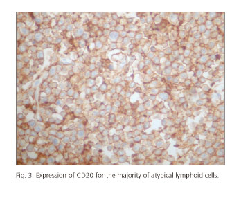
Histopathology of the surgical piece showed a serosa without relevant changes; adjacent to the serosa, an exophytic tumoral lesion of 6.3 x 6 cm that occluded 80% of the intestinal diameter and 95% of the lumen, 5.2 cm of margin of resection, was detected. Microscopy examination of the biopsy smears showed a dense proliferation of atypical lymphoid cells, of median and large-sized, eosinophilic cytoplasm and one or various nucleoli next to the basal membrane, underlying a transmural infiltrate of atypical lymphocytes with extensive areas of ulceration and necrosis (Fig. 2). Histopathology of 15 lymph regional nodes was normal. Immunohistochemistry revealed that neoplastic cells were positive for CD20 (Fig. 3), partial expression of leukocyte common antigen (LCA) and BCL6 with co-expression of BCL2 and MUM1 in 60% of the overall cellularity. Ki67 proliferation index was high (> 90%). A computed tomography (CT) scan of the thorax, abdomen and pelvis was normal and a bone marrow biopsy was negative for atypical neoplastic infiltration. Final histopathological diagnosis was primary diffuse large B cell lymphoma (DLBCL) of the colon (WHO).
Discussion
GI tract is the most common site of extra-nodal NHL, accounting for 5% to 20% of all the cases. However, primary NHL of the gastrointestinal tract is a rare clinical-pathological entity comprising only 1% to 4% of all the gastrointestinal malignancies (4). Although NHL can affect any region of the GI tract, oral cavity, stomach, small intestine and ileocecal region are the most commonly involved sites (5). Primary NHL of the colon is a rare tumor representing only 0.2% to 1.2% of all colonic malignancies (6). These tumors are more frequent in men with a median age of 55 years at diagnosis (6).
Clinical presentation includes insidious abdominal pain, nausea, vomiting, "B" symptoms (fever, night sweats and weight loss), abdominal mass and, most rarely, rectal bleeding (6,7). The most common locations are the cecum and the left colon; colonoscopy, ultrasound and computed tomography scan findings are similar to those of epithelial tumors (6). Only histopathological examination and immunohistochemistry techniques confirm the diagnosis (8). Histologically, 85% of cases are B-cell lymphomas and the most frequent histopathological subtypes, according to the REAL/WHO (Revised European-American Classification of Lymphoid Neoplasms/World Health Organization) are DLBCL, as in our patient, mucosa-associated lymphoid tissue (MALT) lymphoma, mantle-cell lymphoma and Burkitt's lymphoma (6).
In adults, intussusceptions with acute or subacute intestinal obstruction represents only 5% of cases and the small bowel is the most common site of the invagination (3). Intussusceptions can be classified according to the anatomical location into entero-enteric, ileocecal, ileo-colic and colo-colonic (9). In the ileocecal location, the valve acts as the initial point of the invagination (10).
In the last years, a significant number of adult intussusceptions have been reported in AIDS patients, the majority of cases due to Kaposi's sarcoma and NHL of the GI tract (11).
Malignancies are the most common causes for adult intussusceptions of the large bowel, especially adenocarcinoma and secondary NHL. Miscellaneous causes include post-operative adhesions, benign lesions (lipoma, leiomyoma and adenomatous polyps), endometriosis and tuberculosis (9,10).
In a recent review of adult intussusceptions, Kaval et al. (12) analyzed 17 patients assisted between 1998 and 2012. In this series, the median age was 35 years old, the majority of patients were males (11 cases) and intussusception involved the small bowel in 70.5% of cases. In 29.4% of the cases the large bowel was affected. Malignancies were the only cause of intussusceptions of the large bowel, as in the present case.
Abdominal ultrasound is a low cost, non-invasive method with similar sensitivity and specificity to CT scan (10,13). Typical ultra-sonographic features associated with intussusception include the "target sign" or "doughnut sign" on transverse plane and "pseudo-kidney sign" or "hayfork sign" on longitudinal view. In the study carried out by Kaval et al. (12), abdominal ultrasound revealed the diagnosis in 58.8% of cases (10 of 17 patients), and in all cases diagnosis was confirmed by CT scan. In the other 5 cases, diagnosis was only suspected by CT scan. CT scan aided in the pre-surgical diagnosis of intussusception in 15 (88.2%) patients. Enhancement surrounding the intussusception can be seen after the injection of contrast. CT scan may also contribute to define the location, characteristics of the tumor and its relationship to the surrounding tissues. Additionally, it may help to establish the stage of the neoplasm (9).
In our patient, IHQ was positive for CD20 and LCA and negative for cytokeratins, confirming the diagnosis of DLBCL (WHO). The patient had a tumor of the transverse colon without regional lymph nodes or bone marrow infiltration. Laboratory findings and CT scan of the thorax, abdomen and pelvis were also normal. In consequence, our case filled the criteria of Dawson and Richard for the diagnosis of primary NHL of the colon (14).
Primary NHL of the colon is an uncommon disease with a few number of cases published in the medical literature. The optimal treatment of this neoplasm is yet to be established. Surgical treatment is necessary in adult patients, as in our case, due to the frequency of organic lesions. Surgery is the treatment of choice to resolve the intussusception and the obstruction in case of ileocecal or colonic involvement. Surgery is also necessary to obtain a biopsy to establish the diagnosis and to assess the regional extension of the neoplasm. Surgery is the first-line therapy in complications such as perforation (15).
Currently, chemotherapy alone is the gold standard treatment and is complementary to surgery, as in our patient (10). CHOP (cyclophosphamide, doxorubicine, vincristine and prednisone), with or without rituximab, is the most used regimen in both immunocompetent and immunocompromised patients (6).
In conclusion, intussusception is an infrequent cause of mechanical intestinal obstruction in adult patients that should be included in the differential diagnosis of abdominal pain in HIV/AIDS patients. In those cases of colonic involvement, NHL should be considered as a probable diagnosis due to the high frequency of these tumors in the HIV population.
References
1. Agha FP. Intussusception in adults. AJR Am J Roentgenol 1986; 146:527-31. DOI: 10.2214/ajr.146.3.527. [ Links ]
2. Azar T, Berger DL. Adult intussusception. Ann Surg 1997;226:134-8. DOI: 10.1097/00000658-199708000-00003. [ Links ]
3. Huang WS, Changchien CS, Lu SN. Adult intussusception: A 12-year experience, with emphasis on etiology and analysis of risk factors. Chang Gung Med J 2000;23:284-90. [ Links ]
4. Sadiya N, Ghosh M. Primary ALK positive anaplastic large cell lymphoma of T-cell type of jejunum: Report of a rare extranodal entity with review of literature. Arch Int Surg 2014;4:50-3. DOI: 10.4103/2278-9596.136716. [ Links ]
5. Ko YH, Karnan S, Kim KM, et al. Enteropathy-associated T-cell lymphoma - A clinicopathologic and array comparative genomic hybridization study. Hum Pathol 2010;41:1231-7. DOI: 10.1016/j.humpath.2009.11.020. [ Links ]
6. Mahfoud T, Tanz R, Réda Khmamouche M, et al. Primary non-Hodgkin's lymphoma of the colon: A case report and literature review. J Gastrointest Cancer 2012;43:619-21. DOI: 10.1007/s12029-012-9406-1. [ Links ]
7. Corti M, Villafañe Fioti MF, Lewi D, et al. Linfomas del tubo digestivo y glándulas anexas en pacientes con sida. Serie de casos. Acta Gastroenterol Latinoam 2006;36:190-6. [ Links ]
8. Corti M, De Dios Soler M, Baré P, et al. Linfomas asociados con la infección por el virus de la inmunodeficiencia humana: subtipos histológicos y asociación con los virus de Epstein Barr y Herpes-8. Medicina (B Aires) 2010;70:151-8. [ Links ]
9. Weilbaecher D, Bolin JA, Hearn D, et al. Intussusception in adults. Review of 160 cases. Am J Surg 1971;121:531-5. DOI: 10.1016/0002-9610(71)90133-4. [ Links ]
10. Begos DG, Sandor A, Modlin IM. The diagnosis and management of adult intussusception. Am J Surg 1997;173:88-94. DOI: 10.1016/S0002-9610(96)00419-9. [ Links ]
11. Visvanathan R, Nichols TT, Reznek RH. Acquired immune deficiency syndrome-related intussusception in adults. Br J Surg 1997;84:1539-40. DOI: 10.1002/bjs.1800841112. [ Links ]
12. Kaval S, Singhal BM, Kumar S, et al. Adult intussusception: An institutional experience and review of literature. Arch Int Surg 2014;4:25-30. DOI: 10.4103/2278-9596.136706. [ Links ]
13. Cerro P, Magrini L, Porcari P, et al. Sonographic diagnosis of intussusceptions in adults. Abdom Imaging 2000;25:45-7. DOI: 10.1007/s002619910008. [ Links ]
14. Dawson IMP, Comes JS, Morson BC. Primary malignant lymphoid tumors of the gastrointestinal tract. Br J Surg 1961;49:80-9. DOI: 10.1002/bjs.18004921319. [ Links ]
15. Ruskoné-Fourmesestraux A, Aegerter P, Delmer A, et al. Primary digestive tract lymphoma: A prospective multicentric study of 91 patients. Gastroenterology 1993;105:1662-71. [ Links ]
![]() Correspondence:
Correspondence:
Marcelo Corti.
HIV/AIDS Division - Infectious Diseases.
Hospital F. J. Muñiz.
Puán 381, 2o. C1406CQG Buenos Aires, Argentina
e-mail: marcelocorti@fibertel.com.ar
Received: 22-08-2015
Accepted: 27-08-2015


 texto en


