


 Custom services
 English (pdf)
 Article in xml format
 Article references
 Send this article by e-mail
 Cited by SciELO
  Cited by Google
  Similars in
SciELO
Similars in
SciELO  Similars in Google
 Permalink
INTRODUCTION
Intestinal ischemia is caused by several clinical conditions such as trauma, shock, strangulated hernia, intestinal obstruction, and small intestine transplant 1. The intestinal mucosa is injured during ischemia due to the reduction and/or obstruction of blood flow. After reintroduction of blood flow, these injuries may get worse, leading to intestinal ischemia-reperfusion (I/R) injury 2.
Several mechanisms are involved in intestinal I/R injury, such as neutrophil infiltration, intracellular adhesion molecules (ICAM-1), production of proinflammatory cytokines such as interleukin 1β and 6 (IL-1β, IL-6) and tumor necrosis factor alpha (TNF-α), exacerbated generation of reactive oxygen species (ROS), and activation of the nuclear factor kappa-light-chain-enhancer of activated B cells (NF-kB) 3) (4. This condition may damage the integrity of the intestinal barrier, leading to possible bacterial translocation into the bloodstream, thus triggering the onset of the systemic inflammatory response syndrome (SIRS) and the consequent physiological dysfunction of two or more systems, which is known as multiple organ dysfunction syndrome (MODS) 5) (6) (7. The liver is one of the organs that undergoes these changes because of the close relationship between intestinal function and balance of liver function. In case of bowel disease, there is imbalance of the intestinal microbiota, thus causing and worsening liver injuries 8) (9.
Although some molecular aspects of the mechanism of action causing intestinal I/R injuries have not been well defined, studies have shown that inflammatory cytokines and increased ROS generation play an important role in the pathogenesis of intestinal and systemic injuries after an I/R event 10) (11.
Inflammatory mediators, such as IL-6 and TNF-α, are linked to the activation of neutrophils and tissue damage resulting from I/R. Studies have demonstrated that plasma and tissue levels of these cytokines are high in intestinal I/R and MODS. These mediators may be activated by the NF-kB, which plays an important role in the control of adaptive immune responses by regulating the expression of these pro-inflammatory genes 7.
The oxidative processes resulting from the exacerbated generation of ROS may cause damage to proteins, lipids, and DNA. These processes are involved in the progression of the intestinal I/R injury and the integrity and function of other organs. Experimental studies have shown the association of oxidative damage with intestinal I/R injury in organs such as lung, liver, and kidney 7) (11) (12. The antioxidant system may compensate for these damages caused by ROS through enzymatic substances, such as catalase (CAT), superoxide dismutase (SOD), and glutathione peroxidase (GPx), and non-enzymatic substances such as glutathione (GSH). Therefore, recent studies have shown that the administration of antioxidants may contribute to reduce oxidative processes and decrease the local and systemic inflammatory process caused by the intestinal I/R injury 1) (4) (11) (13.
Glutamine (Gln) is a hydrophilic, non-essential amino acid found on the surface of proteins, where it interacts with water. Gln is a precursor for the synthesis of nucleic acids and GSH. In addition, Gln plays a key role in the immune defense of the intestinal mucosal barrier by participating in the formation of immunoglobulins, especially IgA 14) (15. Some mechanisms suggest a protective role of glutamine in the intestinal inflammatory response, namely: increased intestinal antioxidant capacity through the synthesis of GSH, reduced apoptosis, and preserved intestinal integrity, thus reducing the local and systemic inflammatory parameters 14) (16.
Experimental studies have demonstrated the antioxidant role of Gln in reducing ROS generation and pro-inflammatory cytokines, as well as in preserving tissue GSH in experimental models of liver I/R 17. Fillmann et al. (2007), Marques et al. (2001), and Zhang et al. (2011) showed that pretreatment with Gln reduced oxidative and tissue damages in the gut, stomach, and liver. These authors also found a reduction in the inflammatory processes, suggesting a protective effect of Gln against free radicals 14) (15) (18.
In our previous study we demonstrated that pretreatment with Gln reduced intestinal lesions resulting from intestinal ischemia and reperfusion model. We observed a reduction in intestinal tissue damage, decreased oxidative damage as well as reducing inflammation mediators studied 7.
Therefore, the objective of the present study was to investigate the antioxidant and anti-inflammatory effect of Gln based on the evaluation of lipid peroxidation (LPO), DNA damage, activity of CAT, SOD, and GPx, GSH levels, and expression of IL-6 and NF-kB. Our hypothesis suggests that treatment with Gln could reduce oxidative, cell, and inflammatory damage in the liver tissue induced by an intestinal I/R experimental model.
MATERIALS AND METHODS
ANIMALS AND GLUTAMINE ADMINISTRATION
Our procedures involving animals followed the guidelines of the Research and Ethics Committee of the Research and Graduate Studies Group (GPPG), Hospital de Clínicas de Porto Alegre (HCPA), approved with the protocol number 12-0241, and the European Council Directive regarding animal experimentation 19) (20.
We used 20 male Wistar rats. Their mean weight was 300 grams. The animals were divided into four groups: sham operated (SO) (n = 5), glutamine + sham operated (G+SO) (n = 5), intestinal ischemia-reperfusion (I/R) (n = 5), and glutamine + intestinal ischemia-reperfusion (G+I/R) (n = 5). The animals stayed at the Animal Experimentation Unit (UEA) of the Hospital de Clínicas de Porto Alegre. They were kept in plastic boxes lined with wood shavings and measuring 47 x 34 x 18 cm, on a 12:12-hour light/dark cycle (light from 7 am to 7 pm) and temperature between 20 and 25 °C. Water and feed were given ad libitum. The animals were intraperitoneally anesthetized with xylazine hydrochloride (8 mg/kg of body weight) and ketamine hydrochloride (92 mg/kg of body weight). Laparotomy and evisceration were performed to identify the superior mesenteric artery in both groups. In groups SO and G+SO (surgery simulation) the artery was not obstructed and in groups I/R and G+I/R mesenteric artery was occluded for 30 minutes using a microsurgical vascular clamp. After intestinal ischemia was achieved, the microsurgical vascular clamp was removed and the animals were reperfused for 15 more minutes (adapted from Cho et al., 2013) 3. After this period, the liver was removed for histological analysis and other tests. Finally, the animals were killed by exsanguination under deep anesthesia 20.
Gln was administered intraperitoneally at a dose of 25 mg/kg diluted in 1 ml of saline. This dose was given once daily for 48 hours before ischemia induction 14) (15.
Liver function tests based on aspartate aminotransferase (AST), alanine aminotransferase (ALT) and alkaline phosphatase (ALP)
Levels of serum AST, ALT, and ALP were determined by the Biochemistry Center of the Hospital de Clínicas de Porto Alegre. To determine AST and ALT levels in the plasma, we used the commercially available enzymatic method (Boehringer Mannheim, Germany) and the results were obtained by kinetic measurement at 567 nm. Levels of ALP were measured with an automated analyzer by the enzymatic method, using p-nitrophenyl phosphate (pNPP) substrate plus water, forming p-nitrophenol, a compound measured with maximum absorption of 400 nm.
LIVER HOMOGENATE
Liver tissues were homogenized for 30 seconds in an ULTRA-TURRAX(r) disperser (IKA-Werke GmbH & Co. KG) at 4 °C using 1.15% KCl (5 ml/g of tissue) and phenylmethanesulfonylfluoride (PMSF) at a concentration of 100 mM. Next, the homogenates were centrifuged for ten minutes at 3,000 rpm in a refrigerated centrifuge (SORVALL Super T21, Kendro Laboratory Products, USA). The supernatant was removed and stored in microtubes. The samples were stored at -80 °C for later analyses 21.
LPO
The amount of aldehydes generated by LPO is determined by the method that measures the amount of thiobarbituric acid reacting substances (TBARS). Thiobarbituric acid was added to the samples at 0.37%, whereas trichloroacetic acid was added at 15%. The samples were incubated at 100 °C for 15 minutes and centrifuged at 3,000 rpm (1,612 x g) for ten minutes at 4 °C. Absorbency was determined by spectrophotometry at 535 nm 22.
COMET ASSAY
An alkaline comet assay was performed as described by Hartmann and Speit (1999), including some changes suggested by Picada et al. in 2003 23) (24. Blood samples (50 µl) were added to 5 ml of anticoagulant (heparin sodium 25,000 IU/ml solution - Liquemine(r), Roche). Blood cell suspensions (5 µl) were added to 95 ml of 0.75% low melting point agarose (Gilco BRL) and placed on agarose pre-coated microscope slides. After solidification, the slides were placed in lysis buffer (2.5 M NaCl, 100 mM EDTA, 10 mM Tris, pH 10.0). We added 1% Triton X-100 (Sigma) and 10% dimethyl sulfoxide (DMSO) for 48 hours at 4 °C. Next, the slides were subsequently incubated in alkaline buffer (300 mM NaOH and 1 mM EDTA, pH > 13) for 20 minutes at 4 °C. An electric current of 300 mA and 25 V (0.90 V/cm) was applied for 15 minutes to perform DNA electrophoresis. Then the slides were neutralized (0.4 M Tris, pH 7.5), stained with silver, and analyzed by microscope. Images of 100 randomly selected cells (50 cells of each slide) were analyzed from each animal. The cells were also visually classified according to the size of the tail into five categories ranging from undamaged (0) to maximum damage 4, resulting in a single DNA damage score for each animal and for each group. Therefore, the damage index (DI) may range from 0 (completely intact, 100 cells x 0) to 400 (maximum damage, 100 × 4). The damage frequency (%) was calculated based on the number of tailed cells vs tail-less cells.
ACTIVITY OF ANTIOXIDANT ENZYMES: CAT, SOD, GPX AND GSH LEVELS
The analysis of CAT activity was based on the measurement of the reduction of hydrogen peroxide. The activity was detected spectrophotometrically at 240 nm with values expressed as pmol/mg prot 25. The analysis of SOD activity was based on the inhibition of the reaction of epinephrine with superoxide radical, which could be detected spectrophotometrically at 480 nm with values expressed as USOD/mg prot 26. GPx activity was determined based on the consumption of nicotinamide adenine dinucleotide phosphate (NADPH) in the reduction of oxidized glutathione, which could be detected spectrophotometrically at 340 nm within two minutes with the values expressed as nmol/min/mg prot 27. Measurement of GSH levels was detected spectrophotometrically at 412 nm with values expressed as µmol/mg prot, according to the method of Beutler, Duran and Kelly 28.
IMMUNOHISTOCHEMISTRY AND QUANTIFICATION OF THE EXPRESSION OF IL-6 AND NF-KB
The expression of IL-6 and NF-kB in liver tissue was determined by immunohistochemical analysis. Antigen retrieval was performed using buffer at 60 °C, and endogenous peroxidase activity was blocked by incubation in absolute methanol. The slides were incubated with rabbit polyclonal antibody (NF-kB [p65] [SC8008] - Santa Cruz Biotechnology, USA) at 1:100 and (IL-6 [SC1265] - Santa Cruz Biotechnology, USA) at 1:100 overnight at 4 °C. The slides were washed with buffer and incubated with the secondary antibody (anti-mouse IgG-HRP, anti-goat IgG-HRP, Santa Cruz Biotechnology, Santa Cruz, CA, USA) at 1:300 for 30 minutes at room temperature. The slides were examined by a pathologist, who was unaware of the groups, using a microscope equipped with a digital analysis system including a Zeiss Axioskop 40 microscope (Oberkochen, Germany) connected by a Roper Scientific camera (Media Cybernetics, Rockville, USA) to a computer with an image capture software. The Image-Pro Plus version 4.5 software (Media Cybernetics, Rockville, USA) was used to analyze digital images. The expression was determined by multiplying the mean density of the image by the percentage of positively stained areas (brown-stained areas).
WESTERN BLOT
Western blot analysis was performed on nuclear extracts prepared from liver homogenates as previously described. The supernatant fraction was collected and stored at -80 °C in aliquots until use. Lysate proteins were fractionated by sodium dodecyl sulfate-polyacrylamide gel electrophoresis (SDS-PAGE) and transferred to polyvinylidene fluoride (PVDF) membranes. The membranes were then blocked with 5% nonfat dry milk in Tris buffered saline containing 0.05% Tween 20 (TTBS) for one hour at room temperature and probed overnight at 4 °C with polyclonal anti-NF-kB (p65) (SC8008/65kDa) (Santa Cruz Biotechnology, Santa Cruz, CA, USA) at 1:200-1,000 dilution with TTBS in 5% nonfat dry milk. Anti-β-actin antibody (A5060/42kDa) (Sigma Aldrich, St Louis, MO, USA) at 1:2,000 dilution with TTBS in 5% nonfat dry milk. After washing with TTBS, the membranes were incubated for one hour at room temperature with secondary anti-mouse IgG-HRP antibody (Santa Cruz Biotechnology, Santa Cruz, CA, USA, 1:4,000). Protein detection was performed by chemiluminescence using a commercial ECL kit (Amersham Pharmacia Biotech, Little Chalfont, UK). The density of the specific bands was quantified using Scion Image software (Scion Corp., Frederick, MD, USA) 29) (30.
STATISTICAL ANALYSIS
Data are expressed as means ± standard error. Statistical significance was calculated using Graphpad Instat, version 3.0 for Windows. We used one-way analysis of variance (ANOVA) and Student-Newman-Keuls for multiple analysis. Analysis of variance with robust standard errors (Welch) was used to investigate the results of the expression of IL-6 and NF-kB between the groups. Results were considered as statistically significant when the significance level was at least 5% (p < 0.05).
RESULTS
LIVER FUNCTION TESTS BASED ON AST, ALT AND ALP
The serum levels of AST, ALT and ALP were significantly increased in the I/R group as compared with the SO and G+SO groups, whereas there was a reduction in these levels in the I/R+G group in comparison with the I/R group (AST*, #p < 0.001; ALT*, p < 0.001, #p < 0.05; ALP*, #p < 0.01) (Table I).
Table I Effect of glutamine administration on the levels of enzymes of liver cell integrity in the blood of animals subjected to the intestinal ischemia-reperfusion model

Values are expressed as mean ± SEM. *Significant difference between the I/R group and the SO and G+SO groups. #Significant difference between the G+I/R group and the I/R group. Sham operated (SO), glutamine + sham operated (G+SO), intestinal ischemia-reperfusion (I/R), glutamine + intestinal ischemia-reperfusion (G+I/R). AST - *, # p < 0.001; ALT - * p < 0.001, # p < 0.05; ALP - *, # p < 0.01.
LPO AND COMET ASSAY
LPO levels (Fig. 1A) showed a significant increase in the I/R group as compared with the SO and G+SO groups, whereas there was a significant decrease in LPO levels in the G+I/R group as compared with the I/R group (*, #p < 0.001).
The results of the comet assay showed an increase in the damage index (Fig. 1B) and damage frequency (Fig. 1C) in the I/R group as compared with the SO and G+SO groups, whereas there was a reduction in these parameters in the G+I/R group in comparison with the I/R (*p < 0.01, #p < 0.05).

Figure 1 Effect of glutamine administration on levels of lipid peroxidation in the liver tissue and DNA damage in the blood of animals subjected to the intestinal ischemia-reperfusion model. A. TBARS. B. Damage index. C. Damage frequency. Values are expressed as mean ± SEM. A. *Significant difference between the I/R group and the SO and G+SO groups (p < 0.001). #Significant difference between the G+I/R group and the I/R group. (p < 0.001). B and C. *Significant difference between the I/R group and the SO and G+SO groups (p < 0.01). #Significant difference between the G+I/R group and the I/R group (p < 0.05). Sham operated (SO), glutamine + sham operated (G+SO), intestinal ischemia-reperfusion (I/R), glutamine + intestinal ischemia-reperfusion (G+I/R).
ACTIVITY OF CAT, SOD, GPX AND GSH LEVELS
The activities of CAT (Fig. 2A), SOD (Fig. 2B) and GPx (Fig. 2C) decreased significantly in the I/R group when compared with the SO and G+SO groups, whereas these activities increased in the G+I/R group as compared with the I/R group (*p < 0.01, #p < 0.05).

Figure 2 Effect of glutamine administration on the CAT, SOD and GPx activity in the liver of animals subjected to the intestinal ischemia-reperfusion model. A. CAT. B. SOD. C. GPx. Values are expressed as mean ± SEM. *Significant difference between the I/R group and the SO and G+SO groups (p < 0.01). #Significant difference between the G+I/R group and the I/R group (p < 0.05). Sham operated (SO), glutamine + sham operated (G+SO), intestinal ischemia-reperfusion (I/R), glutamine + intestinal ischemia-reperfusion (G+I/R).
The GSH levels (Fig. 3) showed a significant decrease in the I/R group as compared with the SO and G+SO groups, whereas there was a significant increase in the G+I/R group as compared with the I/R group (*p < 0.01, #p < 0.05).
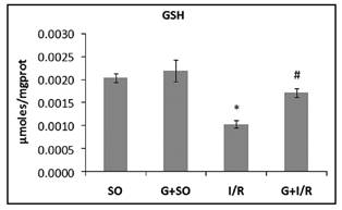
Figure 3 Effect of glutamine administration on GSH levels in the liver of animals subjected to the intestinal ischemia-reperfusion model. Values are expressed as mean ± SEM. *Significant difference between the I/R group and the SO and G+SO groups (p < 0.01). #Significant difference between the G+I/R group and the I/R group (p < 0.05). Sham operated (SO), glutamine + sham operated (G+SO), intestinal ischemia-reperfusion (I/R), glutamine + intestinal ischemia-reperfusion (G+I/R).
Immunohistochemistry and quantification of the expression of IL-6 and NF-kB
The animals of the I/R group showed strong positive staining for IL-6 (Fig. 4) and NF-kB (Fig. 6), which was visible because of the brown staining.
The SO and G+SO groups showed no positive staining. Pretreatment with Gln reduced the staining for IL-6 and NF-kB in the G+I/R group.
Similarly, the quantification of the expression of IL-6 (Fig. 4) and NF-kB (Fig. 5) showed an increase in the expression of inflammatory mediators in the I/R group as compared with the SO and G+SO groups and a significant reduction in the G+I/R as compared with the I/R group (*p < 0.001, #p < 0.05). All images were magnified 400x.

Figure 4 Effect of glutamine administration on the immunohistochemistry and expression of IL-6 in the liver of animals subjected to the intestinal ischemia-reperfusion model. Values are expressed as mean ± SEM. *Significant difference between the I/R group and the SO and G+SO groups (p < 0.001). #Significant difference between the G+I/R group and the I/R group (p < 0.001). Sham operated (SO), glutamine + sham operated (G+SO), intestinal ischemia-reperfusion (I/R), glutamine + intestinal ischemia-reperfusion (G+I/R).

Figure 5 Effect of glutamine administration on the immunohistochemistry and expression of NF-kB (p65) in the liver of animals subjected to the intestinal ischemia-reperfusion model. Values are expressed as mean ± SEM. *Significant difference between the I/R group and the SO and G+SO groups (p < 0.001). #Significant difference between the G+I/R group and the I/R group (p < 0.001). Sham operated (SO), glutamine + sham operated (G+SO), intestinal ischemia-reperfusion (I/R), glutamine + intestinal ischemia-reperfusion (G+I/R).
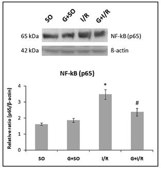
Figure 6 Effect of glutamine administration on the expression of NF-kB (p65) by western blot analysis in the liver of animals subjected to the intestinal ischemia-reperfusion model. Values are expressed as mean ± SEM. *Significant difference between the I/R group and the SO and G+SO groups (p < 0.01). #Significant difference between the G+I/R group and the I/R group (p < 0.05). Sham operated (SO), glutamine + sham operated (G+SO), intestinal ischemia-reperfusion (I/R), glutamine + intestinal ischemia-reperfusion (G+I/R).
WESTERN BLOT NF-KB (P65)
The expression of nuclear factor NF-kB (p65) (Fig. 6) showed an increase in the I/R group as compared with the SO and G+SO groups, and a significant decrease in the G+I/R as compared with the I/R group (*p < 0.01, #p < 0.05).
DISCUSSION
Intestinal I/R is often associated with small intestine transplant, strangulated hernia, aortic aneurysm surgery, and necrotizing enterocolitis, with high rates of mortality and morbidity in recent decades. Some studies suggest that this injury may trigger the involvement of other organs and it may be the main mechanism in the pathogenesis of MODS 6) (13.
The small intestine is susceptible to ischemic injury, and the main factors involved are adhesion molecules, nitric oxide, pro-inflammatory cytokines, and reactive oxygen species. Some studies suggest that exacerbated generation of free radicals is one of the major causes of local and systemic injuries 8) (9) (12.
The gut-liver axis plays a key role in the deleterious effects induced by intestinal I/R because the intestinal bloodstream is connected to liver vascularization. Intestinal I/R leads to the production and subsequent release of deleterious substances, such as ROS and inflammatory mediators, which reach the liver through the portal vein causing severe liver injuries 31) (32) (33.
Gln has been studied as a possible treatment option for injuries associated with ROS and inflammatory substances because it plays an antioxidant and anti-inflammatory role, and may reduce these oxidative and inflammatory damages 7) (18.
In our study, the liver damage was also evaluated based on the function of the hepatic enzymes AST, ALT, and ALP. Our results showed an increase in these enzymes in the I/R group, and pretreatment with Gln reduced these levels. Inan et al. (2012) found an increase in the activity of AST and ALT in animals subjected to an ischemia-reperfusion model. The administration of sildenafil reduced the levels of these enzymes 32.
Some studies have linked the involvement of oxidative processes, such LPO and DNA damage 34, oxidative damage, inflammatory process and DNA damage, to the progression of tissue and cell injuries caused by intestinal I/R in other organs 7) (10) (12. We measured the levels of LPO by TBARS and DNA damage by comet assay. We found an increase in LPO levels and DNA damage index and frequency in the I/R group; however, there was a significant decrease in terms of oxidative damage in the group pretreated with Gln, possibly due to its antioxidant activity.
Our results corroborate other studies showing similar results in the reduction of LPO levels when there is use of a substance with antioxidant effect. Shafik (2012) found an increase in the serum levels of malondialdehyde of animals with I/R. After the administration of febuxostat, these levels were reduced, thus demonstrating a possible antioxidant action of this substance 35. Cámara-Lemarroy et al. (2011) evaluated the possible antioxidant and anti-inflammatory effects of triflusal, S-adenosylmethionine and dextromethorphan, suggesting that the administration of these substances reduced the serum levels of malondialdehyde of animals with I/R 36. Oliveira et al. (2013) showed that glutamine reduced DNA damage in animals receiving cisplatin chemotherapy, a medication that affects the DNA synthesis, suggesting a protective effect of glutamine against damage of DNA bases 37) (38.
The antioxidant system is a compensation mechanism for the oxidation process involving enzymatic substances, such as catalase, superoxide dismutase and glutathione peroxidase, and non-enzymatic substances, such as GSH. These substances play an important role against oxidative damage 10) (11. In our study, we found a significant reduction in the activity of CAT, SOD and GPx, as well as in the levels of GSH in the I/R group, whereas there was a significant increase in the antioxidant parameters in the group pretreated with Gln.
Zhang et al. (2011) found that pretreatment with Gln was able to enhance the antioxidant capacity in an ischemia-reperfusion model. The animals treated with different doses of Gln showed an increase in SOD activity, suggesting a protective role of Gln against the oxidative damage caused by hepatic I/R-mediated liver injury 18.
Similar results were found in the study by Zhao et al. (2010), where a reduction in the activity of SOD and GPx and in the levels of glutathione in the liver of animals subjected to an intestinal I/R model was observed. The administration of sulforaphane compound (SFN), which is present in several plants, was able to increase the antioxidant capacity of the liver, suggesting a protective role against oxidative damage 39.
Intestinal I/R may lead to increased generation of free radicals and to the consequent production of pro-inflammatory cytokines (IL-1β, IL-6, TNF-α) that may be mediated by NF-kB 5) (40. We measured the expression of IL-6 and NF-kB by immunohistochemistry and western blot analysis in the liver. Increased expression of inflammatory markers (IL-6 and NF-kB) was observed in the I/R group, whereas it was significantly reduced in the group pretreated with Gln, suggesting a possible anti-inflammatory effect of this amino acid.
Cho et al. (2013) evaluated the systemic inflammatory response based on the IL-6 plasma level of animals subjected to an intestinal I/R model. They found an increase in these levels in the intestinal I/R group; conversely, these levels were reduced with the administration of remifentanil, suggesting an inhibitory action on NF-kB activity thus reducing the production of IL-6 3. Ma et al. (2014) found a positive expression of NF-kB in the liver tissue of the intestinal I/R group, whereas, when protocatechuic acid was administered, a polyphenolic compound with antioxidant and anti-inflammatory effect, there was a reduction in the expression of this inflammatory mediator 11. Similar results were found by Yao et al. (2009), showing that the treatment with carnosol, a compound with antioxidant effect, inhibited the expression of NF-kB, thereby reducing intestinal I/R-induced liver injuries 40.
In conclusion, our findings suggest that Gln inhibits LPO, restoring the activity of antioxidant enzymes, as well as reducing the IL-6 and NF-kB expression, possibly due to its antioxidant and anti-inflammatory effect. Therefore, these results support the use of Gln to treat liver injuries caused by intestinal ischemia-reperfusion. Nevertheless, further studies should be conducted to validate its use in clinical practice.

