


Mi SciELO
 Servicios personalizados
Servicios Personalizados
Revista
Articulo
 texto en
  Inglés (pdf)
 Articulo en XML
 Referencias del artículo
 Enviar articulo por email
Indicadores
-
 Citado por SciELO
 -
 Accesos
Links relacionados
 Citado por Google
 -
 Similares en
SciELO
Similares en
SciELO  Similares en Google
Compartir

 Permalink
Medicina Oral, Patología Oral y Cirugía Bucal (Ed. impresa)
versión impresa ISSN 1698-4447
Med. oral patol. oral cir. bucal (Ed.impr.) vol.9 no.5 nov./dic. 2004
Fine-needle aspiration cytology in the diagnosis of cervicofacial actinomycosis:
report of 15 cases
CUSTAL-TEIXIDOR M, TRULL-GIMBERNAT JMª, GARIJO-LÓPEZ G, VALLDOSERA-ROSELLO M..FINE-NEEDLE ASPIRATION CYTOLOGY IN THE DIAGNOSIS OF CERVICOFACIAL ACTINOMYCOSIS: REPORT OF 15 CASES. MED ORAL PATOL ORAL CIR BUCAL 2004;9:464-70.
SUMMARY
Objectives:
Actinomycosis is quite an infrequent bacterial infection nowadays.
However it can be considered in cases with a persistent cervicofacial disease.
Although it is a bacterial infection, microbiologic cultures are
frequently not diagnoses, therefore histopathologic studies and image
studies are essential.
Our interest is to explain our experience with cervicofacial
actinomycosis; the clinical behaviour, evolution and treatment,
always assisted by their elected diagnostic technique: the FNAC.
Study design:
In the last 16 years, 15 patients have been diagnosed with
cervicofacial actinomycosis by FNAC, treated by Maxillofacial,
Internal Medicine and Paediatrics units.
Clinical course, evolution, anatomical space situation, antibiotic
treatment, and surgical treatment have been studied.
Results and conclusions:
The fine-needle aspiration cytology (FNAC) is an easy, safe and
rapid method, with a high effect, that has made the final diagnosis
in 15 cases in our Hospital.
All the patients have had a good clinical evolution, only in one
case did we need a new treatment for recidive.
In all the cases treatment has been definitive.
Our interest is to explain our experience in the treatment of
cervicofacial actinomycosis, its clinical presentation and evolution,
together with its elected method of diagnosis, FNAC.
Key words: Cervicofacial actinomycosis, fine needle aspiration cytology, FNAC.
INTRODUCTION
Actinomycosis is a chronic granulomatous infectious disease, caused by a Gram positive bacteria genus Actinomyces. Although before they were related to fungus because of their filamentous aspect, and we stain them with Gomori silver methenamine(1), it is an anaerobic or microanaerobic bacteria, which lives in the digestive tract (mouth and colon), and in the female sexual tract (2).
Actinomyces israelii is the most frequent germ, although other species of actinomyces can be found such as A. naeslundii, A. odontolyticus, A. viscosus, A. meyeri, Propionibacterium propionicum.
Actinomycosis is a polymicrobial disease associated with other microorganisms such as Actinobacillus actinomicetemcomitans, Eikenella corrodens, Fusobacterium, Bacteroides and others depending of the site of infection. (2,3)
Cervicofacial actinomycosis can exist at any age, it is more frequent in males than females with a ratio of 3:1. This relationship disappears when a traumatic etiologic cause exists. (4)
They have not found a favourite race or geographic factor, but are found among socio-economic and hygienic habits, so their appearance has decreased along the years with the improvement in oral hygiene and early antibiotic treatment. (2)
All these lesions, are obliged to a differential diagnosis between primary or metastatic neoplasm, mycobacterium infection (tuberculosis or atypical), brachial cyst, sialadenitis and tiroiditis (5).
Our work is based on 15 patients with cervicofacial actinomycosis, treated in our hospital, with diverse clinical forms and to who a FNAC has been made.
PATIENTS AND METHODS
A retrospective review of 15 cases of cervicofacial actinomycosis, diagnosed in the Hospital Universitari de Girona Doctor Josep Trueta during a 16year period (1986 to 2002) by fine-needle aspiration cytology. With the FNAC material we made smears and cellular block study.
We have studied the clinical behaviour, location, timing and extension of the lesions using frequency tables, also the use of long term antibiotic therapy in all the patients, with the need for surgical treatment in some of them. We made microbiological cultures in 9 cases, only one of them being positive.
We do not consider as actinomycosis, those patients with a histological diagnosis and presence of actinomyces but without clinical disease. This situation is due to a non- pathogen oral flora colonisation.
RESULTS
Of the 15 patients, 10 were men (66:7%) and 5 women (33.3%); ranging in age from 6 to 81 years, the average age was 45.67 years.
53.3% of the patients were smokers.
The most frequent location of the infection was mandibular, 9 cases (60%). (Figure 1) Other locations were cervical lateral, parotid gland, floor of the mouth, masseter muscle, parapharyngeal space and maxilla. (Table 1)
The clinical presentation was a localised tumour in 10 patients (66.7%), local tumour with fistula in 3 patients (20%) and trismus with disphagia in 1 patient (6.7%). (Table 2)
The evolution time was less than a month in 6 patients (40%), between 1 and 6 months in 5 patients (33.3%) and 6 or more months in 4 patients (26.7%).
Among the etiological factors we found: previous tooth extraction, days or weeks, before the onset of infection in 4 patients (26.7%), presence of dental caries in 4 patients (26.7%), cysts in 2 patients (13.3%) and the swallowing of a foreign body (fish bone) a month before without any other cause in 1 patient (6.6%). No etiologic factor was found in 4 cases (26.7%).
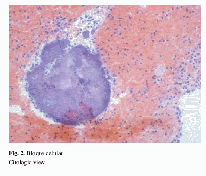
In all the patients we made a FNAC with cytologyc study of the material obtained, and that was the diagnosis in 100% of the cases. The cytopathologic findings are acute inflammatory infiltrate with the presence of lymphocytes and histiocytes. Also we identified colonies of actinomyces formed by masses of filaments extending in a radiating fashion. Those colonies are surrounded by polymorphonuclear leukocytes. (Figure 2)
We cultured the obtained material in 9 of the 15 cases, and which was positive in only 1 case. The difficulty of in vitro growing of the germ was the main cause of the low diagnostic sensitivity.
Among there is a patognomonic sign of the lesion, we have only seen one patient with the presence of sulphur granules, characteristic of actinomyces infection (figure 3), from a maseter abscess opened to the skin.
We made image studies (TAC, MNR) to determine the extension of the lesions, with good results in 9 cases.
All cases were treated with antibiotics, we used penicillin as an elected treatment, that is indicated in all cases of actinomycosis. Penicillin as an elected antibiotic was used in 11 of our patients (73.3%).
We began with 12 to18 million units administered intravenously in adults (in boys we used doses adjusted by weight) followed with a long treatment of 1.5 to 3 gm. of amoxicyllin daily.
In 3 patients we used penicillin intravenously followed by a macrolid orally.
One patient with a proved allergy to penicillin, was treated with erythromycin intravenously followed by clarithromycin orally until the finish of the treatment.
The total treatment duration was variable: 5 patients (33.3%) received 6 moths of treatment; 4 patients 7 months; 4 patients 12 months and 1 case 9 months. In one case we don't know the duration of treatment because he went to live in another country.
We associated surgical intervention to medical treatment in 7 patients. We did teeth extraction of some teeth with periapical granuloma - usually the last molars - abscess incision, and sequestrectomy or enucleation of bone lesions.
The patient progress was followed until the total outcome of the disease.
In 93.3% of the cases, we solved them with the described treatment. Only 1 case (6.7%) relapsed, and he recovered with a tooth extraction and a new antibiotic treatment without complications.
DISCUSSION
Actinomycosis is a relatively infrequent disease nowadays, that oblige us to maintain a high level of clinical inspection, especially when we are facing a long cervicofacial process without response to previous treatments.
The main etiologic factors are poor dental hygiene and oro-facial trauma. Actinomyces are common inhabitants of the oral cavity and pharyngeal region and can be isolated on dental plaque, dental caries, periodontal sockets, tonsillar crypt, gingival crevices, saliva, salivary glands and periodontal pockets (4), that explains the presence of dental treatment in the medical history. Periapical actinomycosis has also been seen with a radicular cyst (6).
Other patients explain foreign bodies, oral surgery previous to infection, radiotherapy for other pathology (7) or systemic diseases such as diabetes mellitus. It has been seen in a patient with the AIDS virus (8).
In our sample a high incidence of males stands out, this can be explained by a high level of hygiene and dental care in females in our country.
The clinical course can be diverse, from slow chronic progressive evolution - the most frequent clinical presentation - to acute form - less frequent.
In clinical exploration the presence of sulphur granules is characteristic (Figure 3), which appears with low frequency; also with the presence of fistula that does not follow the anatomic spaces of involved tissues (4,9,10).
It can be spread by continuity to near structures such as blood ducts, the skull, trachea, thorax, or begin as a neurologic focality if the lesion is at the inferior and posterior cervical level (11). Actinomycosis often involves lymphatic nodes but by the direct extension of a primary lesion; it is uncommon as a lymphadenopaty (12).
The soft tissues are often involved, presenting oedema, soft tissue swelling and skin abscess, and can be associated with general symptomatology such as fever or weight loss.
When the masticatory muscles are involved trismus and dysfagia appear, also if the base or tongue is involved (13); a clinical history of snoring and sore throat can get worse with a mass in piriform sinus (13); we can see osteomyelitis at different levels of the mandible (14); or an ulcerated palatal mass with involvement of bone and soft tissue (15).
The most frequent clinical behaviour in our cases, was a painful and localised tumour in 66'7% of patients, as in other series of the literature (16), although in others revisions the most frequent presentation was a painless tumour (17). The perimandibular level was the most frequent localisation, 60% of the cases, which was in agreement with the literature (2,4).
For actinomycosis infection diagnosis we use exfoliative cytology in cervicovaginal infections on patients using IUD, and histopathology of affected tissue at other levels, as cervicofacial.
For the etiologic diagnoses we use local histologic studies, usually FNAC, or by smears obtained by surgical puncture of the abscess. With our work we have seen that FNAC allows the morphologic identification of bacteria in a similar form to a biopsy. With the FNAC material, we can also do microbiologic culture, but the results aren't positives in a great proportion, which is due to a overgrowing of other associated bacteria, previous antibiotic therapy or inadequate anaerobic conditions in the culture (5,18,19).
In the bibliographic review we have found studies that confirm FNAC has a high level of diagnostic results as a procedure, which allows differentiation between benign and malign lesions, in an easy, quick and safe method (with minimal risk of haemorrhage and bacteriological contamination). Also it allows an ambulatory diagnosis (20,21). FNAC is also a very useful method of diagnosis in salivary gland pathology (22) where actinomycosis can appear.
Only in a case of 9 (11.1%) has the culture been positive, for this reason we did not consider the culture as a good method of diagnosis.
For serious cases of actinomycosis, in treatment we use penicillin intravenously for 4 to 6 weeks followed by penicillin orally for 6 to 12 months more. In proved cases of penicillin allergy, tetracycline, erythromycin and clindamycin can be good substitutes (23,10).
The good course obtained in all the patients of our sample is the result of the efficacy of antibiotic treatment, maintained for enough time to eradicate the micro-organism accompanied by the surgical approach to drain and clean the affected tissues in many cases.
CONCLUSIONS
- The FNAC not only allows the morphologic identification of germ as a biopsy, but is also an effective method to obtain material to culture.
- The FNAC is an easy, safe and precise method, it allows a quick diagnosis in suspected cases of actinomycosis, to provide a premature treatment.
- Facing a cervicofacial tumour FNAC, where we see abundant acute inflammation, it is necessary to think about this entity, and look for actinomyces filaments.
- The medical treatment, associated or not, with surgical treatment is effective.
REFERENCES
1. Silverman JF. Guides to Clinical Aspiration Biopsy: Infectious and inflammatory diseases and other nonneoplastic disorders. New York:IGAKU-SHOIN Medical Publishers, Inc;1991. p. 49-51,183. [ Links ]
2. Russo TA. Agents of actinomycosis, en Mandell GL, Bennett JE, Dolin R, eds. Mandell, Douglas and Bennett's principles and practice of infectious disease. New York: Churchill Livingstone; 2000. p. 2645-54. [ Links ]
3. Somacarrera ML, Hernández Vallejo G, López A, Aracil L, Arriba L, Lucas-Tomás M. Estudio clínico de seis casos de actinomicosis cervicofacial. Archivos de Odontoestomatología 1996;l12:547-61. [ Links ]
4. Miller M, Haddad AJ. Cervicofacial actinomycosis. Oral Surg Oral Med Oral Pathol Radiol Endod 1998;85:496-508. [ Links ]
5. Vera-Alvarez J, Marigil-Gomez M, Abascal-Agorreta M. Fine needle aspiration cytology of cervicofacial actinomycosis. Acta Cytol 1993;37:109-11. [ Links ]
6. Hirshberg A, Tsesis I, Metzger Z, Kaplan I. Periapical actinomycosis associated with radicular cyst: A clinicopathologic study. Oral Surg Oral Med Oral Pathol Oral Radiol Endod. Abstract. 2002;94:208. [ Links ]
7. Liu CJ, Chang KM, Ou CT. Actinomycosis in a patient treated for maxillary osteoradionecrosis. J Oral Maxillofac Surg 1998;56:251-3. [ Links ]
8. Casariego Z, Pérez H. Actinomicosis oral en paciente VIH positivo. Med Oral 1997;2:102-4. [ Links ]
9. Nagler R, Peled M, Laufer D. Cervicofacial actinomycosis. A diagnostic challenge. Oral Surg Oral Med Oral Pathol Radiol Endod 1997;83:652-6. [ Links ]
10. Smego RA, Foglia G. Actinomycosis. Clin Infec Dis 1998;26:1255-61. [ Links ]
11. Stiefel P, Miranda ML, Martín Sanz V, Gutiérrez JL, Villar J, Carneado J. Actinomicosis cervical comenzando como monoparesia de miembro superior izquierdo. Rev Clin Esp 1990;187:343-5. [ Links ]
12. Shatrughan Prasad Sah, Asim Mishra, Sudha Rani, Ramachandran VG: Cervicofacial Actinomycosis: Diagnosis by Fine Needle Aspiration Cytology. Acta Cytol 2001;45: 665-7. [ Links ]
13. Belmont MJ, Behar PM, Wax MK. Atypical presentations of actinomycosis. Head & Neck 1999;21:264-8. [ Links ]
14. Bartkowski SB, Zapala J, Heczko P, Szuta M. Actinomycotic osteomyelitis of the mandible: review of 15 cases. Journal of Cranio-Maxillofacial Surgery 1998;26:63-7. [ Links ]
15. Herman WW, Whitaker SB, Williams M F, Sangueza OP. Acute Actinomycosis presenting as an ulcerated palatal mass. J Oral Maxillofac Surg 1998;56:1098-101. [ Links ]
16. Samuels RH, Martin MV. A clinical and microbiological study of Actinomycetes in oral and cervicofacial lesions. Br J Oral Maxillofac Surg 1988:26:458-63. [ Links ]
17. Aguirrebengoa K, Romaña M, López L, Marín J, Montejo M, González de Zárate P. Actinomicosis orocervicofacial. Presentación de 5 casos clínicos. Enferm Infecc Microbiol Clin 2002:20:53-6. [ Links ]
18. Neville BW, Damm DD, Allen CM, Bouquot JE, eds. Oral and Maxillofacial Pathology. Philadelphia:WB Saunders Company; 1995. p. 156-7. [ Links ]
19. Das DK, Bhatt NC, Khan VA, Luthra UK. Cervicofacial Actinomycosis: Diagnosis by Fine Needle Aspiration Cytology. Acta cytol 1989;33:278-80. [ Links ]
20. August M, Faquin WC, Ferraro NF, Kaban LB. Fine-needle aspiration biopsy of intraosseous jaw lesions. J Oral Maxillofac Surg 1999;57:1282-6. [ Links ]
21. Gerbino G, Bernardi M, Secco F, Sapino A, Pacchioni D. Diagnosis of actinomycosis by fine-needle aspiration. Letters to the editor. Oral Surg Oral Med Oral Pathol Oral Radiol Endod 1996;81:381. [ Links ]
22. Costas A, Martín-Granizo R, Castro P, Monje F, Marrón C, Diaz F et al. Punción-aspiración con aguja fina (PAAF) en las lesiones de glándulas salivales. Med Oral 1999;4:519-27. [ Links ]
23. Henderson HM, Chapman SW. Infections due to Fungi, Actinomyces and Nocardia. En: Betts RF, Chapman SW, Pen RL, eds. Reese and Betts' a practical approach to infectious diseases. Fifth edition. Philadelphia: Lippincott Williams and Wilkins; 2003. p. 588-648. [ Links ]

