




 Servicios personalizados

 Inglés (pdf)
 Articulo en XML
 Referencias del artículo

 Traducción automática
 Enviar articulo por email
 Citado por SciELO
 Citado por Google
 Similares en
SciELO
 Similares en Google

 Permalink

INTRODUCTION
Endovascular techniques, such as endovascular aneurysm repair (EVAR) and thoracic endovascular aortic repair (TEVAR), often require large-bore vascular access. In patients with small, calcified, or diseased femoral arteries, traditional femoral access becomes infeasible. In these cases, the iliac conduit offers a viable alternative. The modification of omitting the distal anastomosis in the first stage simplifies the procedure further and ensures a safer, faster approach with improved outcomes. We describe this technique to optimize the approach for challenging vascular access.
INDICATIONS
The iliac conduit technique is indicated when the native iliac artery cannot accommodate the endovascular device due to size, calcification, or stenosis. Common clinical scenarios include:
Large-bore prosthesis requirements in EVAR or TEVAR procedures.
Aortoiliac occlusive disease with infeasible traditional accesses.
Hostile groin anatomy, such as prior surgical procedures, radiation damage, or extensive scar tissue.
Avoiding the femoral artery, this approach mitigates complications in patients at higher surgical risk.
ADVANTAGES OF THE ILIAC CONDUIT WITHOUT DISTAL ANASTOMOSIS
The iliac conduit without distal anastomosis, performed as a first step, offers several significant advantages over traditional femoral access or standard iliac conduit procedures:
Reduced Operative Time: omitting distal anastomosis eliminates the need for extensive suturing, streamlining the surgical process.
Avoidance of Groin Incision and Dissection: by not accessing the femoral artery, the groin remains untouched, thus reducing the risk of surgical site infections, particularly in high-risk patients.
Simplified Access for Future Interventions: if any re-interventions were required, the iliac conduit can be ligated distally and advanced to the inguinal ligament, which facilitates further endovascular access through a simple groin incision.
Lower Risk of Complications: by reducing the operative exposure and bypassing the femoral artery, the likelihood of hematoma, infection, and groin wound complications is minimized.
SURGICAL TECHNIQUE
A/ First stage
Preoperative Planning: Preoperative imaging modalities (CCTA) are essential to assess the iliac anatomy, vessel caliber and disease. The appropriate conduit material needs to be properly selected as well. (Dacron 10 mm).
Exposure of the Common Iliac Artery: We perform a standard retroperitoneal approach to expose the common iliac artery with careful dissection to further minimize injury to surrounding structures.
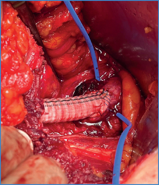
Proximal Anastomosis: After systemic heparinization and clamping, an end-to-side anastomosis of the conduit to the common iliac artery should be performed. This is the only anastomosis required (Fig. 1).
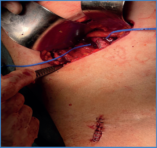
Place the conduit subcutaneously at inguinal level: The conduit is placed subcutaneously and at inguinal level to ensure hemostasia and patency. Finally, the end is ligated and sutured (prolene 5/0) to prevent bleeding (Fig. 2).
B/ Second stage
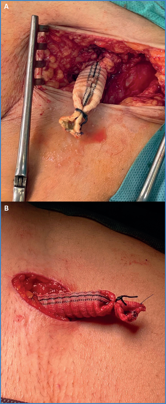
Conduit Management: Several days later, we look for the prothesis subcutaneously at inguinal level. It is easy because it has not been yet integrated. Logically, the conduit is thrombosed, but via manual thrombectomy with a Fogarty catheter the prothesis is reopened, and can be used as an access point to introduce endovascular devices (Fig. 3).
After finishing the procedure, the conduit is again distally ligated, sutured and placed subcutaneously. Finally, the wound is closed.
DISCUSSION
The iliac conduit without distal anastomosis provides a safe and effective alternative for patients requiring large-bore vascular accessess while presenting complex anatomies or significant comorbidities. This change reduces procedural time, avoids unnecessary groin complications, and streamlines management of future interventions.
Our experience has demonstrated favorable outcomes with this approach, particularly in high-risk patients undergoing EVAR and TEVAR.
CONCLUSIONS
The iliac conduit without distal anastomosis as the index step is a significant advancement in endovascular treatment, particularly for patients with limited vascular access options. By simplifying the conduit creation process and eliminating the need for extensive distal suturing, this technique improves procedural efficacy, reduces procedural risk, and enhances patient outcomes.
Further studies and long-term follow-up data will continue to validate its role in contemporary vascular surgery.

