Mi SciELO
Servicios Personalizados
Revista
Articulo
Indicadores
-
 Citado por SciELO
 -
 Accesos
Links relacionados
-
 Citado por Google
 -
 Similares en
SciELO
 -
 Similares en Google
Compartir
Revista Española de Enfermedades Digestivas
versión impresa ISSN 1130-0108
Rev. esp. enferm. dig. vol.102 no.8 Madrid ago. 2010
PICTURES IN DIGESTIVE PATHOLOGY
Primary omental infarction as cause of non-surgical acute abdomen: imaging diagnose
Infarto omental primario como causa de abdomen agudo no quirúrgico: diagnóstico por imagen
C. L. Fernández-Rey
Service of Radiodiagnosis. Hospital El Bierzo. Ponferrada, León. Spain
Introduction
Omental infarction is a relatively uncommon and benign condition, which usually involves the right side omentum and, as its name indicates, it results from an omental focal fat infarction (1,2). Frequently, the omental infarction occurs after recent abdominal surgery. But, cases of idiopathic or primary omental infarction have also been described, and these are more frequent in obese patients. The etiology and pathogenesis are uncertain. A redundant omentum or vascular anomalies have been postulated as possible causes that makes the omentum susceptible to torsion and infarction (1). Other hypothesis indicates that the origin of this disorder is a vascular congestion associated with increased intra-abdominal pressure or abundant foods (1,2).
The omental infarction represents a self-limiting and benign disorder, which does not require surgery but, its symptoms may simulate a surgical acute abdomen. Imaging diagnose of omental infarction is determinant to guide the management of patients and to avoid unnecessary surgery.
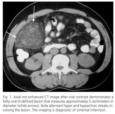
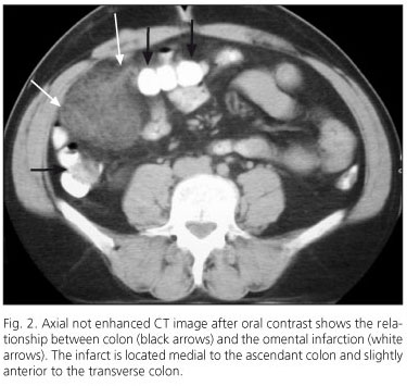
Ultrasonography only suggests the diagnosis (1-3). Although, computer tomography (CT) constitutes a essential diagnostic tool that may rule out other surgical acute abdomen causes and may provide a certain diagnosis of omental infarction (1-3). Its aspect on CT is characteristic and distinctive. CT images demonstrate a fat density lesion, located medial to the ascending colon or just anterior to the transverse colon, with ill-defined borders and internal hyperattenuating streaks (2,3). Lack of internal high density ring and the size (superior to 3 centimeters) may distinguish omental infarction from epiploic appendicitis, which is also a benign and self limiting course disease (3,4).
Case report
The reported case corresponds to a 43-year-old man who is overweight and has not remarkable medical history. The patient presents with intense right flank pain within the previous 48 hours. The physical exploration reveals peritoneal irritation on the right flank. There is no fever, there is no leukocitosis. With a presumptive diagnose of acute diverticulitis, an abdominal ultrasound is performed showing a hyperechoic oval painful lesion on the right that affects the largest omentum. Next, abdominal CT demonstrates a fatty lesion involving the right side omentum that contains internal hyperattenuating streaks and measures approximately five centimeters in diameter (Figs. 1 and 2). The findings are diagnostic of omental infarction. Conservative management is decided, showing good clinical evolution and final resolution on follow-up imaging studies.
References
1. Sierra P, Cabrera R, Fuentealba IM, Soto G, Abud M. Caso clínico radiológico para diagnóstico. Rev Chil Radiol 2009; 15(3): 155-8. [ Links ]
2. Varela C, Fuentes M, Rivadeneira R. Procesos inflamatorios del tejido adiposo intraabdominal, causa no quirúrgica de dolor abdominal agudo: hallazgos en tomografía computada. Rev Chil Radiol 2004; 10(1): 28-34. [ Links ]
3. Miguel A, Ripollés T, Martínez MJ, Morote V, Ruiz A. Apendicitis epiploica e infarto omental: hallazgos en ecografía y tomografía computarizada. Radiología 2001; 43(8): 495-501. [ Links ]
4. Castro García FJ, Santos Sánchez JA, García Íñigo P, Díez Hernández JC. Apendicitis epiploica. Rev Esp Enferm Dig 2006; 28(2): 732-3. [ Links ]


 texto en


