


Mi SciELO
 Servicios personalizados
Servicios Personalizados
Revista
Articulo
 texto en
  Inglés (pdf)
 Articulo en XML
 Referencias del artículo
 Enviar articulo por email
Indicadores
-
 Citado por SciELO
 -
 Accesos
Links relacionados
-
 Citado por Google
 -
 Similares en
SciELO
Similares en
SciELO -
 Similares en Google
Compartir

 Permalink
Archivos Españoles de Urología (Ed. impresa)
versión impresa ISSN 0004-0614
Arch. Esp. Urol. vol.62 no.3 abr. 2009
CT scan as a predictor of composition and fragility of urinary lithiasis treated with extracorporeal shock wave lithotripsy in vitro
Tomografía computada como predictor de composición y fragilidad de la litiasis urinaria al tratamiento con litotricia extracorporea por ondas de choque in vitro
Patricio García Marchiñena, Nicolás Billordo Peres, Juan Liyo, Jorge Ocantos1, Mariano González, Alberto Jurado and Francisco Daels.
Lithiasis Department. Urology Service and Ultrasound Department. Italian Hospital. Buenos Aires. Buenos Aires. Argentina.
SUMMARY
Objectives: To evaluate the ability of non contrast computed tomography (NCCT) to predict stone composition and fragility for treatment with extracorporeal shock wave lithotripsy (ESWL).
Methods: 27 stones of about 10 mm from patients who had undergone different endourological procedures were collected. All patients had been evaluated with NCCT. To perform in vitro ESWL an experimental device was designed. Three thousand pulses were applied with 17.2 Kv intensity using an electromagnetic generator (Lithostar) to all stones. Composition of each fragment was studied with crystallographic study. Results were statistically analyzed with Student Test, Chi2 Test and multivariate study.
Results: In vitro ESWL had a success rate of 59.26%. Average stone HU, grouped by composition: cistine 1015 HU, Calcium monohydrate oxalate 1193 HU, uric acid 419 HU, dihydrate calcium oxalate 2122 HU, struvite 1543 HU and basic phosphate magnesium 1517 HU. A statistically significant relationship was found between values which were lower than 500 HU and uric acid composition (p=0.0006), as well as values higher than 2000 HU and composition of dihydrated calcium acid (p=0.0244). In the group of stones with less than 1000 HU (n=11) efficacy was 81.1%, whereas it was 43.75% in the others (p=0.0479). We found a statistically significant relationship between uric acid and effectiveness (p=0.021). There was not statistically significant relationship between size and treatment effectiveness.
Conclusions: The use of NCCT will allow predicting stone composition and fragility.
Key words: Extracorporeal lithotripsy. Urinary stones. Cristalography. Hounsfield units.
RESUMEN
Objetivo: Desde la aparición de la litotricia extracorpórea por ondas de choque (LEOC), esta se ha convertido en el tratamiento de elección para los cálculos renales menores de 2 cm. La tasa de éxito de la misma varía entre el 60 al 99 % dependiendo de factores tales como la composición, tamaño, tipo de generador, localización, entre otros. El objetivo de este trabajo es evaluar si la densidad del cálculo en unidades Hounsfield (UH) mediante una tomografía computada sin contraste (TCSC) es capaz de predecir composición y fragilidad de la litiasis al tratamiento con LEOC.
Métodos: Prospectivamente fueron recolectados 27 litos de alrededor de 10 mm provenientes de pacientes sometidos a diferentes procedimientos endourológicos (19 litotricias percutaneas, 2 litotricias ureterales y 6 litotricias vesicales), los cuales habían sido evaluados tomográficamente, midiéndose en UH la densidad de sus cálculos. Los litos fueron sometidos a litotricia extracorporea por ondas de choque "in vitro", para lo cual se conformó un dispositivo experimental. Se aplicaron 3000 pulsos a una intensidad de 17.2 Kw utilizando un generador electromagnético (Lithostar) a todos los cálculos. Se evaluó la composición de cada uno de los fragmentos mediante un estudio cristalográfico. Los resultados obtenidos fueron analizados estadísticamente utilizando el test de Student, test de Chi2 y análisis multivariado.
Resultados: La LEOC "in vitro" fue efectiva en 16 casos (59.26 %). Del total de los cálculos estudiados, 11 fueron puros y 16 tuvieron una composición mixta. Las UH promedio de los cálculos, agrupados por composición fue: cistina 1015 UH, oxalato de calcio monohidratados 1193, ácido úrico 419 UH, oxalato de calcio dihidratado 2122 UH , estruvita 1543 UH y fosfato básico de magnesio 1517 UH. Se encontró una relación estadísticamente significativa entre valores menores de 500 UH y composición de ácido úrico (p=0.0006), así como también valores mayores a 2000 UH y composición de oxalato de calcio dihidratado (p=0.0244). En el grupo de cálculos con menos de 1000 UH (n=11) la efectividad fue del 81.8 %, mientras que en el resto fue del 43.75 % (p=0.0479). Al asociar composición del cálculo con efectividad, encontramos una relación estadísticamente significativa entre presencia de ácido úrico y efectividad (p=0.021). No se encontraron relaciones estadísticamente significativas entre tamaño y efectividad del tratamiento.
Conclusiones: El uso de la TCSC permitiría predecir la composición de oxalato de calcio dihidratado y ácido úrico. Valores de UH menores a 1000, aumentan significativamente el éxito del tratamiento.
Palabras clave: Litotricia extracorpórea. Litiasis. Cristalografía. Unidades Hounsfield.
Introduction
Since its introduction in the early eighties by Chaussy et al., ESWL has been accepted as first election procedure to treat renal stones smaller than 2cm. Analyzing different series its success rate varies from 60 to 99%. ESWL failure increases sanitary costs. Stone shape, fragility, location and composition, obstructive uropathy, urinary infection, shock wave's physical properties and the distance from skin to stone are described as related to therapeutic result. Nevertheless, after Dretler's stone fragility studies, it seems that stone composition is the main factor that determines ESWL efficacy.
The aim of this paper is to evaluate if the stone density measured in Hounsfield Units with a non contrast-enhanced CT scan (NCCT) is able to predict its composition and its fragility if (sometido) to a standard ESWL session
Material and methods
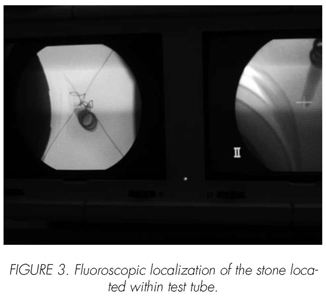
Prior to their surgery, all these patients were evaluated with a NCCT(1mm cuts) where the density of each stone was measured in Hounsfield Units.
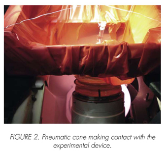
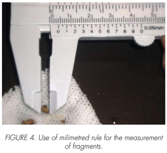
After surgery each stone was broken in two and one of the fragments was studied crystallographically and the other exposed to a standard in vitro ESWL session (3000 pulses, 17,2 KV, electromagnetic lithotripter (Lithostar Siemens*)). Abscence of fragmentation or presence of residual fragments larger than 5mm were considered treatment failures.
Results were stathistically analized with tehe Statistix 7.0 program, using Student test, Chi square test and multivariated analisys.
Results
Eleven out of the total studied stones were pure and 16 had a mixed composition. Table I shows distribution according to cristallographic composition.
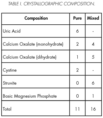
The average HU values of the different stone compositions were as following:
• Calcium Oxalate (monohydrate) pure and mixed: 1193 HU (range 496- 1865)
• Struvite: 1543 HU (790.-2143)
Calcium Oxalate (dihydrate) pure and mixed: 2122 HU (1853-2536)
• Uric Acid: 419 HU (65-769)
• Basic Magnesium Phosphate (1 single case): 1517 HU (Graphic 1).
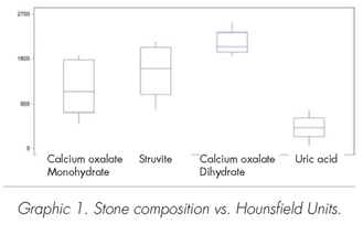

We observed that Uric Acid stones had the lowest UH values and that dihydrate Calcium Oxalate, the highest
ESWL in vitro was effective in 8 of nine stones with density lower than 1000 HU (81,8%) and only in 7 of sixteen (43,7%)of those with density values higher than 1000 HU.
Considering stone composition, only the Uric Acid stones demonstrated to respond significantly well to in vitro ESWL (Table II).
Analyzing pure and mixed stone composition it was observed that treatment was effective in 8 of eleven cases (72,7%) of pure stones and only in 8 of the 16 cases of the mixed stones (50%).
Discussion
ESWL success depends on the stone composition. If this composition can be diagnosed, the best therapeutic option can be offered in each case.
Since 1980, NCCT scan has been studied as a possible useful tool to predict stone composition through density measurements (Hounsfield Units) (Table III).
Newhouse et al (12) and Hillman et al (11) stated that NCCT scan could only differenciate the uric acid stones from the rest. On the other hand, Nakada et al (12) reasures that this instrument can identify uric acid and calcium oxalate stones (In this paper, the calcium oxalate average HU measured was 690, significantly lower than the value obtained in our series (1193 HU and 2122 HU for oxalate calcium stones, mono and dihydrate respectively). Table IV
Zarse et al, explains that the NTCT incapacity to identify stone composition could be due to:
1) 3-5mm slices are unable to distinguish different mineral components.
2) Commonly, soft tissue windows are used that increase stone brightness changing the HU values, therefore the results might not be so accurate.
3) The area selected to measure stone density is too wide and the value obtained is not representative considering the amount of mixed stones.
Nevertheless, HU are in vitro ESWL effectiveness predictors.
Gupta et al (2) concludes that stone density, not the size, predicts better ESWL effectiveness. A stone with less than 750 HU, should be treated with ESWL as first choice of treatment, no matter its size.
As well as Joseph et al (5), we observed that the best ESWL results are obtained with stone densities lower than 1000 HU.
Conclusion
Non contrast-enhanced CT scan predicts dihydrate Calcium Oxalate and Uric Acid stones.
ESWL success increases significantly with stones with a density lower than 1000 HU.
 Correspondence:
Patricio García Marchiñena
Centro de Litiasis
Servicio de Urologia
Hospital Italiano de Buenos Aires
Buenos Aires. (Argentina).
patricio.garcia@hospitalitaliano.org.ar
Accepted for publication: October, 21th 2008.
References and recomended readings (*of special interest, **of outstanding interest)
**1. Chaussy C, Brendel W, et al. Extracorporeally induced destruction of kidney stones by shock waves. Lancet, 1980; 2: 1265-68. [ Links ]
*2. Gupta N, Ansari M, et al. Role of computed tomography with no contrast medium enhancement in predicting the outcome of extracorporeal shock wave lithotripsy for urinary calculi. BJU Int, 2005; 95: 1285-88. [ Links ]
3. Wolf S. Treatment selection and outcomes: ureteral calculi. Urol Cl N Am, 2007; 34: 421-30. [ Links ]
4. Dretler S. Stone fragility - a new therapeutic distinction. J Urol, 1988; 139: 1124-27. [ Links ]
*5. Joseph P, Mandal A, et al. Computed tomography attenuation value of renal calculus: can it predict successful fragmentation of the calculus by extra-corporeal shock wave lithotripsy? A preliminary study. J Urol, 2002; 167: 1968-71. [ Links ]
**6. Pareek G, Armenakas N, et al. Extracorporeal shock wave lithotripsy success based on body mass index and hounsfield units. Urology, 2005; 65:33-36. [ Links ]
*7. Pareek G, Armenakas N, et al. Hounsfield units and computerized tomography predict stone-free rates after extracorporeal shock wave lithotripsy. J Urol, 2003; 169:1679-81. [ Links ]
8. Yoshida S, Hayashi T, et al. Role of volume and attenuation value histogram of urinary stone on non contrast helical computed tomography as predictor of fragility by extracorporeal shock wave lithotripsy. Urology, 2006; 68:33-37. [ Links ]
*9. Weld K, Montiglio C, et al. Shock wave lithotripsy success for renal stones based on patient and stone computed tomography characteristics. Urology, 2007; 70:1043-47. [ Links ]
**10. Newhouse J, Prien E, et al. Computed tomographic analysis of urinary calculi. Am J Roentgenol, 1984; 142: 549-52. [ Links ]
11. Hillman B, Drach G, et al: Computed tomographic analysis of renal calculi. Am J Roentgenol, 1984; 142: 549-552. [ Links ]
*12. Nakada S, Hoff D, et al. Determination of stone composition by noncontrast spiral computed tomography in the clinical setting. Urology, 2000; 55: 816-19. [ Links ]
*13. Zarse C, McAteer J, et al. Helical computed tomography accurately reports urinary stone composition using attenuation values: in vitro verification using high-resolution micro-computed tomography calibrated to fourier transform infrared microspectroscopy. Urology, 2004; 63: 828-33. [ Links ]
*14. Deveci S, Coskun M, et al. Spiral computed tomography: role in determination of chemical compositions of pure and mixed urinary stones - an in vitro study. Urology, 2004; 64:237-40. [ Links ]
15. Yoshida S, Hayashi, et al. Three-dimensional assessment of urinary stone on non-contrast helical computed tomography as the predictor of stones-treet formation after extracorporeal shock wave lithotripsy for stones smaller than 20 mm. Int J Urol, 2007; 14:665-67. [ Links ]
*16. El-Nahas A, El-Assmy A, et al. Aprospective multivariate analysis of factors predicting stone disintegration by extracorporeal shock wave lithtripsy: the value of high-resolution noncontrast computed tomography. Eur Urol, 2007; 51:1688-93. [ Links ]
17. Abdel-Khalek M, Sheir K, et al. Prediction of success rate after extracorporeal shock-wave lithotripsy of renal stones. A multivariate analysis model. Scand J Urol Nephrol, 2004; 38:161-7. [ Links ]
**18. Zarse C, Hameed T, et al. CT vesible internal stone structure, but not Hounsfield unit value, of calcium oxalate monohydrate (COM) calculi predicts lithotripsy fragility in vitro. Urol Res, 2007; 35:201-6. [ Links ]
19. Bellin M, Renard-Penna R, et al. Helical CT evaluation of the chemical composition of urinary tract calculi with a discriminant analysis of CT-attenuation values and density. Eur Radiol, 2004; 14:2134-40. [ Links ]
**20. Hurtado F, Gutierrez J, et al. In vivo relation between CT Attenuation Value and Shockwave fragmentation. J Endourology, 2007; 21:343-46. [ Links ]
*21. Oh K, Chae H, et al. Noncontrast CT in predicting the outcome of extracorporeal shock wave lithotripsy. Eur Urol Suppl, 2008; 7(3): 79. [ Links ]
22. Motley G, Dalrymple N, et al. Hounsfield unit density in the determination of urinary stone composition. Urology, 2001; 58: 170. [ Links ]
23. Lavin V, Pathak S, et al. Stone density determined by computed tomography: does it predict the success of extracorporeal shock wave lithotripsy?. Eur Urol Suppl, 2008; 7(3):79. [ Links ]
**24. Saw K, McAteer, et al. Calcium stone fragility is predicted by Helical CT attenuation values. J Endourol, 2000; 14: 471-74. [ Links ]
*25. Elbahanasy M, Elnady M, et al. The value of renal stone attenuation by non-contrast computed tomography in predicting fragmentation and stone free rate after extracorporeal shock wave lithotripsy. Eur Urol Suppl, 2008; 7(3):79. [ Links ]
**26. Mostafavi M, Ernst R, et al. Accurate determination of chemical composition of urinary calculi by spiral computerized tomography. J Urol, 1998; 159:673. [ Links ]

