


Meu SciELO
 Serviços customizados
Serviços Personalizados
Journal
Artigo
 texto em
  Inglês (pdf)
 Artigo em XML
 Referências do artigo
 Enviar este artigo por email
Indicadores
-
 Citado por SciELO
 -
 Acessos
Links relacionados
-
 Citado por Google
 -
 Similares em
SciELO
Similares em
SciELO -
 Similares em Google
Compartilhar

 Permalink
Revista Española de Enfermedades Digestivas
versão impressa ISSN 1130-0108
Rev. esp. enferm. dig. vol.97 no.11 Madrid Nov. 2005
| PICTURES IN DIGESTIVE PATHOLOGY |
Pancreatic intraductal papillary mucinous tumor
E. Marín Serrano, M. A. Macías Rodríguez, P. Rendón Unceta, J. Pérez Requena1, P. Guillén Mariscal and L. Martín Herrera
Services of Digestive Diseases and 1Pathology. Hospital Puerta del Mar. Cádiz, Spain
 |  |
A 56-year-old woman presented with belt-like radiated epigastric pain and elevated pancreatic enzymes (amilase: 850 U/L; lipase: 135 U/L). The patient had been cholecystectomized two years before after suffering from two acute episodes of pancreatitis of biliary origin. Since then, the patient reported postprandial epigastric discomfort.
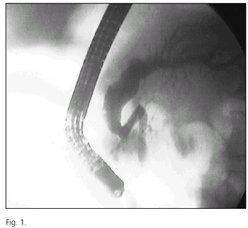
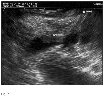
An abdominal ultrasonography showed a dilated Wirsung's duct with a nodular filling defect of 10.8 mm in size. ERCP demonstrated a severely dilated primary pancreatic duct and secondary branches at the pancreatic head, inner filling defects (Fig. 1), and a negative cytology for malignity. CT and MRI findings were suggestive of chronic pancreatitis. Echoendoscopy disclosed a heterogeneous pancreatic parenchyma and a dilated primary pancreatic duct that was maximal at the isthmus (7 mm), where a hypoechoic, 12.5-mm wall nodule was identified (Fig. 2).
The patient was then diagnosed with a pancreatic intraductal papillary mucinous tumor (IPMT), and underwent duodenopancreatectomy. Histology of the surgical specimen confirmed an intraductal papillary adenoma with no stromal invasion at the pancreatic isthmus. After 15 months of follow-up the patient remains symptom-free and has no evidence of tumor relapse.
IPMT is an uncommon pancreatic neoplasm that is difficult to diagnose because of its slow growth and non-specific manifestations. It has been primarily reported in males aged between 60 and 70 years (1,2). Most of these tumors are located in the pancreatic head. Their site is the primary pancreatic duct in 75% of cases, and they involve secondary ducts and even the papilla as they extend along the former duct (3). Although rare, suspicion should be raised by the presence of idiopathic pancreatitis with duct dilation, or of pancreatic cystic lesions (2).
Both ERPC and MRI detect pancreatic duct changes with a high sensitivity; however, IPMT-related findings may be difficult to differentiate from those of chronic pancreatitis (4). EUS is a safe technique for the diagnosis of IPMT, as it provides high-resolution images of the pancreatic duct, wall nodules, and neighboring structures, and is useful for loco-regional tumor staging (5).
The treatment of choice for pancreatic IPMT is surgical resection. Prognosis is excellent for non-invasive cases, but close patient surveillance is needed after surgery as tumor relapse is common (4,5).
REFERENCES
1. Loftus Jr. EV, Olivares-Pakzad BA, Batts KP, Adkins MC, Stephens DH, Sarr MG, et al. Intraductal papillary-mucinous tumors of the pancreas: clinicopathologic features, outcome, and nomenclature. Gastroenterology 1996; 110: 1909-18.
2. Azar C, Van de Stadt J, Rickaert F, Devière J, Delhaye M, Baize M, et al. Intraductal papillary mucinous tumours of the pancreas. Clinical and therapeutic issues in 32 patients. Gut 1996; 39: 457-64.
3. Farrell J.J, Brugge W.R. Intraductal papillary mucinous tumor of the pancreas. Gastrointest Endosc 2002; 55: 701-13.
4. Brugge W.R. Evaluation of pancreatic cystic lesions with EUS. Gastrointestinal Endoscopy 2004; 59: 698-707.
5. Sugiyama M, Atomi Y, Saito M. Intraductal papillary tumors of the pancreas: evaluation with endoscopic ultrasonography. Gastrointest Endosc 1998; 48: 164-71.

