


Meu SciELO
 Serviços customizados
Serviços Personalizados
Journal
Artigo
 texto em
  Inglês (pdf)
 Artigo em XML
 Referências do artigo
 Enviar este artigo por email
Indicadores
-
 Citado por SciELO
 -
 Acessos
Links relacionados
-
 Citado por Google
 -
 Similares em
SciELO
Similares em
SciELO -
 Similares em Google
Compartilhar

 Permalink
Revista de Osteoporosis y Metabolismo Mineral
versão On-line ISSN 2173-2345versão impressa ISSN 1889-836X
Rev Osteoporos Metab Miner vol.15 no.3 Madrid Jul./Set. 2023 Epub 08-Mar-2024
https://dx.doi.org/10.20960/revosteoporosmetabminer.00016
CASE REPORT
Heterotopic ossification after hip arthroplasty: role of bone SPECT/CT scintigraphy
1Department of Nuclear Medicine. Hospital Universitario Juan Ramón Jiménez. Huelva, Spain
2Department of Nuclear Medicine. Hospital Universitari Vall d’Hebron. Barcelona, Spain
3Department of Rehabilitation. Hospital Universitario Juan Ramón Jiménez. Huelva, Spain
Heterotopic ossification is a limiting condition that predominantly affects the hip. Because of its association with post-traumatic/postoperative pathology, bone SPECT/CT scintigraphy has proven to be especially useful regarding differential diagnosis involving prosthetic mobilization, even in the absence of radiological abnormalities. Additionally, it is an effective tool for surgical planning, considering the degree of bone maturation and the possibility of creating biomodels using 3D printing.
Keywords: Heterotopic ossification; Bone scintigraphy; SPECT/CT; Biomodel; 3D
CASE REPORT
This is the case of a 55-year-old man treated with right total hip arthroplasty (THA) two and a half years ago with persistent pain and limited mobility, but without any significant abnormalities according to the X-rayimages (Fig. 1). A three-phase bone scintigraphy using 99mTc-diphosphonates was requested to assess prosthetic mobilization. The early-phase images did not show any significant changes (Figs. 2 A and B). However, in the late bone phase, increased tracer uptake was seen in the right femur proximal third (Fig. 2C, arrow) that in the SPECT/CT images (Fig. 3) was consistent with an enhanced osteogenic activity in bone islands (up to 1.8 cm) inside the soft tissues adjacent to the greater trochanter. These findings are consistent with heterotopic ossifications (HO), and rule out the presence of right THA mobilization.

Figure 1. Plain hip X-ray after 2 years and 4 months following right THA implantation showing no signs of mobilization.
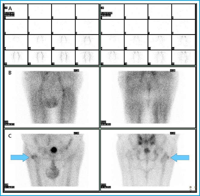
Figure 2. Anterior (left column) and posterior sections (right column) of bone scintigraphy, revealing no significant abnormalities to the early flow (A) and vascular pool phases (B). In the late bone phase (C), a focal and irregular increase in osteoblastic activity can be identified in the right femur proximal third (arrows).

Figure 3. Axial slice of fused SPECT/CT image (A), 3D reconstruction (B), and segmentation using 3Dslicer software (9) (C) of heightened osteoblastic activity (arrows) in periarticular heterotopic ossification that rules out the presence of significant right THA mobilization. The semiautomatic segmentation of the osteoblastic activity area could be a useful tool for surgical planning.
DISCUSSION
HO is a limiting condition that causes pain and reduced joint range of motion due to abnormal mature lamellar bone formation inside the soft tissues adjacent to periarticular bone (1,2). Of variable prevalence (ranging from 10 % to 53 %) (3), the hip joint is the most widely affected one. Despite its uncertain etiology, HO is associated with a previous congenital/post-traumatic/postoperative pathology that activates osteoblast and chondroblast progenitor cells, leading to calcium salt deposits inside the connective tissue (4). Also, former studies show the presence of elevated serum levels of inflammatory cytokines (TNF, IL-1, IL-6, and monocyte chemotactic protein), and alkaline phosphatase during the early phases of this bone formation that are also present in post-traumatic repair processes (5). Scintigraphy allows the diagnosis of HO early even before radiographic changes become apparent, and even before the findings become evident on the CT/MRI (6). Additionally, SPECT/CT acquisition is particularly useful to differentiate HO from prosthetic mobilization and myositis ossificans, and enable surgical planning through 3D printing of biomodels (7,8). Although the early management of HO is conservative, scintigraphy can provide insights on the degree of bone maturation, thus determining the optimal timing for surgery if indicated (9).
REFERENCES
1. Schmidt J, Hackenbroch MH. A new classification for heterotopic ossifications in total hip arthroplasty considering the surgical approach. Arch Orthop Trauma Surg 1996;115:339-43. DOI:10.1007/BF00420328 [ Links ]
2. Shehab D, Elgazzar AH, Collier BD. Heterotopic ossification. J Nucl Med 2002;43(3):346-53. [ Links ]
3. Romero-Muñoz LM, Barriga-Martin, A, DeJuan-García, J. Cirugía de la anquilosis de cadera por osificación heterotópica secundaria a lesión medular. Rev Esp Cir Ortop Traumatol 2018;62:458-66. DOI:10.1016/j.recot.2018.01.003 [ Links ]
4. García-Arpa M, Flores-Terry MA, Franco-Muñoz M, Villasanti-Rivas N, González-Ruiz L, Banegas-Illescas ME. Report of a man with heterotopic ossification of the legs. Reumatol Clin 2020;16:300-2. DOI:10.1016/j.reuma.2018.03.004 [ Links ]
5. Zagarella A, Impellizzeri E, Maiolino R, Attolini R Castoldi MC. Pelvic heterotopic ossification: when CT comes to the aid of MR imaging. Insights Imaging.2013;4:595-603. DOI:10.1007/s13244-013-0265-5 [ Links ]
6. Purcell KF, Lachiewicz PF. Heterotopic Ossification After Modern Total Hip Arthroplasty: Predisposing Factors, Prophylaxis, and Surgical Treatment. J Am Acad Orthop Surg 2023;31:490-6. DOI:10.5435/JAAOS-D-22-01070 [ Links ]
7. Ballard DH, Wake N, Witowski J, Rybicki FJ, Sheikh A;RSNA Special Interest Group for 3D Printing Abdominal, Hepatobiliary, and Gastrointestinal Conditions Voting Group. Radiological Society of North America (RSNA) 3D Printing Special Interest Group (SIG) clinical situations for which 3D printing is considered an appropriate representation or extension of data contained in a medical imaging examination: abdominal, hepatobiliary, and gastrointestinal conditions. 3D Print Med 2020;8:6-13. DOI:10.1186/s41205-020-00065-6 [ Links ]
8. Van den Wyngaert T, Paycha F, Strobel K, Kampen WU, Kuwert T, van der Bruggen W, et al. SPECT/CT in Postoperative Painful Hip Arthroplasty. Semin Nucl Med 2018;48:425-38. DOI:10.1053/j.semnuclmed.2018.05.002 [ Links ]
9. Nieto Morales ML, Lara Martínez MF, Luna Gómez C, Bello Báez A, Allende Riera AJ. Osificación heterotópica en paciente con SARS-CoV-2: imágenes gammagráficas y radiológicas [Heterotopic ossification in SARS-CoV-2:Scintigraphic and radiological images]. Rehabilitacion 2022;56:399-403. DOI:10.1016/j.rh.2021.09.003 [ Links ]
10. Fedorov A, Beichel R, Kalpathy-Cramer J, Finet J, Fillion-Robin J-C, Pujol S, et al. 3D Slicer as an Image Computing Platform for the Quantitative Imaging Network. Magnetic Resonance Imaging 2012;30:1323-41. DOI:DOI:10.1016/j.mri.2012.05.001 [ Links ]
Moreno-Ballesteros A, de Bonilla-Candau M, Cabaleiro-Burguillos B, Custodio Rebollo-Aguirre Á, Sánchez-de Mora E, Jiménez-Heffernan A. Heterotopic ossification after hip arthroplasty: role of bone SPECT/CT scintigraphy. Rev Osteoporos Metab Miner 2023;15(3):125-128
Received: May 04, 2023; Accepted: July 10, 2023
 This is an open-access article distributed under the terms of the Creative Commons Attribution License
