


Meu SciELO
 Serviços customizados
Serviços Personalizados
Journal
Artigo
 texto em
  Inglês (pdf)
 Artigo em XML
 Referências do artigo
 Enviar este artigo por email
Indicadores
-
 Citado por SciELO
 -
 Acessos
Links relacionados
-
 Citado por Google
 -
 Similares em
SciELO
Similares em
SciELO -
 Similares em Google
Compartilhar

 Permalink
Medicina Oral, Patología Oral y Cirugía Bucal (Internet)
versão On-line ISSN 1698-6946
Med. oral patol. oral cir.bucal (Internet) vol.11 no.3 Mai./Jun. 2006
Removal torque and physico-chemical characteristics of dental implants etched with hydrofluoric and nitric acid. An experimental study in Beagle dogs
Torque de desinserción y propiedades fisico-químicas de implantes dentales grabados con ácidos fluorhídrico y nítrico. Estudio experimental en perros Beagle
José María Martínez González 1, Francisco García Sabán 2, Javier Ferrándiz Bernal 2,
Juan Carlos Gonzalo Lafuente 2, Jorge Cano Sánchez 2, Cristina Barona Dorado 3
(1) Assistant Professor of Maxillofacial Surgery. Madrid
Complutense University. Dental School. Madrid (Spain)
(2) Implantologist in private practice
(3) Collaborating Professor in Oral Surgery. Madrid
Complutense University. Dental School. Madrid (Spain)
ABSTRACT
Objective: To study the composition, surface
characteristics and response to removal torque of an implant surface subjected
to hydrofluoric acid etching and posterior passivating with hydrofluoric and
nitric acid.
Study design: Twelve implants were initially selected and
their physico-chemical characteristics were evaluated by means of energy-dispersive
X-rays (EDS), scanning electron microscopy (SEM) and photoelectron spectroscopy
(XPS). In addition, 24 implants 12 measuring 8 mm and 12 measuring 10 mm in
length were implanted in 6 Beagle dogs. Twelve implants were removed after a
recovery period of 6 weeks, followed by removal of the remaining 12 implants
after 12 weeks, using a torque calibrator (Gauge Tonichi® model
BGT150CN-S) with a force registry range of 0-150 Ncm.
Results:
EDS analysis of the surface chemical composition
only revealed the presence of titanium in the etched surfaces. In the same way
as with the surfaces of other dental implants, XPS analysis revealed traces of
other elements present in the surface, fundamentally carbon. Following
dual acid etching, the surface showed the roughness resulting from acid action,
with a morphology that proved to be quite homogeneous. The roughness values
obtained exceeded 1 mm.
The mean removal torque values after 6 weeks were 79.7 Ncm for the 8 mm implants
and 115 Ncm for the 10 mm implants. After 12 weeks, these values increased to
101.2 Ncm and 139.7 Ncm, respectively.
Conclusions:
Key words: Dental implants, surface treatment, hydrofluoric and nitric acid, roughness, removal torque.
RESUMEN
Objetivo: Estudiar la composición, características
superficiales y respuesta al torque de desinserción de una superficie
implantaria tratada inicialmente con ácido fluorhídrico y posterior pasivado con
ácidos fluorhídrico y nítrico.
Diseño del estudio: En una primera fase, se seleccionaron
12 implantes en los que se estudiaron las características fisico-químicas
mediante mediciones de energía dispersa de rayos X (EDS), microscopio
electrónico de barrido y análisis de XPS (espectrometría de fotoelectrones).
Asimismo, se colocaron 24 implantes doce de 8 mm y doce de 10 mm de longitud-,
en seis perros beagle, en los que tras un período de reposo, se procedió a la
retirada de 12 implantes a las seis semanas y los 12 restantes a las doce
semanas, mediante un calibrador de torque Gauge TonichiR modelo
BGT150CN-S -con un rango de registro de fuerza de 0 a 150 Ncm.
Resultados: El análisis de la composición química
superficial mediante EDS sólo mostró la presencia de titanio en las superficies
grabadas. En el análisis mediante XPS, al igual que sucede con las superficies
de otros implantes dentales, aparecieron trazas de otros elementos presentes en
la superficie, fundamentalmente de carbono.
La morfología de la superficie tras el doble grabado con
ácido, permitió observar la rugosidad creada por el ataque ácido, con una
morfología bastante homogénea. Los valores de rugosidad obtenidos fueron
superiores al micrómetro.
Los valores medios encontrados para el torque de desinserción,
a las seis semanas, fueron de 79,7 Ncm para los implantes de 8 mm de longitud y
115 Ncm para los implantes de 10 mm. A las doce semanas, estos valores
incrementaron hasta 101,2 Ncm para los implantes de 8 mm y 139,7 Ncm para los
implantes de 10 mm de longitud.
Conclusiones: El grabado con ácido fluorhídrico y
nítrico, posee características superficiales óptimas y comparables al de otras
superficies. Los valores de torque de desinserción abren la posibilidad para su
aplicación en clínica humana para procedimientos de carga precoz o inmediata.
Palabras clave: Implantes dentales, tratamiento superficial, ácidos fluorhídrico y nítrico, rugosidad, torque de desinserción.
Introduction
The concept of osseointegration proposed by Branemark more than 30 years ago has been the reference point for the use of osseointegrated implants as rehabilitation treatment for partially and totally edentulous patients (1).
The initial principles proposed by this author have since undergone a series of transformations due to the development of new implant designs, surface treatment modalities, and improved knowledge of bone biology.
Of the key factors underlying effective osseointegration, mention must be made of the characteristics of the material used, the design of the implant, and the type of surface. At present, the use of titanium implants with a threaded macrostructure and rough surface is widely accepted (2).
As regards the quantitative assessment of osseointegration, the scientific literature offers different studies that compare integration using biomechanical tests - including the determination of removal torque (3-6). Such testing is carried out in experimental animal models the rabbit tibia and femur being the most frequent bone components cited in the literature, followed by Beagle dogs and, to a lesser extent, minipigs, goats and monkeys. In most such studies the typical post-implantation recovery time is three months, or even as little as 1-2 months. This short healing period prior to assessment of the different surface characteristics reflects the current tendency in implantology of shortening the time to implant loading.
Different instruments are used to measure removal torque, expressed in Ncm - one of the most commonly used devices being the Tonichi torque calibrator (MFG Co., Ltd, Japan).
The present study explores the physico-chemical characteristics (composition, morphology and roughness) of an implant surface treated with an aqueous solution of hydrofluoric acid, followed by passivating of the surface with a solution of hydrofluoric and nitric acid, and determination of the corresponding removal torque values.
Material and methods
The present study made use of 12 Defcon TSA® implants (Impladent S.L., Sentmenat, Spain) with a rough surface prepared by hydrofluoric acid etching with posterior passivating using a combination of hydrofluoric and nitric acid.The chemical composition of the titanium was assessed using a flame and combustion atomic absorption system (LECO) capable of determining the quantitative chemical composition of the interstitial elements contained in the titanium including hydrogen with a sensitivity in the parts per million (ppm) range. The surface chemical composition was determined by an energy-dispersive X-ray (EDS) system (EDS Link-Inca) connected to a scanning electron microscope (SEM)(Leica Electroscan 360)(Servicio cientifico-técnico, Barcelona, Spain). This system is able to detect atoms with an atomic weight equal to or greater than that of boron, and allows semiquantitative determination of the composition of a surface within a thickness range of about 1 µm with high lateral resolution.
Photoelectron spectroscopy (XPS) in turn was carried out with a Physical Electronics 5500 system operating with a X-ray monochromator equipped emission source in the K-band of aluminum, in an ultrahigh vacuum of 5.10-9 mmHg (0.6.10-6 Pa). The detection angle was 90º for all samples.
Roughness was measured in three dimensions (3D), using a Sensofar® Plu white-light confocal microscope (Sensofar, Terrassa, Spain). The measurements were made using a Nikon L 150 microscope with an SL WD20x eyepiece, vertical resolution < 20 nm, and lateral resolution 0.91 m. Based on the measurements obtained with the confocal microscope, calculations were also made of profile roughness with a Gaussian filter and cut-off value of 800 µm according to DIN 4768 specifications.
At the same time a study was made involving 6 Beagle dogs in the animal experimentation center of Gómez Ulla Hospital (Madrid, Spain), with adherence to the specifications of royal Decree 223/1998, regarding the protection of animals used for experimentation purposes.
For the evaluation of removal torque, surgery was carried out under general anesthesia (induction with medetomidine (20-40 mg/kg), butorphanol (0.2-0.4 mg/kg) and 0.5 ml atropine; maintenance with isoflurane, protoxide and oxygen). During the operation, each animal was administered 2 ml of Vetione® (penicillin G) and 0.5 ml of Flunixin Meglumine® (a nonsteroidal antiinflammatory drug).
Twenty-four Defcon TSA® implants were placed in the internal aspect of the tibia of the animals (two in each limb), with the following distribution: 12 implants measuring 10 mm in length in the proximal tibial epiphysis; and 12 implants measuring 8 mm in length in the distal tibial epiphysis.
A tibial incision was made in the aforementioned locations, with raising by layers and using the drilling sequence recommended by the manufacturer. The bed was prepared and the implant positioned under physiological saline irrigation. Posteriorly, the implant was left submerged, followed by layered suturing with Vicryl® 2/0 for the periosteum and single-strand nylon 3/0 for the skin.
In a second surgical step 6 weeks later, and following anesthesia of three of the animals, an incision was again made in the implant zone, with raising by layers until the heads of the 12 implants were exposed. After 12 weeks the same procedure was carried out with the remaining 12 implants in the other three animals.
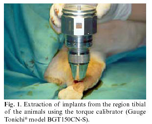
In both study intervals the locking screw was removed, and the torque calibrator (Gauge Tonichi® model BGT150CN-S, force registry range 0-150 Ncm) was positioned in the most coronal portion and in the same direction as the implant axis, for measurement of the removal torque (Fig. 1).
Results
The chemical composition of the material from which the implants were machined is reported in Table 1, compared with the composition limits specified by the ISO 5832-2 standard for grade 2 titanium. All the requirements for clinical implantation were seen to be met.
EDS analysis of the surface chemical composition only revealed the presence of titanium in the etched surfaces. In the same way as with the surfaces of other dental implants, XPS analysis revealed traces of other elements present in the surface, fundamentally carbon (Table 2).
Following dual acid etching, the surface showed the roughness resulting from acid action, with a morphology that proved to be very characteristic and quite homogeneous, in the form of blocks attributable to preferential hydrofluoric acid action (Fig. 2).
The mean roughness values of the etched implant surfaces are shown in Table 3, along with the mean roughness values measured for the machined surfaces before treatment. In order to allow measurements according to the defined standard, the calculated values for a linear profile were included in Table 4. In both cases, the roughness values obtained, Ra and Sa, exceeded 1 µm. The mean distance between peaks (Sm) was likewise within the adequate range (8.06) thus allowing for optimum osteoblast anchoring.
The mean removal torque values after 6 weeks were 79.7 Ncm for the 8 mm implants (range 75-90 Ncm) and 115 Ncm for the 10 mm implants (range 105-120 Ncm). During removal after 12 weeks, these values increased to 101.2 Ncm (range 86-120 Ncm) and 139.7 Ncm (range 116-150 Ncm), respectively (Fig. 3).
Discussion
Implant surfaces are the subject of continuous studies to favor increasingly faster and safer clinical consolidation of the implants. At present, machined or additioned surfaces clearly have been surpassed by newer techniques (7). Among the latter, special mention should be made of sandblasting procedures with or without etching (SLA and Tioblast surfaces), anodic oxidation (TiUnite surface), and acid etching (Osseotite surface).
While the SLA surface uses acids such as hydrochloric and sulfuric acid, the Osseotite surface involves first etching with hydrofluoric acid, followed by second etching with a combination of hydrochloric and sulfuric acid.
In this same line, the present study evaluates the results obtained with a surface initially treated with an aqueous solution of hydrofluoric acid, followed by chemical passivating with hydrofluoric and nitric acid.
The initial studies conducted to determine the composition of the titanium used confirmed that the latter satisfies the requirements for implantological purposes.
Photoelectron spectroscopy identified the presence of carbon and other elements in lesser proportions. This is also observed in surfaces corresponding to other implants, and according to Massaro et al. (8) is attributable to surface hydrocarbon contamination secondary to atmospheric exposure, that tends to disappear after light sanding for one minute.
Etching with hydrofluoric and nitric acid increases surface roughness compared with machined surfaces this being one of the fundamental principles for ensuring the improved osseointegration of the current surfaces (9).
The quantitative results obtained regarding roughness coincide with the requirements proposed by other authors such as Rodríguez (10), Martin (11), Wennenberg (12), Orsini (13) and Peñarrocha (14), who advocated values in excess of 1 µm to ensure good osteoblastic stability.
The application of removal torque testing for implant extraction in turn provides information on the percentage bone-implant contact. According to the literature, one of the main inconveniences of this technique is that test performance and the values recorded are not very homogeneous, due to differences between the animals used in each experimental series.
In 1997, Klokkevold (15) in the rabbit femur studied the biomechanical behavior of implants after 8 weeks. The implant surfaces had been subjected to acid etching (hydrochloric / sulfuric acid; Osseotite surface), and comparisons were made with smooth-surfaced implants. The results showed four-fold higher removal torque values for the etched surfaces (20.30 Ncm versus 4.85 Ncm in the case of smooth surfaces).
Among other surfaces, Cordioli et al. (4) compared those obtained by sandblasting with titanium oxide (Tioblast) versus acid-etched surfaces (Osseotite) in a study involving rabbit tibias. Twelve implants corresponding to each type of surface were placed, and the results relating to removal torque were evaluated after 5 weeks. The values obtained were significantly greater for the acid-etched surfaces (Osseotite: 40.85 Ncm, versus titanium oxide sandblasted surfaces: 26.85 Ncm). The histomorphometric study made parallel to the torque tests corroborated these findings.
Gottlow et al. (16) studied the TiUnite surface with acid etching (Osseotite) in the rabbit tibia, and after 6 weeks recorded superior removal torque values for the TiUnite surface (35 Ncm versus 25 Ncm).
In our study, the results obtained after 6 weeks ranged from mean values of 79.7 to 115 Ncm for the implants measuring 8 and 10 mm in length, respectively. In the case of implant removal after 12 weeks, these figure increased to mean values of 101.2 and 139.7 Ncm.
These results are clearly superior to those reported to date, and are justified by the use of animals with a larger bone volume thus allowing the use of surfaces of greater length and width. In this context, our findings may be correlated to those of the first studies by Buser et al. (17) in 1998, involving the SLA surface in comparison with smooth surfaces in minipigs. These authors recorded removal torque values 8 to 10 times greater for the SLA surface (139 Ncm) versus the smooth surfaced implants (13-26 Ncm).
These data indicate that etching with hydrofluoric and nitric acid may be included among the new implant surface treatments, with optimum results comparable to those afforded by other surfaces, and paving the way for possible clinical application in the context of early and immediate implant loading.
![]() Correspondence:
Correspondence:
Prof. José María Martínez González
Facultad de Odontología.
Ciudad Universitaria.
28040. Madrid
E-mail: jmargo@odon.ucm.es
Received: 30-04-2005
Accepted: 30-11-2005
References
1. Branemark P-I, Zarb GA, Albrektson T. Tissue-integrated prostheses. Osseointegration in clinical dentistry. Germany: Quintessence books Edit; 1990. p. 11-71. [ Links ]
2. Sykaras N, Lacopino AM, Marker VA, Triplett RG, Woody RD. Implant materials. Designs, and surface topographies: their effect on osseointegration. A literature review. Int J Oral Maxillofac Impl 2000;15:675-90. [ Links ]
3. Gotfredsen K, Nimb L, Hjorting-Hansen E, Jensen JS, Holmen A. Histomorphometric and removal torque analysis of titanium implant blasted Ti O2. Clin Oral Impl Res 1992;3:77-84. [ Links ]
4. Cordioli G, Majzoub Z, Piatelli A, Scarano A. removal torque and histomorphometric investigation of 4 different titanium surfaces: An experimental study in the rabbit tibia. Int J Oral Maxillofac Impl 2000; 15:668-74. [ Links ]
5. Klokkevold P, Johnson P, Dadgostari S, Caputo A, Davies J, Nishimura R. Early endosseous integration enhanced by dual acid etching of titamium: a torque removal study in the rabbit. Clin Oral Impl Res 2001; 12:350-7. [ Links ]
6. Piatelli A, Manzon L, Scarano A, Paolantonio M, Piatelli M. Histologic and histomorphometric analisis of the bone response to machined and sandblasted titanium implants: And experimental study in rabbits. Int J Oral Maxillofac Impl 1998;13:805-10. [ Links ]
7. Cano Sánchez J, Martínez-González JM, Gonzalo Lafuente JC, Cantero Álvarez M, Barona Dorado C. Superficie de los implantes dentales: estado actual. Quintessence 2004;5:301-8 [ Links ]
8. Massaro C, Rotolo P, de Riccardis F, Milella E, Napoli A, Wieland M et al. Comparative investigation of the surface properties of comercial titanium dental implants. Part I:chemical composition. J Mat Sci: Mater in Med 2002;13:536-48. [ Links ]
9. Martínez-González JM, Cano J, Campo J, Martínez MJS, García-Sabán F. Diseño de los implantes dentales: Estado actual. Av Periodon Implantol 2002;14:129-36 [ Links ]
10. Rodríguez Rius D, García Sabán F. Caracterización fisico-química de la superficie de 9 implantes dentales con 3 distintos tratamientos de superficies. Med Oral Pat Oral Cir Bucal 2005;10:58-65. [ Links ]
11. Martin JY, Schwartz Z, Hummert TW, Schraub DM, Simpson J, Lankford J et al. Effect of titanium surface roughness on proliferation, differentation, and protein synthesis of human osteoblast-like cells. J Biomed Mater Res 1995;29:389-401. [ Links ]
12. Wennenberg A, Ektessabi A, Albrektsson T, Johansson C, Andersson B. A 1-year follow-up of implants of differing surface roughness placed in rabbit bone. Int J Oral Maxillofac Impl 1997;12:486-94. [ Links ]
13. Orsini G, Assenza B, Scarano A, Piattelli M Piattelli A. Surface analysis of machined versus sandblasted and acid-etched titanium implants. Int J Oral Maxillofac Impl 2000;15:779-84. [ Links ]
14. Peñarrocha M, Guarinos J, Sanchís JM, Balaguer J. A retrospective study (1994-1999) of 441 ITI ® implants in 114 patients followed-up during an average of 2.3 years. Med Oral 2002;7:144-55 [ Links ]
15. Klokkevold P, Nishimura R, Adachi M, Caputo A. Osseointegration enhanced by chemical etching of the titanium surface. A torque removal study in the rabbit. Clin Oral Impl Res 1997;8:442-7 [ Links ]
16. Gottlow J, Johansson C, Albrektson T, Lundgren AK. Biomechanical and histologic evaluation of the TiUnite and Osseotite implant surfaces in rabbits afters 6 weeks of healing. Applied Ossointegrated Research 2000;1:255-77. [ Links ]
17. Buser D, Nydegger T, Hirt P, Cochran D, Nolte L. Removal torque values of titanium implants in the maxilla of miniature pigs. Int J Oral Maxillofac Impl 1998;13:611-9. [ Links ]

